Case 1 : Oral Lichen Planus
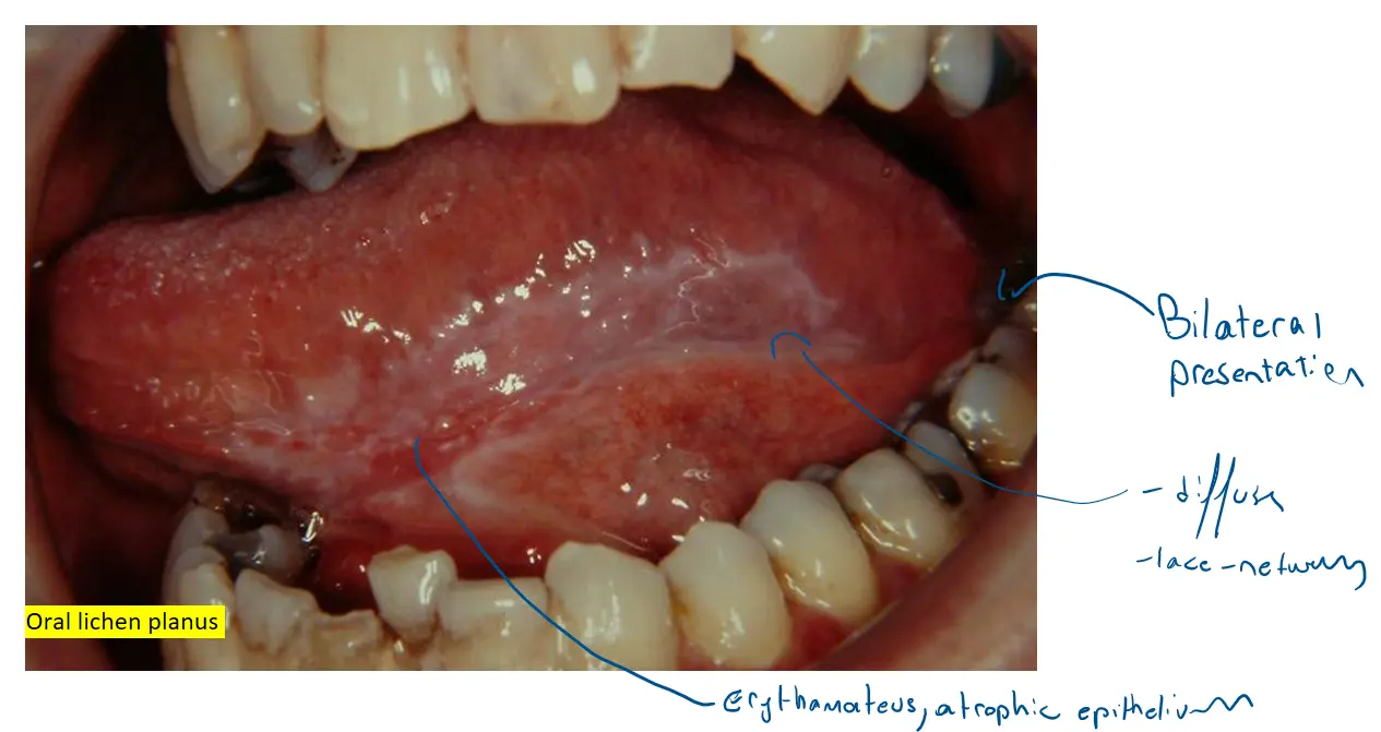
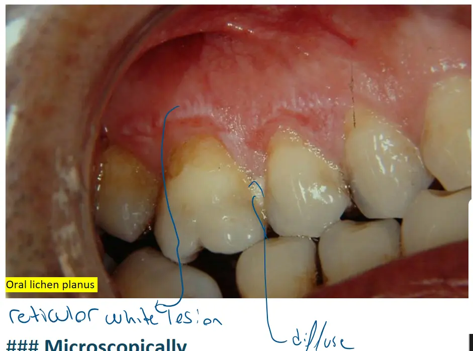
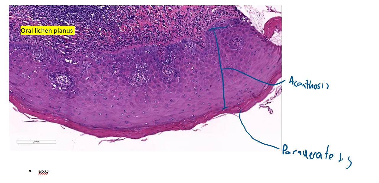
Of course. These slides present a textbook example of oral lichen planus. This is a chronic autoimmune condition that causes inflammation at the interface between the oral mucosa’s epithelium and the underlying connective tissue.
Let’s locate each of the features you’ve listed on your slides.
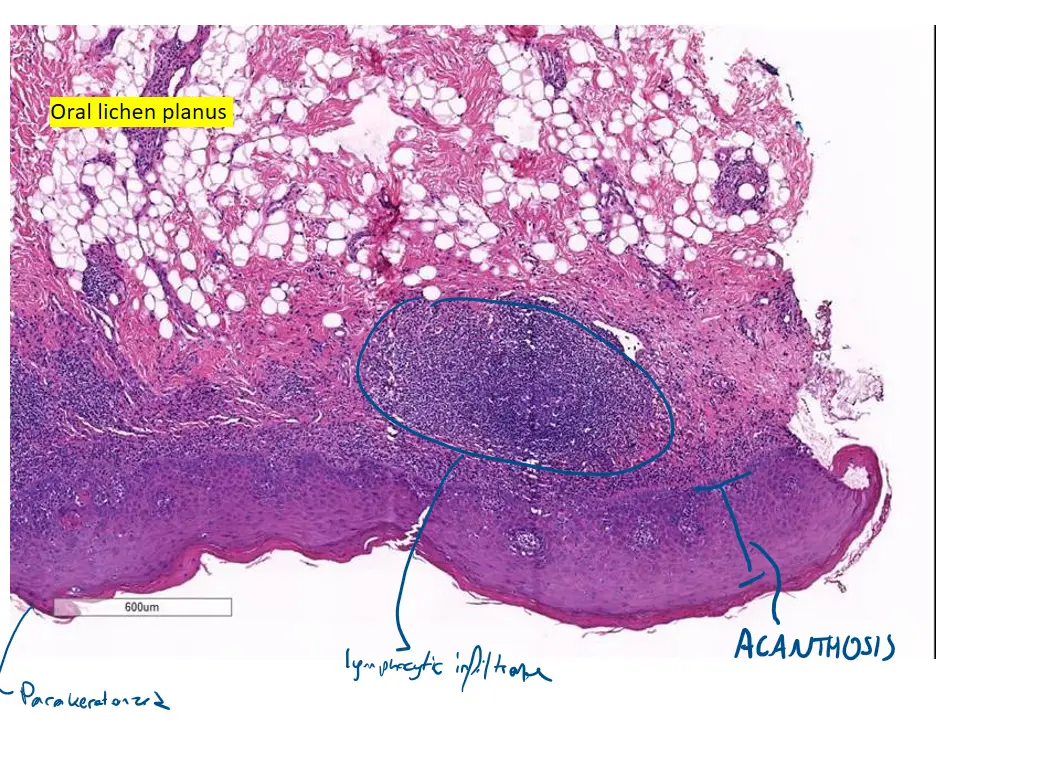
🔬 Low Power View (Top Image)
This view is excellent for observing the overall architecture of the lesion.
-
Band-like lymphocytic infiltrate at the interface: This is the most striking feature at low power. Notice the dense, continuous, dark purple band of inflammatory cells that runs horizontally across the slide. It is sharply defined and is located directly underneath the pink epithelium, “hugging” its base.
-
Acanthosis: The entire pink epithelial layer is clearly thickened. The downward projections of the epithelium, known as rete ridges, are often described as having a pointed, “saw-tooth” appearance in lichen planus, which you can appreciate here.
🔬 Medium & High Power Views (Middle and Bottom Images)
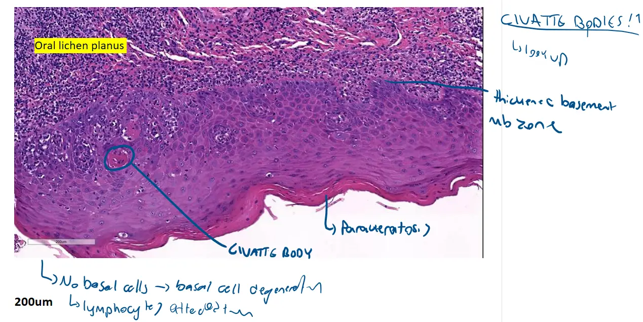
These closer views are perfect for examining the details at the crucial interface zone.
-
Degenerated basal cells: This is a key diagnostic feature. The dense lymphocytic infiltrate attacks the bottom layer of the epithelium (the basal cells). As a result, the normally neat, single row of basal cells is disrupted, damaged, and difficult to identify. This process is called liquefactive degeneration. You can also sometimes see small, round, bright pink dead keratinocytes, known as Civatte bodies, at the interface.
-
Parakeratosis: Look at the very top surface of the epithelium. You can see small, dark, flattened nuclei retained within the most superficial keratin layer. In normal oral mucosa (orthokeratosis), this layer would be pure pink with no nuclei.
-
Thickened basement membrane zone: The chronic inflammation and damage can cause the basement membrane (the thin line separating the epithelium from the connective tissue) to thicken, though this is often subtle and better visualized with special stains.
In summary, your images perfectly demonstrate the defining features of oral lichen planus: a dense, band-like infiltrate of lymphocytes causing damage to the basal cell layer, which in turn leads to reactive changes like acanthosis and parakeratosis in the overlying epithelium.
Would you like to discuss the clinical appearance of oral lichen planus or its potential causes?
Case 2 : Capillary Hemangioma
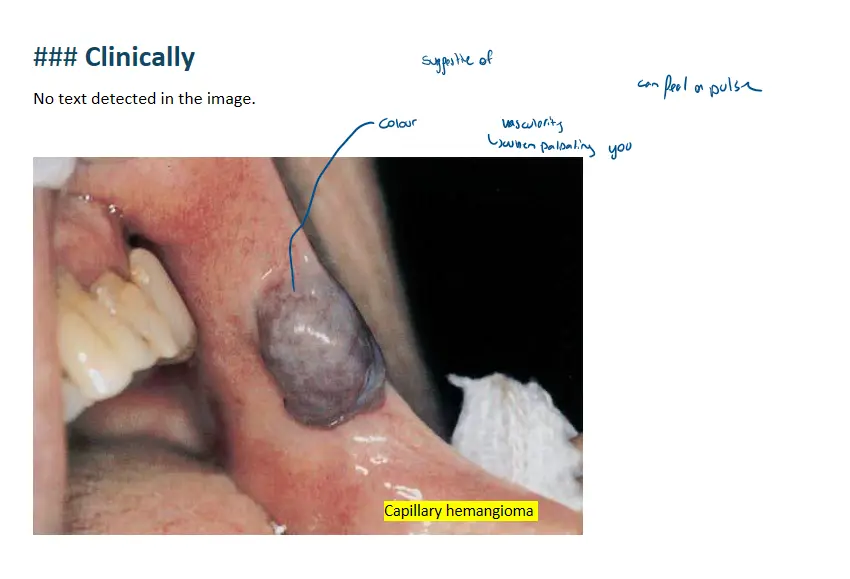
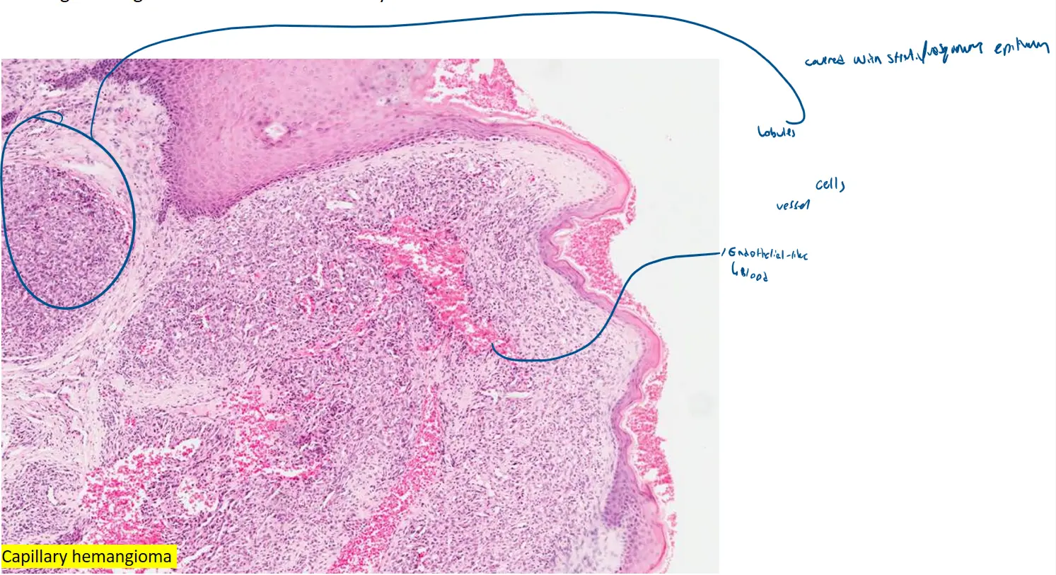

Based on the provided images of a capillary hemangioma, you can find the key features in the following locations:
🔬 High-Magnification View (Bottom Image)
This image is the best for observing the fine details.
-
Small Capillaries: The small capillaries are the numerous, small, circular, or irregularly shaped spaces that are filled with pinkish-red material (red blood cells). Look for these throughout the entire field of view. They vary in size and shape but are generally small and clustered together.
-
Single Layer of Endothelial Cells: Each of these capillary spaces is lined by a single layer of endothelial cells. These cells are characterized by their flattened, elongated, and dark purple nuclei. You can see these purple nuclei forming a thin border directly around the red-filled capillary lumens. In some areas, the endothelial cells may appear plumper, but they still form a single, distinct layer.
-
Connective Tissue Stroma: The connective tissue stroma is the pink, fibrous material that surrounds and supports the entire network of capillaries. It contains scattered, spindle-shaped nuclei of fibroblasts and other stromal cells. This stroma acts as the “scaffolding” in which the capillaries are embedded.
🔬 Low-Magnification View (Top Image)
This view shows the overall architecture. While the individual cells are less clear, you can still identify:
-
The dense proliferation of small capillaries, which collectively make up the pink and purple nodules of the hemangioma.
-
Areas of hemorrhage (the brighter red patches) where blood has leaked out of the capillaries.
-
The entire lesion is supported by the connective tissue stroma, which is the background pink tissue holding the vascular structures together.
In summary, to see the specific features you asked about, focus on the bottom, higher-magnification image. Identify the small, blood-filled circles (capillaries), note the thin purple nuclei lining them (endothelial cells), and recognize the surrounding pink tissue as the supportive stroma.