Case 1
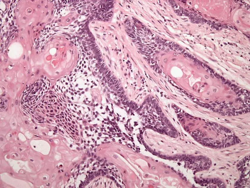
Of course. This image shows the classic microscopic features of an ameloblastoma, a benign but locally aggressive tumor of odontogenic (tooth-forming) origin.
🔬 Annotated Histology
Let’s walk through the features one by one:
-
Islands of Odontogenic Epithelium in Fibrous Connective Tissue:
-
The distinct, purple, multi-lobed structures are the islands of odontogenic epithelium (the tumor cells).
-
These islands are embedded in the surrounding pink, less cellular material, which is the fibrous connective tissue stroma.
-
-
Peripheral Palisading & Hyperchromatic Nuclei:
-
Look at the outermost layer of the epithelial islands. You’ll see that the cells are tall and elongated (columnar).
-
They are arranged in a neat, fence-like row, which is called peripheral palisading.
-
Their nuclei (the dark purple dots within the cells) are intensely stained, which is what hyperchromatic means.
-
-
Reverse Polarization:
-
This feature is also seen in the outer palisading cells. Normally, the nucleus of a columnar cell sits at the base, near the connective tissue.
-
Here, the nuclei are positioned at the end of the cell that is away from the basement membrane (the border with the pink connective tissue). This is known as reverse polarization.
-
-
Suprabasal Cells (Stellate Reticulum-like):
-
Now, look at the central area within the epithelial islands, just inside the palisading layer.
-
The cells here are loosely arranged and appear star-shaped, forming a delicate, web-like network. This morphology is a recapitulation of the stellate reticulum, a group of cells found in the developing tooth enamel organ.
-
In summary, the image perfectly illustrates the classic “follicular” pattern of ameloblastoma, with nests of epithelium showing palisading, reverse polarization, and central stellate reticulum-like cells.
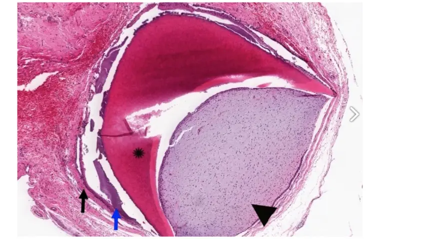 Of course. This image displays the microscopic features of an odontoma, which is a benign tumor composed of disorganized but mature dental tissues.
As the description states, it’s histologically similar to the layering of a normal tooth. Here’s where you can find those layers on your image:
🦷 Identifying the Tooth Tissues
The pointers on your image highlight the key components that mimic a developing tooth:
-
Pulp (Black Arrowhead): The area indicated by the black arrowhead is the dental pulp. You can see it’s a cellular, soft connective tissue, just like the pulp found in the center of a normal tooth.
-
Enamel Matrix (Asterisk): The dark pink, acellular (cell-free) hard tissue marked with the asterisk (*) is the enamel matrix.
-
Dentine: While not explicitly marked with a symbol, dentine is the hard tissue that would be located between the pulp and the enamel. In this image, it would be the layer of pink material immediately adjacent to the pulp-like tissue.
-
Odontogenic Epithelium (Blue Arrow): The blue arrow points to the layer of epithelial cells responsible for producing the enamel.
-
Connective Tissue Capsule (Black Arrow): The black arrow indicates the outer fibrous connective tissue that encloses the entire structure.
In essence, you are looking at a disorganized, tooth-like structure with a central pulp, surrounded by layers of dentine and enamel, all contained within a fibrous sac.
Would you like to know more about the difference between a complex and a compound odontoma?