 Of course. Here is the corrected explanation for that image.
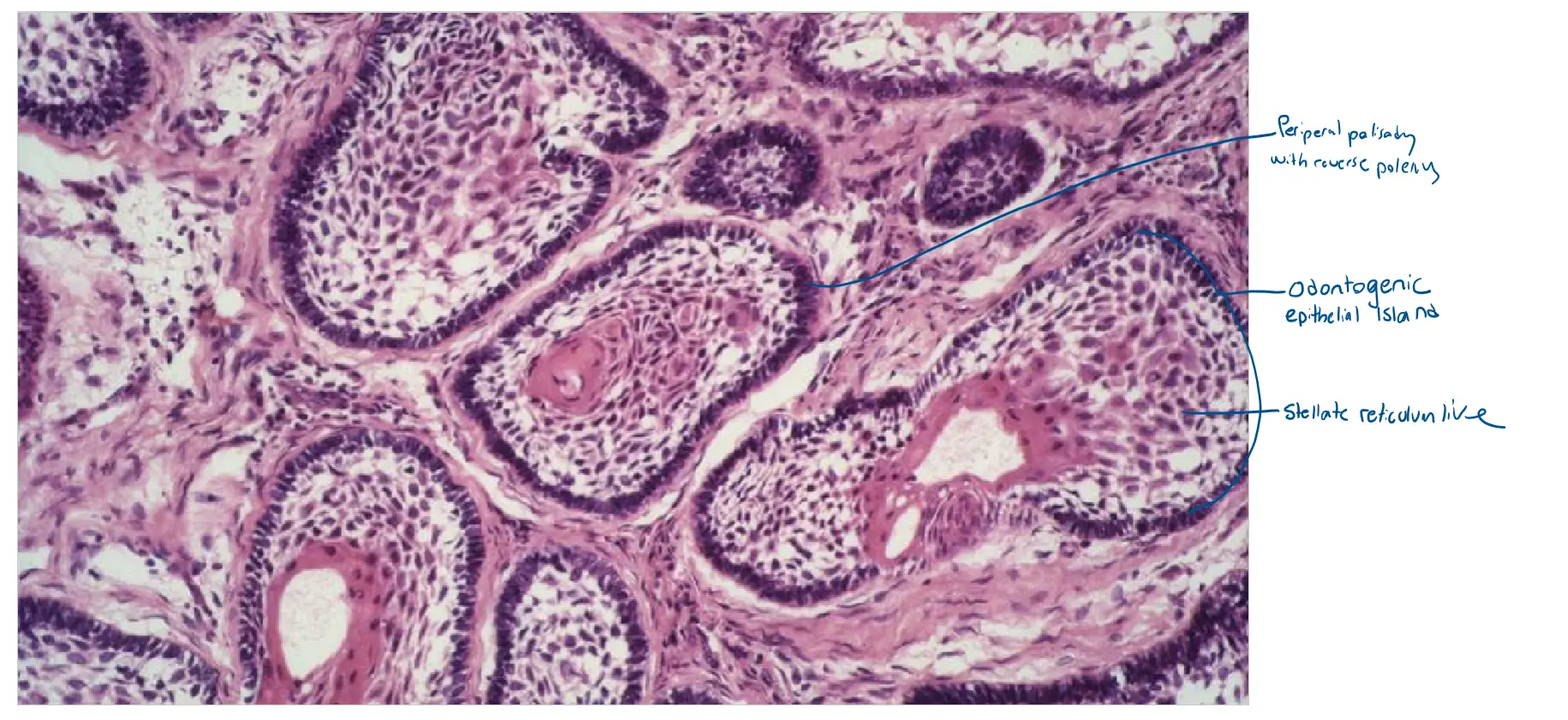
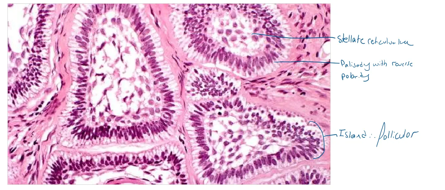
You are correct; the image shows an ameloblastoma where the primary architectural pattern is the follicular subtype. The other features present are specific changes occurring within that follicular framework.
🔬 Primary Follicular Pattern
The tumor’s fundamental structure is follicular, which is the most common subtype of ameloblastoma. You can see this in the overall architecture:
-
Odontogenic Epithelial Islands: The tumor is composed of numerous, well-defined islands of epithelial cells.
-
Peripheral Palisading: The outer border of these islands is lined by a single layer of tall, columnar cells with hyperchromatic (dark) nuclei, arranged neatly like a fence.
-
Stellate Reticulum-like Cells: The central core of the islands contains a loose, web-like network of cells that mimics the stellate reticulum of a developing tooth.
Acanthomatous Features
Now, looking closely at the island in the center of the image, you can see secondary changes that are described as “acanthomatous”:
-
Squamous Metaplasia: The central stellate reticulum-like cells have transformed into larger, pinker squamous cells that are more solidly packed together.
-
Keratinization: At the very heart of that central island, there is a swirled, pink structure known as a “keratin pearl.”
Therefore, the most accurate and complete description is a follicular ameloblastoma exhibiting acanthomatous features.
Would you like to explore the microscopic features of another ameloblastoma subtype, such as the plexiform variant?
Case 2 - Adenomatoid Odontogenic tumour
 Excellent. These three images provide a perfect low-to-high magnification view of an Adenomatoid Odontogenic Tumor (AOT). Let’s break down where to find each microscopic feature from your list.
🔬 Architectural Features (Top & Middle Images)
These features are best seen at low to medium magnification.
-
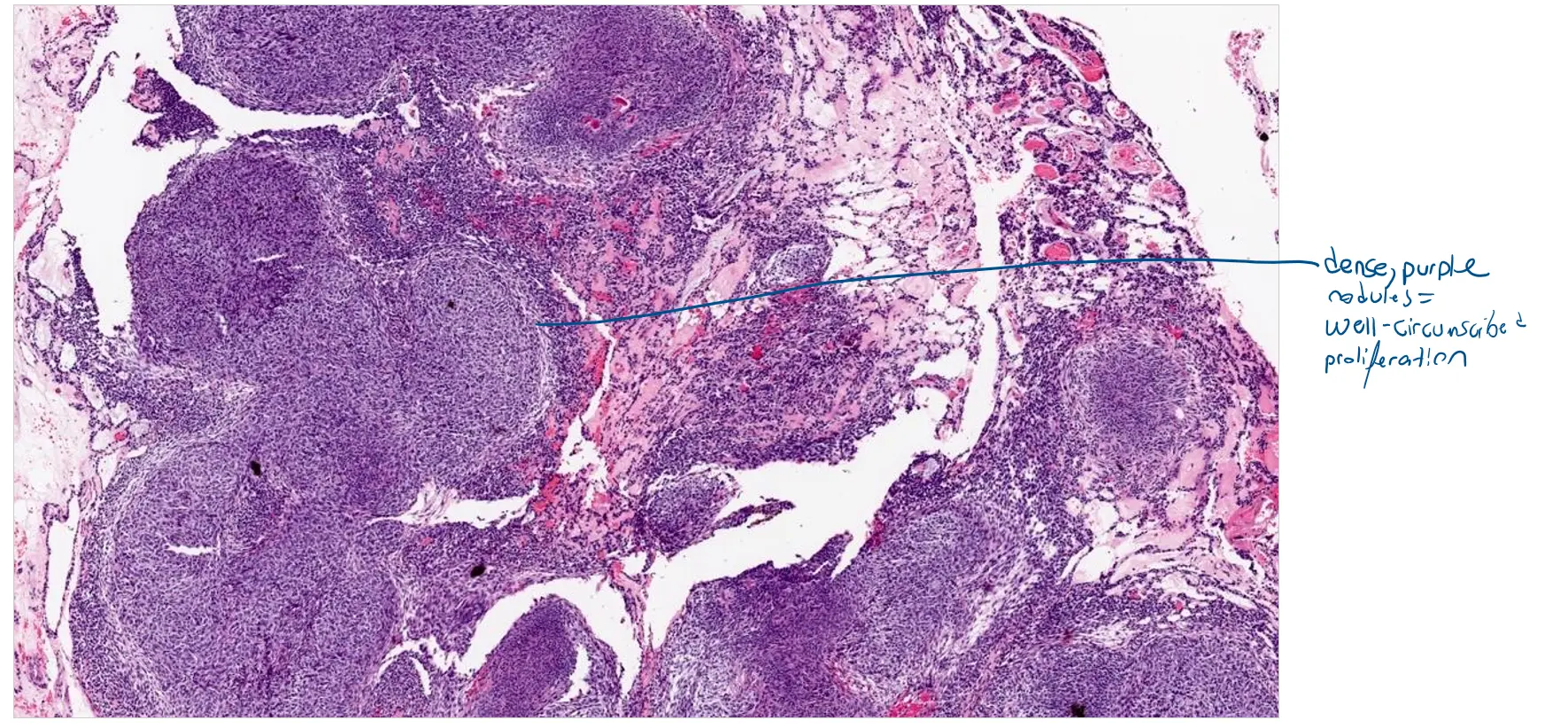
Well-Circumscribed Proliferation: The top image shows the overall architecture. You can see how the tumor is composed of dense, purple nodules that form a well-defined mass, distinct from the surrounding tissue.
-
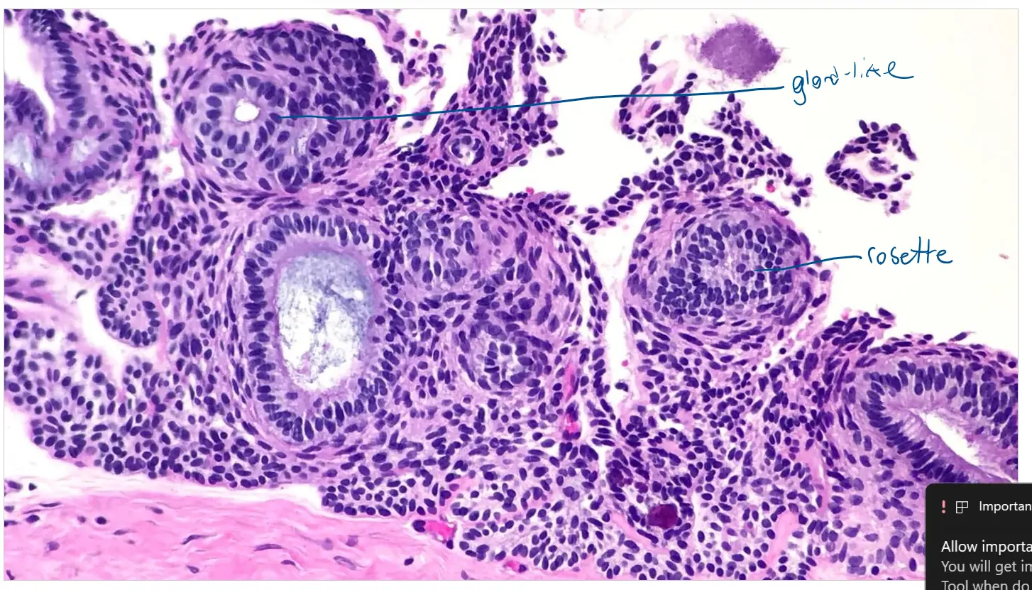
Ductlike Structures & Rosettes: The middle image clearly shows the key patterns. The tumor cells are arranged in circular, gland-like configurations that are the characteristic ductlike structures. In other areas, the cells form small, whorled nests or rosettes.
🔬 Cellular Features (Bottom Image)
The high-magnification bottom image allows us to see the specific details of the cells and the material they produce.
-
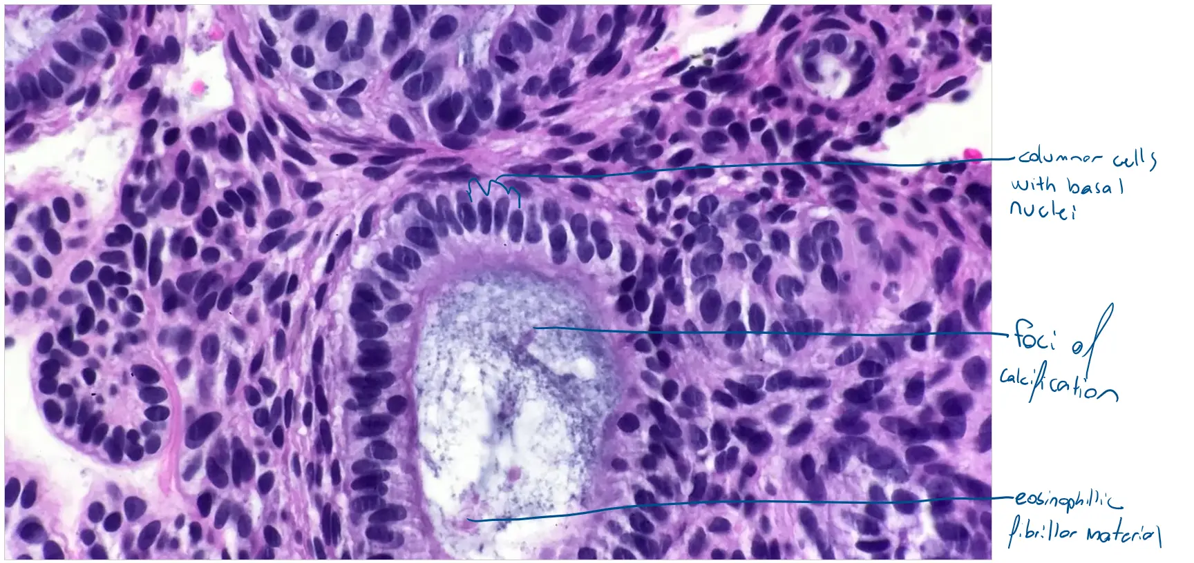
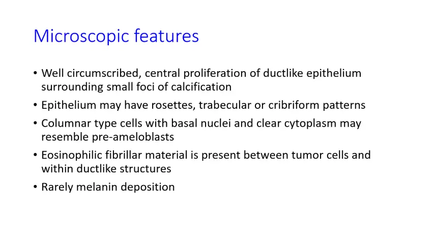
Columnar Cells with Basal Nuclei: Look at the cells lining the ductlike structure. They are distinctly tall and rectangular (columnar). Their nuclei are pushed to the base, away from the central lumen, which is what basal nuclei refers to. This arrangement strongly resembles the pre-ameloblasts of a developing tooth.
-
Eosinophilic Fibrillar Material: Inside the duct and between the surrounding tumor cells, you can see wisps of pink, stringy material. This is the eosinophilic fibrillar material, often referred to as “tumor droplettes.”
-
Foci of Calcification: In the very center of the ductlike structure, there is a cluster of amorphous, bluish-purple material. These are the small foci of calcification, another classic finding in AOT.
In summary, the images beautifully illustrate the classic features of an AOT: a well-defined tumor forming ducts and rosettes lined by columnar cells, with deposits of eosinophilic material and central calcifications.
Would you like to discuss the typical clinical presentation of an Adenomatoid Odontogenic Tumor?