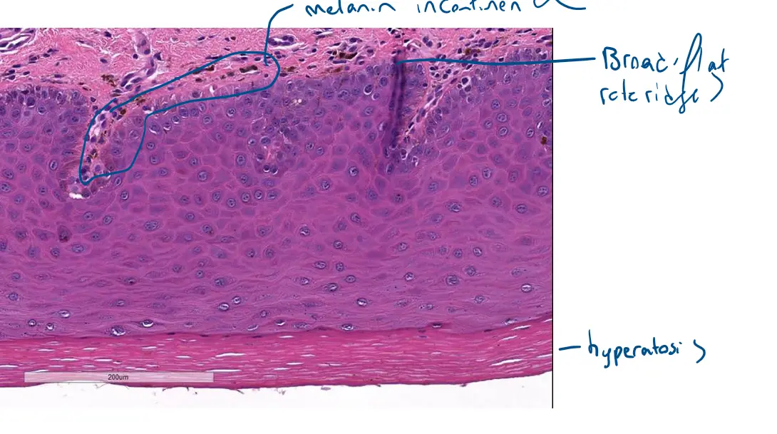
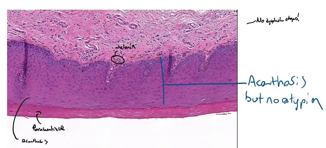
Case 1 : Keratosis with Mild Dysplasia
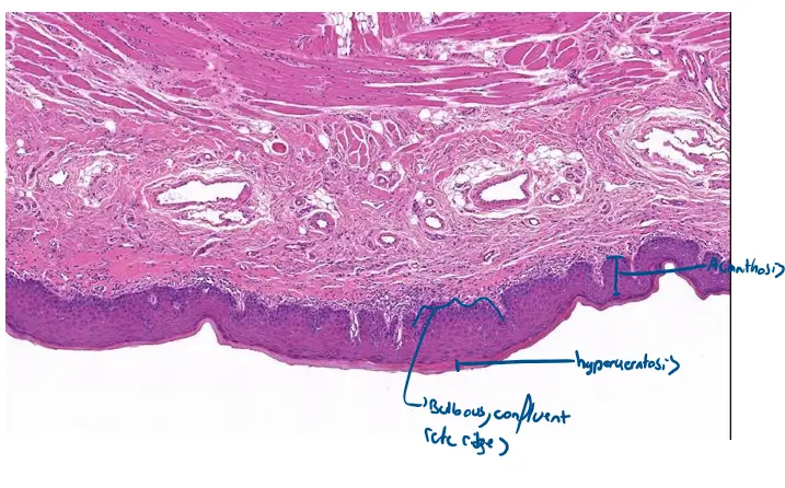
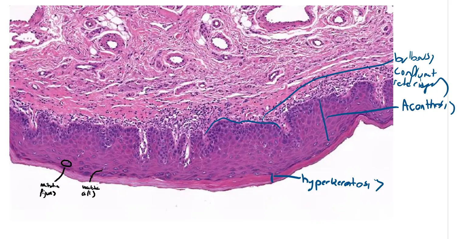
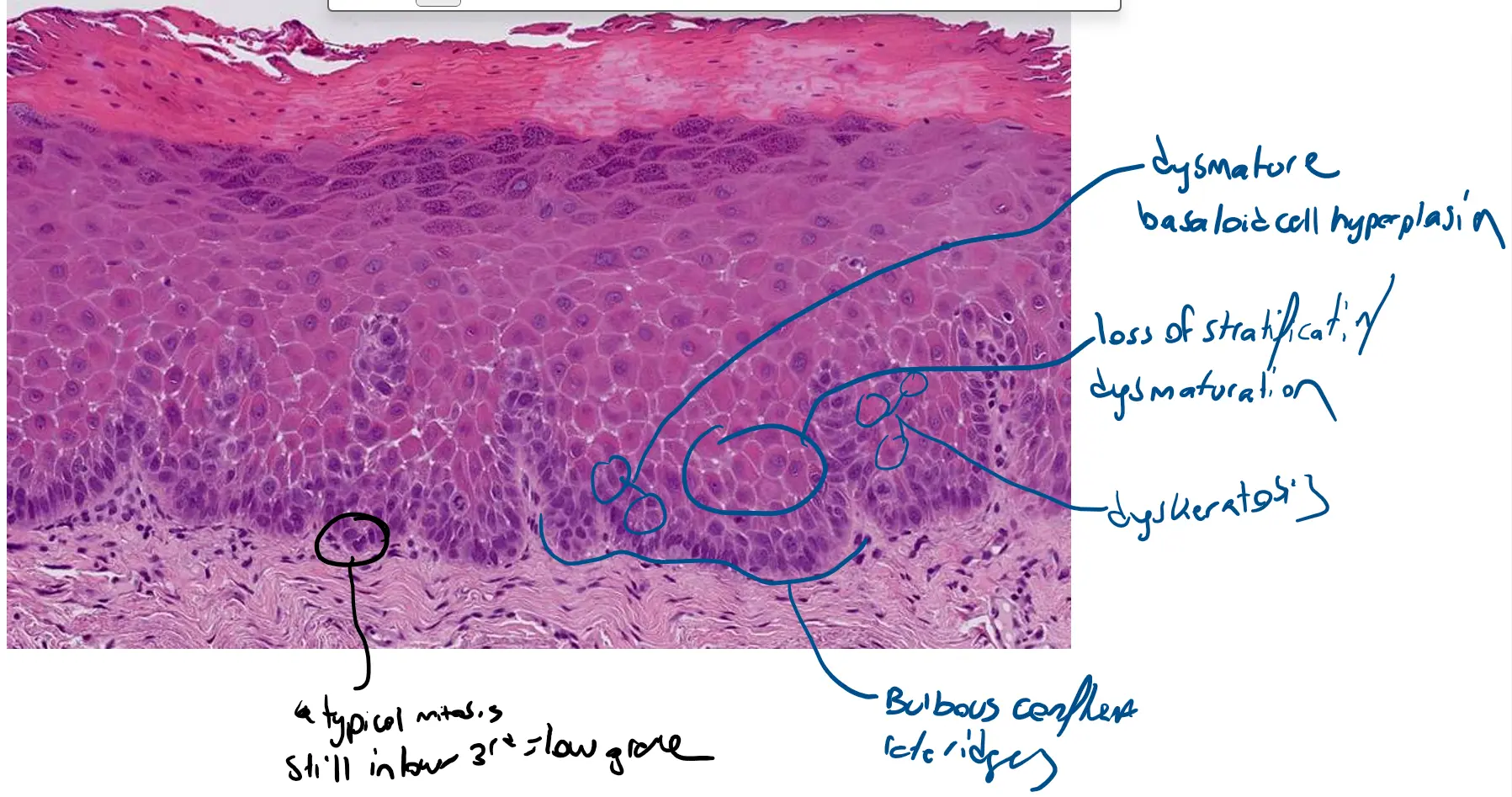
🔬 Locating the Microscopic Features
Hyperkeratosis and Acanthosis
-
Hyperkeratosis: This is the thickened, dense, pink layer on the very top surface of the epithelium. It represents a thickened layer of dead, scaly cells (the stratum corneum). You can see this most clearly in the bottom, high-magnification image.
-
Acanthosis: This refers to the overall thickening of the entire living (purple) portion of the epithelium. It is evident in all three images, giving the tissue a robust, expanded appearance.
Bulbous Confluent Rete Ridges
- This architectural feature is best observed in the top and middle images. The rete ridges are the downward projections of the epithelium into the underlying connective tissue. Here, they are not thin and tapered as they normally would be; instead, they are abnormally broad, deep, and blunt-ended (bulbous). Confluent means they are starting to fuse together at their bases.
Loss of Normal Stratification with Dysmaturation and Dyskeratosis
-
This is best appreciated in the bottom, high-magnification image.
-
Loss of Stratification / Dysmaturation: Normally, epithelial cells mature and flatten as they move from the bottom to the top. Here, that orderly process is lost. The cells remain large, crowded, and disorganized almost all the way to the surface, giving the epithelium a jumbled, chaotic appearance.
-
Dyskeratosis: Look for small, isolated, bright pink cells scattered amongst the purple “basaloid” cells. These are individual cells that have keratinized prematurely and abnormally, a sign of severe cellular disarray.
Dysmature Basaloid Cell Hyperplasia
-
This feature is the primary cause of the abnormal appearance seen in the bottom image. The epithelium is filled with immature-looking cells that resemble basal cells (basaloid).
-
These cells have very large, dark-staining nuclei and very little cytoplasm, which is described as an increased nuclear-to-cytoplasmic ratio. This is why the epithelium looks so crowded and purple.
Increased Number of Mitotic Figures
-
To find these, you would need to carefully scan the bottom image. Mitotic figures are cells caught in the act of division and appear as small, dark, “spidery” or star-shaped chromatin clumps.
-
In this condition, not only is their number increased, but more importantly, they are found abnormally high up in the epithelium. In normal tissue, they are restricted to the single bottom-most layer.
Case 2 : Frictional keratosis