Case 1: Moderately differentiated squamous Cell carcinoma
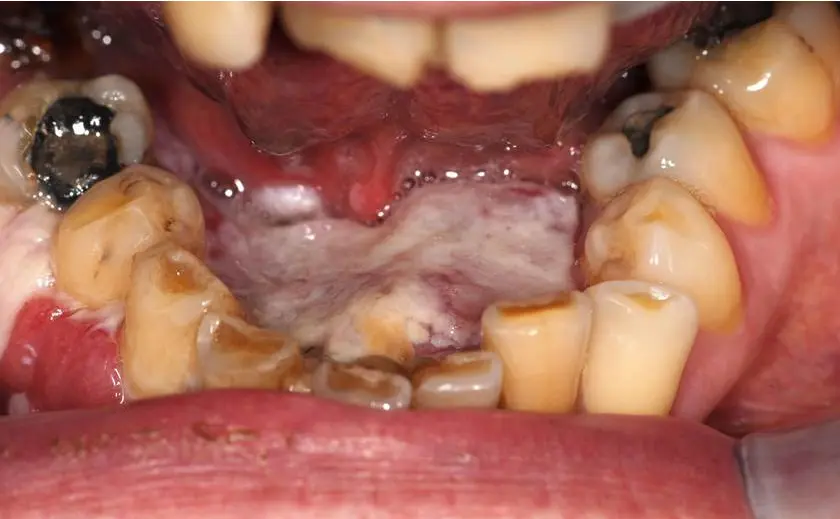
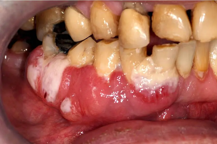

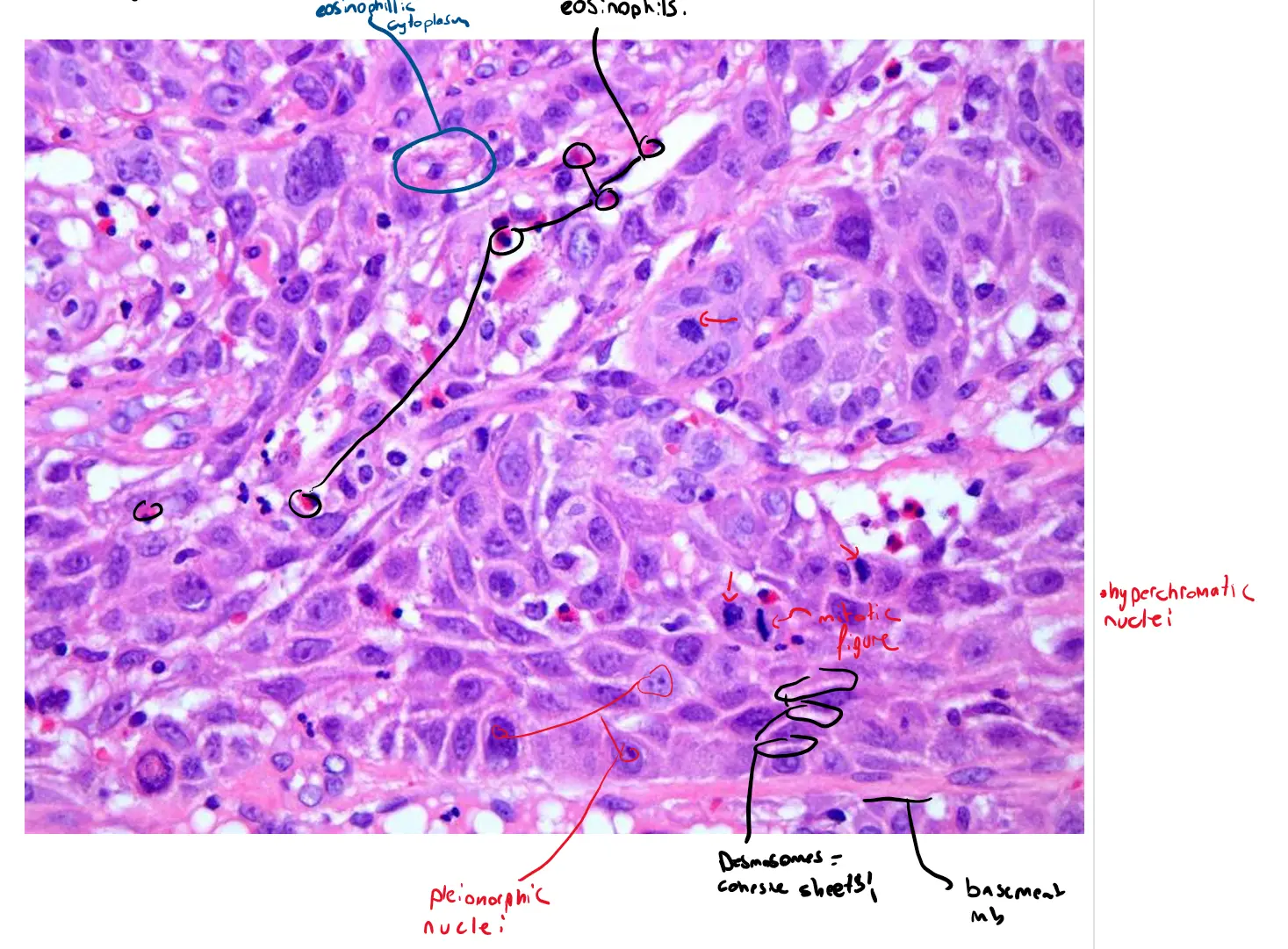
Locating the Histological Features
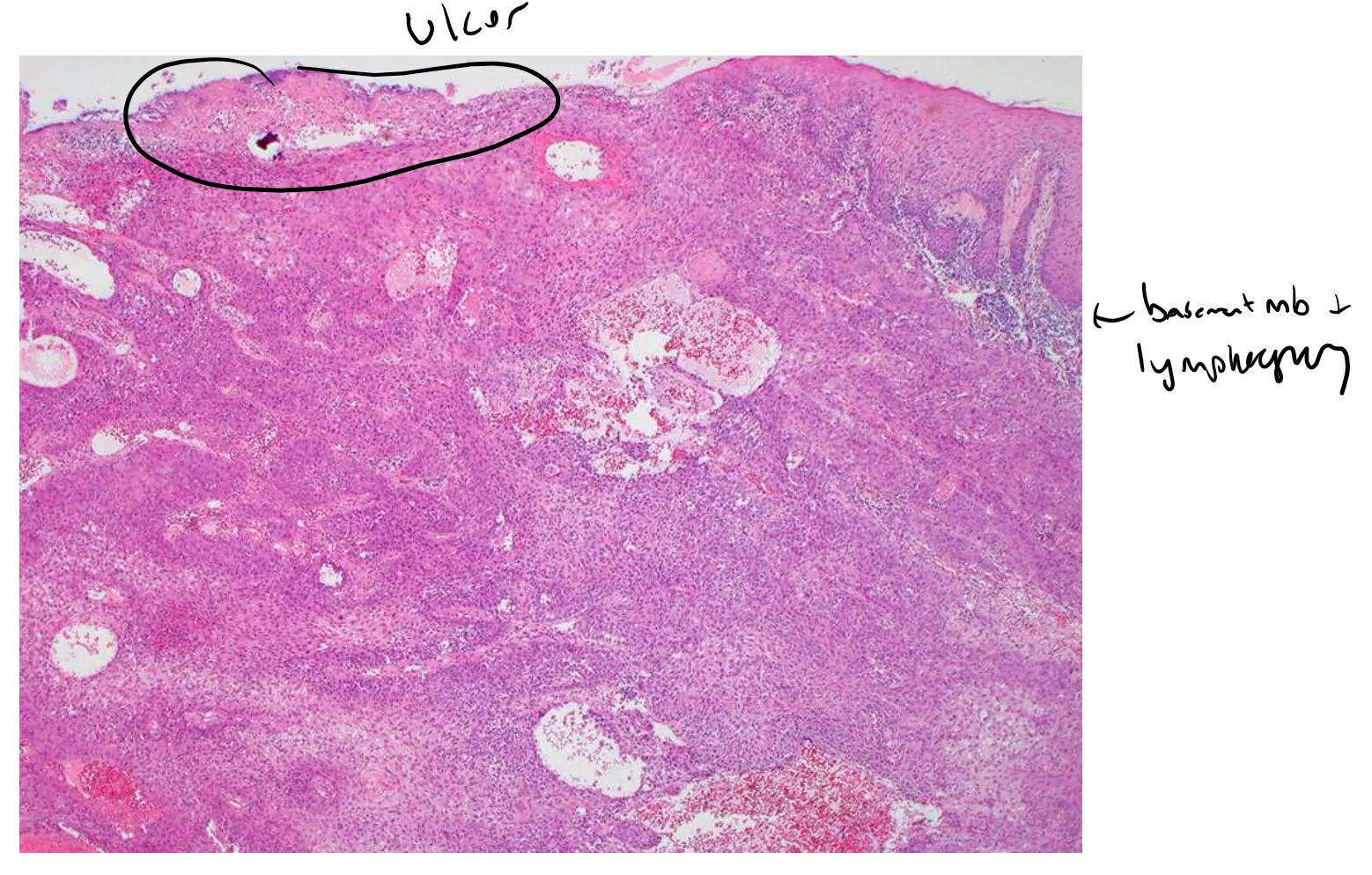
1. Invasion into Connective Tissue
-
Where to look: Top Image (Low Power)
-
What you’re seeing: In this view, you can see large, irregular islands and nests of pink-staining (eosinophilic) epithelial cells that have breached their normal boundary (the basement membrane) and are infiltrating the underlying bluish/pink fibrous connective tissue. This is the hallmark of invasive carcinoma. The disorganized growth pattern is evident even at this low magnification.
2. Malignant Epithelial Cells in Islands and Cords
-
Where to look: Middle Image (Medium Power)
-
What you’re seeing: This image zooms in on the invasive nests seen in the top image. You can clearly see the cancer cells growing in large, interconnected groups. Notice the distinct boundary between the packed malignant cells and the surrounding connective tissue, which contains smaller, dark blue dots representing inflammatory cells (lymphocytes) that have been recruited to fight the cancer.
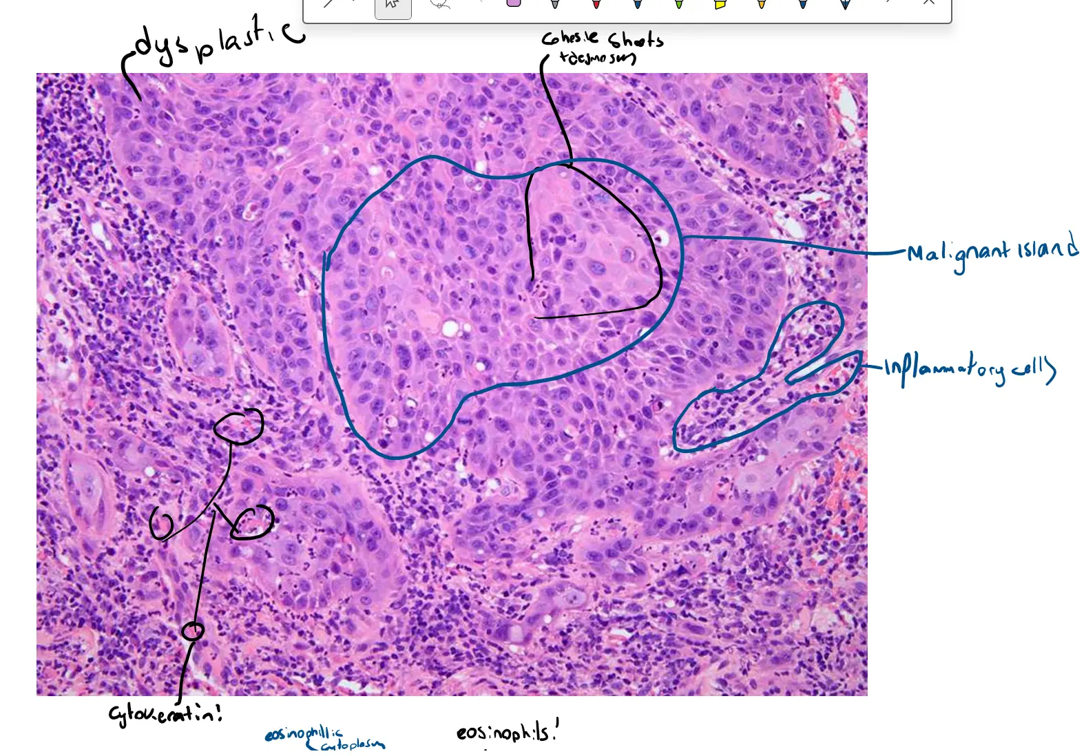
3. Malignant Cellular Features
-
Where to look: Bottom Image (High Power)
-
What you’re seeing: This high-magnification view is essential for observing the specific characteristics of individual cancer cells as described in your text:
-
Eosinophilic Cytoplasm: The cytoplasm (the area of the cell outside the nucleus) of most cells is a bright pink color.
-
Hyperchromatic and Pleomorphic Nuclei: The nuclei (the dark purple centers of the cells) are much darker than normal (hyperchromatic). They also vary significantly in size and shape (pleomorphism). Some are round, some are oval, and some are irregularly shaped.
-
Mitotic Activity: Look for dark, condensed figures within the nuclei. These are mitotic figures, indicating that the cells are dividing rapidly and uncontrollably. Finding abnormal mitotic figures is a strong indicator of malignancy.
-
Intercellular Bridging: In some areas, you might be able to see very fine, spiny projections connecting adjacent cancer cells. These are called intercellular bridges (or desmosomes) and are a feature of squamous cells, both normal and malignant. They can be difficult to spot without very high magnification but are often present in well to moderately differentiated SCC.
-
Keratinization**
-
Where to look: Middle and Bottom Images
-
What you’re seeing:
-
Individual Cell Keratinization: In the bottom image, you may see some individual cells that have very dense, bright pink cytoplasm and a small, shrunken nucleus. This is a sign of premature and abnormal keratin production within a single cell.
-
Keratin Pearls (if present): While not definitively visible in these specific images, you would look for them in the center of the larger nests seen in the middle image. A keratin pearl is a circular, layered deposit of keratin that looks like an onion cut in cross-section, with concentric rings of bright pink. The presence of these pearls is characteristic of well-differentiated tumors, but they can also be found in moderately differentiated ones.
-
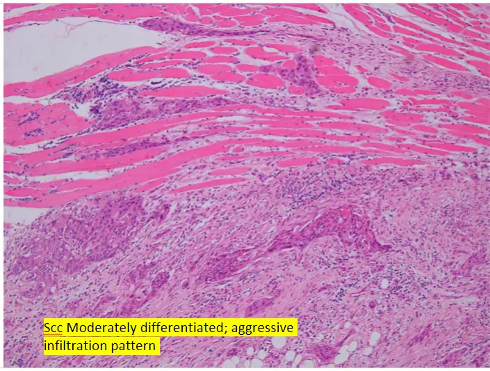
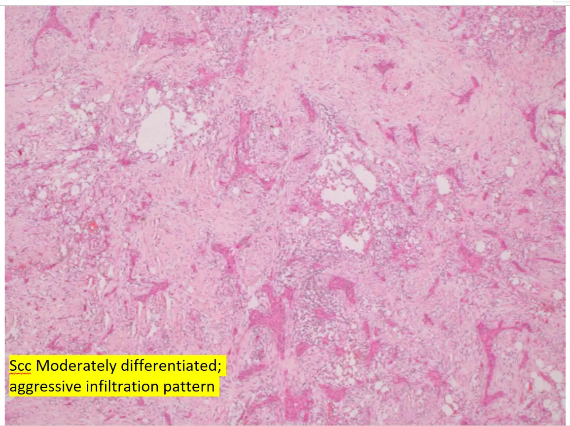
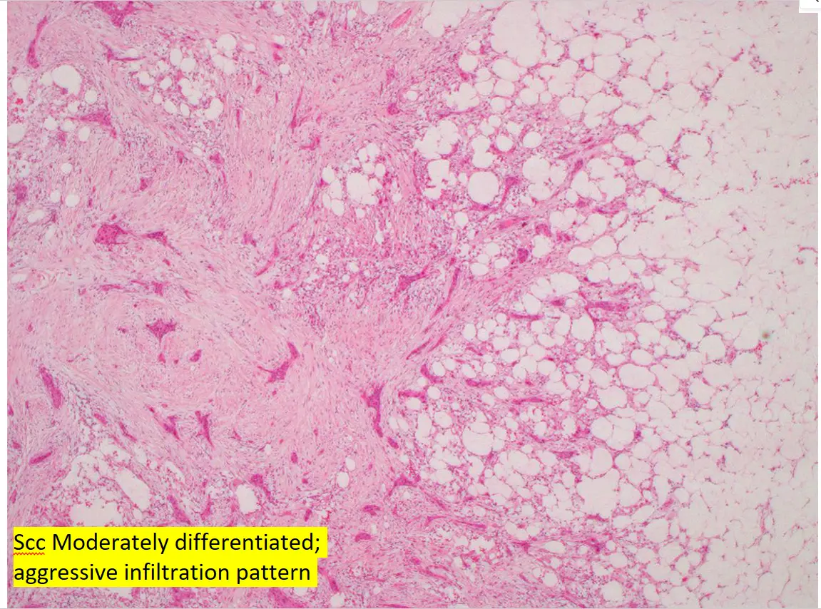
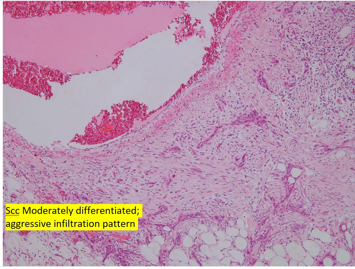
Case 2 SCC Moderately differentiated ; aggressive infiltration pattern
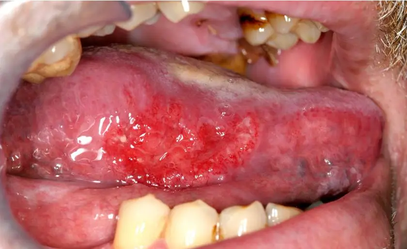

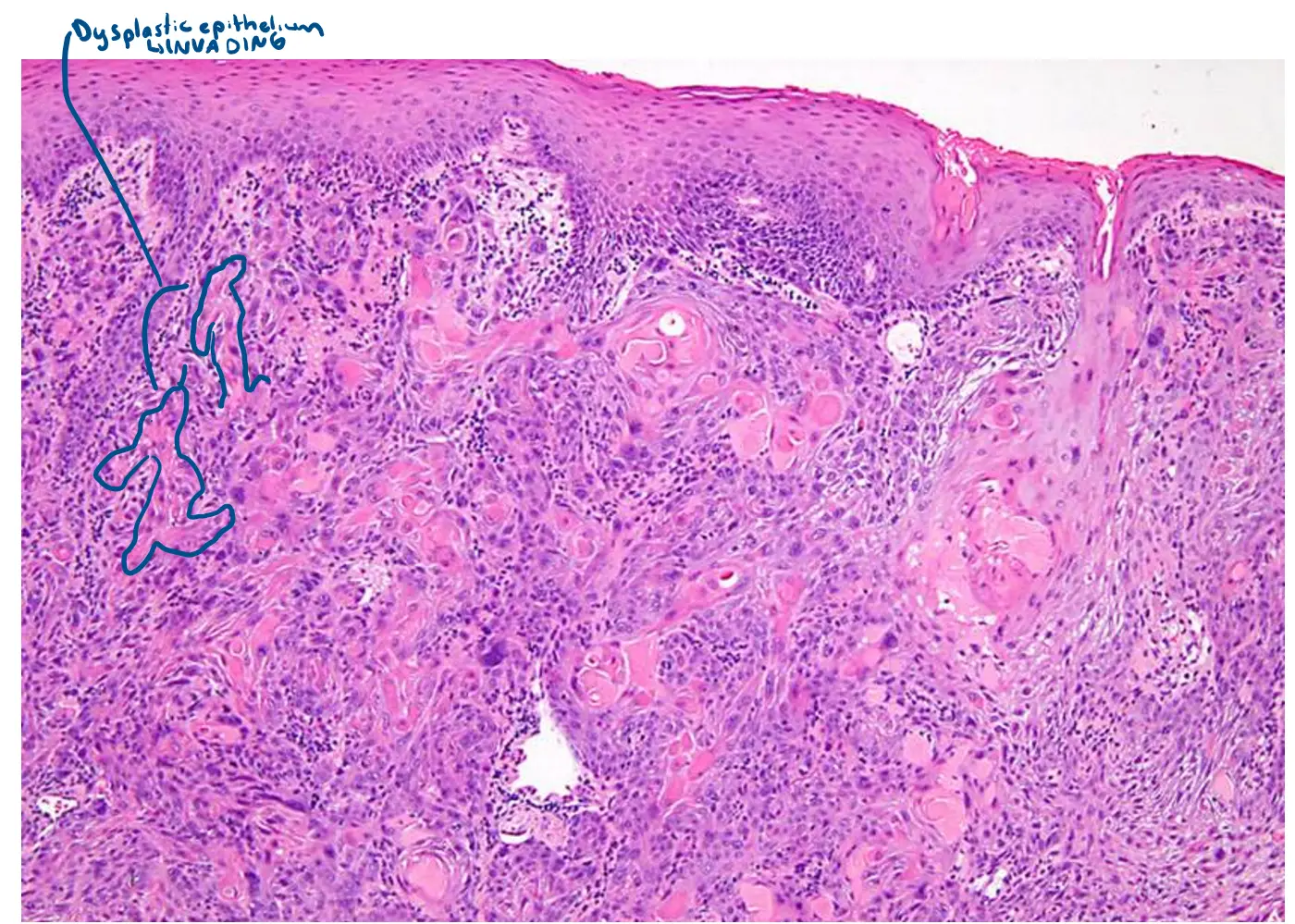
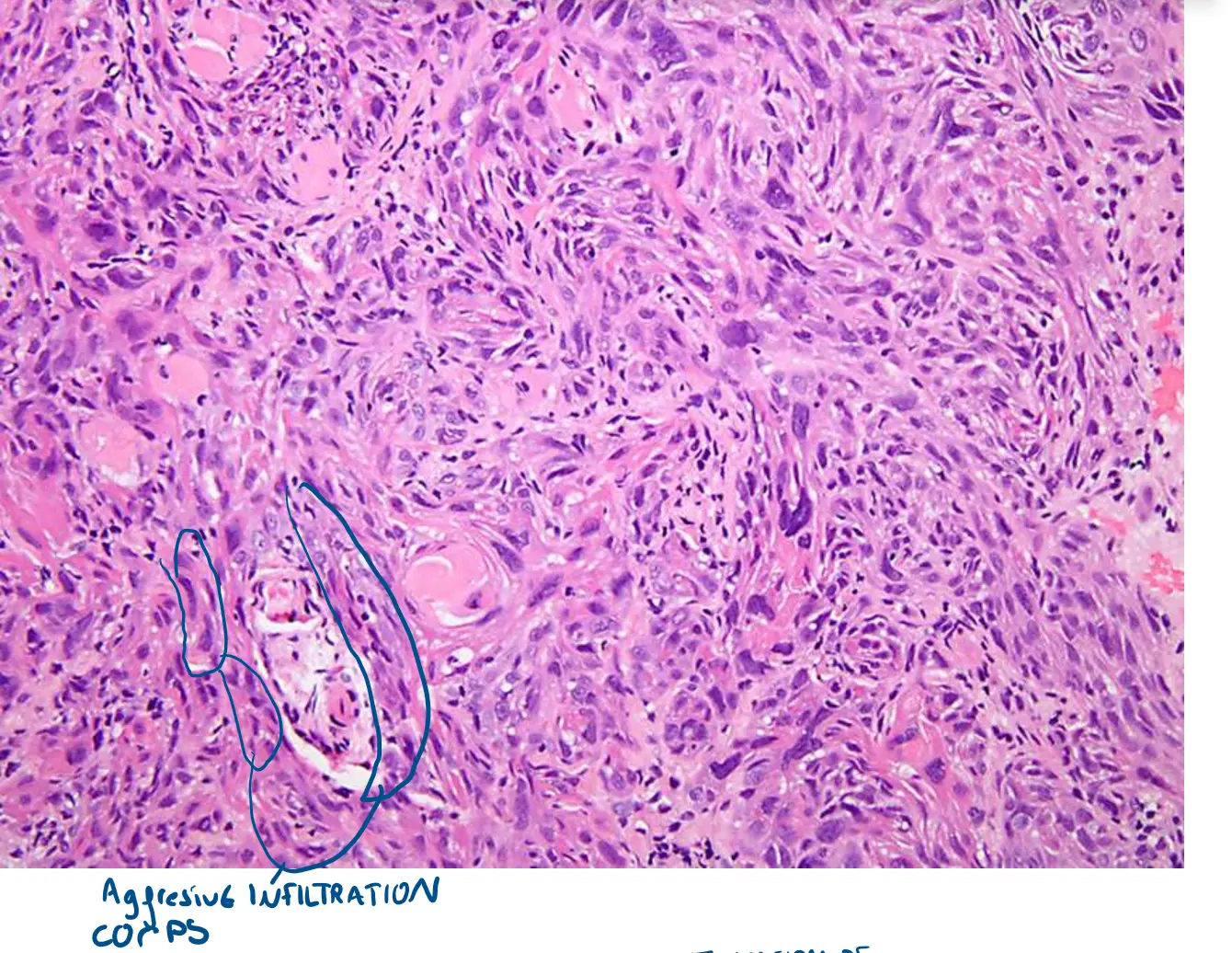
Explanation of Microscopic Features
This case demonstrates a more aggressive growth pattern compared to the previous example. Here is where you can see the key features:
1. Dysplastic Epithelium and Invasion
-
Where to look: Top Image (Low Power)
-
What you’re seeing: At the very top, you can see the original surface epithelium. Below this, there is a massive infiltration of pink (eosinophilic) malignant cells flooding the underlying fibrous connective tissue. Unlike the previous example with more rounded nests, the invasion here appears more widespread and less organized, which is a sign of aggressive behavior. The tumor cells are seen growing in irregular, jagged islands and cords.
2. Aggressive Infiltration Pattern: Islands, Cords, and Individual Cells
-
Where to look: Middle Image (Medium Power)
-
What you’re seeing: This view clearly shows that the tumor is not forming well-defined, rounded nests. Instead, the malignant cells are arranged in long, streaming cords and smaller, more separated islands. Many of the cells have an elongated, spindle-like shape. This growth pattern, with smaller, more disconnected groups of cells, allows the tumor to more easily infiltrate through tissue planes, which is a hallmark of an aggressive tumor.
3. Malignant Cellular Features & Invasion of Deeper Structures
-
Where to look: Bottom Image (High Power)
-
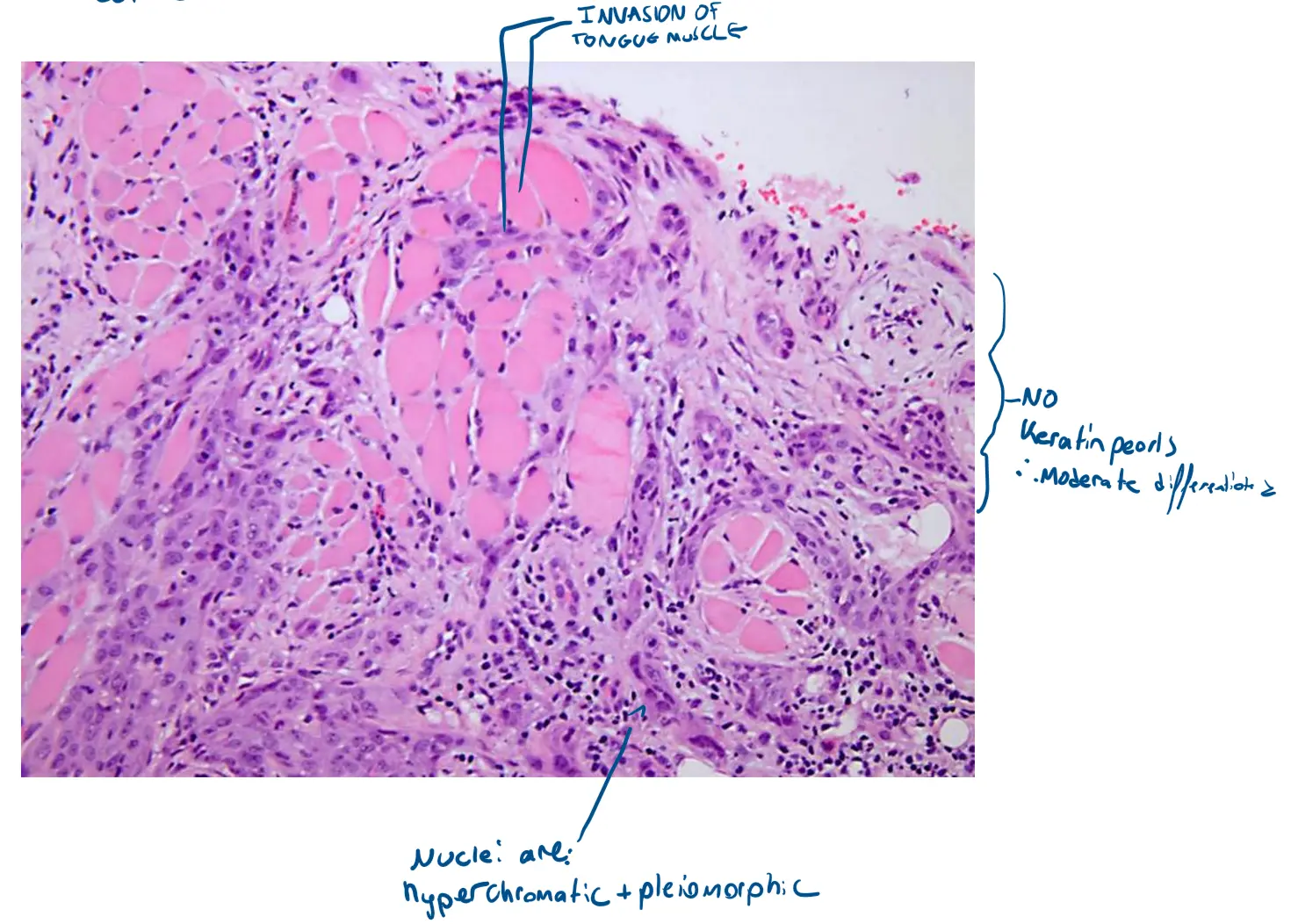
What you’re seeing: This high-power view reveals two critical aspects: the features of the cancer cells themselves and their invasion into other tissues.
-
Malignant Cellular Details: You can clearly see the key features described in your list. The cells have abundant eosinophilic (pink) cytoplasm. The nuclei are hyperchromatic (dark purple) and exhibit significant pleomorphism (variation in size and shape).
-
Invasion of Muscle: The most striking feature in this image is the direct invasion of skeletal muscle. The large, bright pink, structured fibers are muscle bundles. You can see the smaller, darker, and more disorganized malignant epithelial cells infiltrating between and around these muscle fibers. This is a definitive sign of deep and aggressive invasion.
-
Lack of Keratin Pearls: In contrast to more well-differentiated tumors, there are no keratin pearls visible here. The primary features are the infiltrative growth and the malignant characteristics of the individual cells, which are consistent with a moderately differentiated, aggressive SCC.
-