case 1 : Squamous cell carcinoma; well-differentiated
Explanation of Microscopic Features
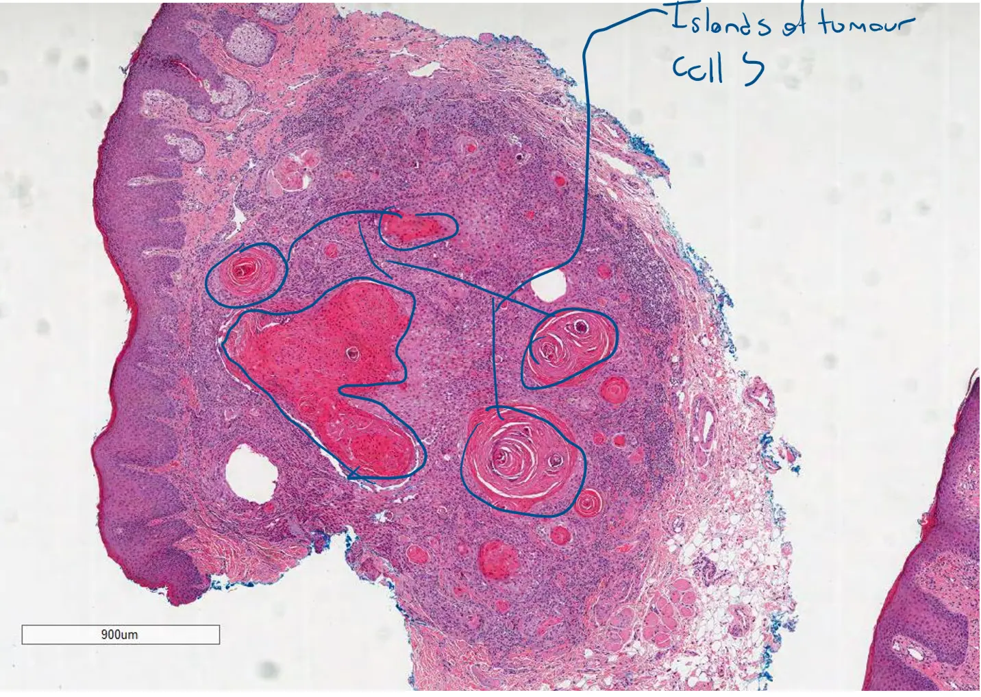
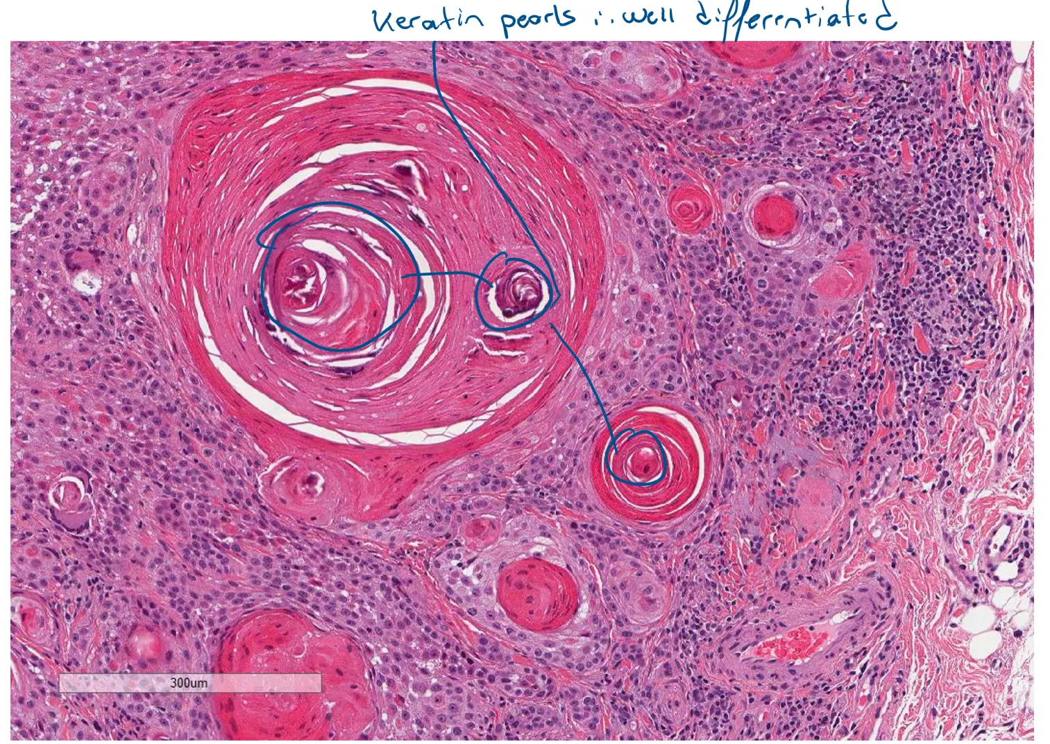
Here’s where to see the key features described as “Islands and stands of atypical squamous epithelial cells with keratin pearls invading the connective tissue.”
1. Multiple Tissue Fragments
-
Where to look: Top Image (Scanner View)
-
What you’re seeing: This view shows that the biopsy consists of several separate pieces of tissue. Pathologists examine all fragments to get a complete picture of the lesion. You can already see the abnormal pink and purple areas even at this distance.
2. Invasive Islands of Atypical Squamous Cells
-
Where to look: Second Image (Low Power)
-
What you’re seeing: In this view, you can clearly see the “islands and stands” of tumor cells. These are large, well-defined, irregular nests of pink (eosinophilic) cells that are invading the underlying bluish connective tissue (stroma). The surface epithelium is visible at the top. The large size and rounded, pushing borders of these islands are characteristic of a well-differentiated tumor.
3. Prominent Keratin Pearls
-
Where to look: Third Image (Medium Power)
-
What you’re seeing: This is the most defining feature of well-differentiated SCC. You can see numerous keratin pearls. These are the circular, layered structures with bright pink centers. They resemble an onion sliced in half. A keratin pearl is a focus of abnormal, excessive keratin production in the center of a tumor island. The presence of many large, well-formed keratin pearls is the hallmark of a well-differentiated squamous cell carcinoma.
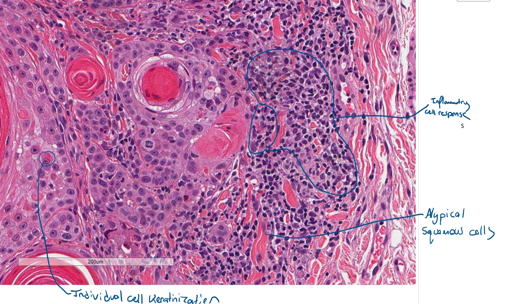
4. Atypical Squamous Cells and Inflammation
-
Where to look: Bottom Image (High Power)
-
What you’re seeing: This high-magnification view shows:
-
Atypical Squamous Cells: The tumor cells are large with abundant pink cytoplasm and large, dark nuclei. While they are cancerous, they still retain features of squamous cells, such as intercellular bridging (fine lines connecting the cells).
-
Keratinization: You can see smaller keratin pearls and individual cells with dense, pink cytoplasm (individual cell keratinization).
-
Inflammatory Response: Notice the dense collection of small, dark blue dots surrounding the tumor islands. These are lymphocytes, which are immune cells that have been recruited to attack the cancer. A brisk inflammatory response is very common in SCC.
-
Case 2 Squamous Cell Carcinoma Moderately differentiated
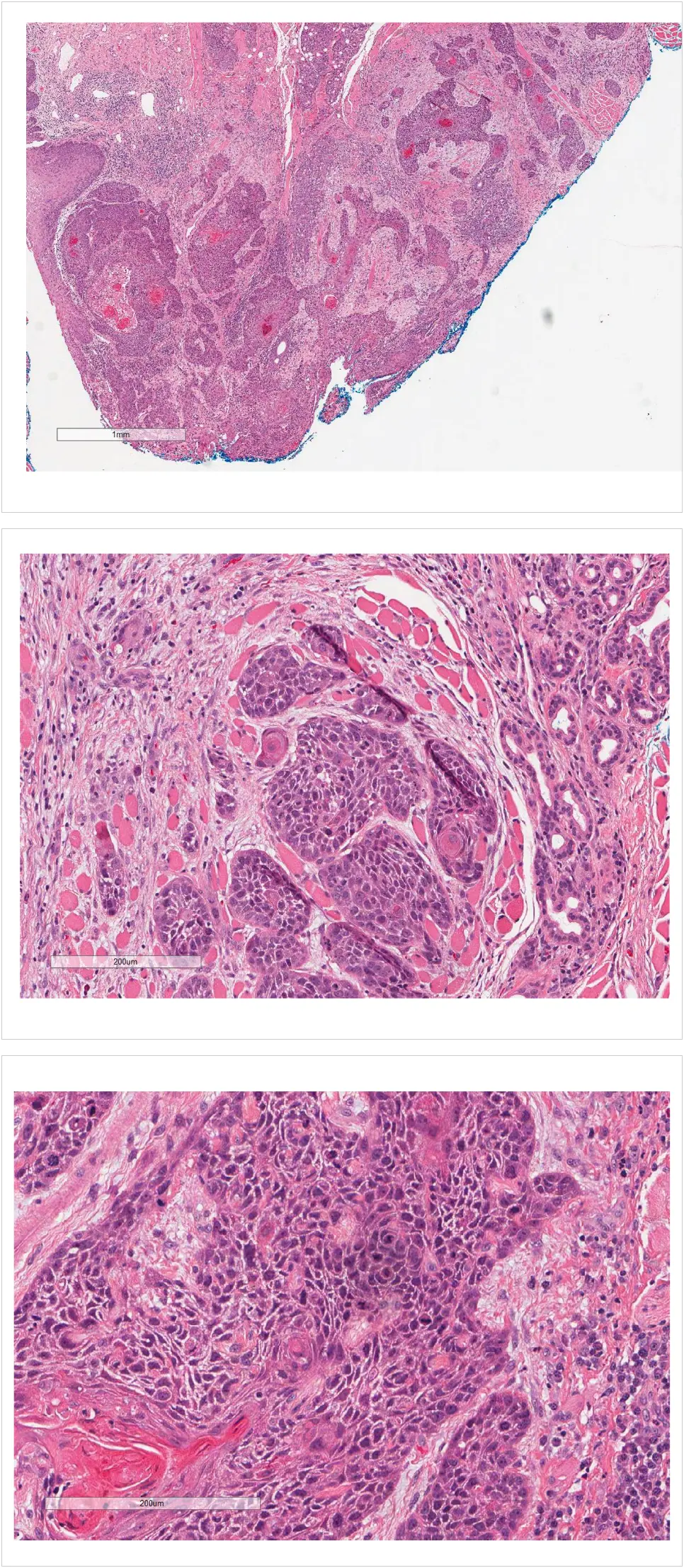 Of course. Let’s break down the microscopic features of this new case of moderately differentiated Squamous Cell Carcinoma (SCC).
This set of images serves as an excellent intermediate example between the well-differentiated and the more aggressive, infiltrative cases we looked at previously.
Explanation of Microscopic Features
Here is how this specimen demonstrates the features of a moderately differentiated SCC.
1. Infiltrative Growth Pattern
-
Where to look: Top Image (Low Power)
-
What you’re seeing: This view shows large, irregular nests of tumor cells deeply invading the connective tissue. Unlike the very well-differentiated example, the borders of these nests are more jagged and infiltrative rather than rounded and “pushing.” The organization is less uniform, indicating a more aggressive nature.
2. Irregular Nests and Limited Keratinization
-
Where to look: Middle Image (Medium Power)
-
What you’re seeing: This magnification clearly shows the tumor growing in solid, interconnected nests. A key feature of moderate differentiation is visible here: there is no significant keratin pearl formation. While the cells are recognizably squamous, they are not mature enough to produce the large, organized keratin pearls seen in well-differentiated tumors. You can also see the surrounding pink fibrous stroma and some normal glandular structures on the right, which the tumor is invading.
3. Increased Atypia and Mitotic Activity
-
Where to look: Bottom Image (High Power)
-
What you’re seeing: The features that firmly place this in the moderately differentiated category are evident at high power:
-
Greater Pleomorphism: Compared to a well-differentiated tumor, the nuclei (the dark purple centers) show more variation in size and shape. They are more angular and irregular.
-
Prominent Nucleoli: Many of the nuclei contain a small, distinct red dot inside called a nucleolus. Prominent nucleoli are a sign of active cell growth and are more common in moderately differentiated tumors.
-
Increased Mitotic Activity: You are more likely to see cells in the process of division (mitotic figures) in this grade of tumor. These appear as small, dark, condensed clumps of chromatin.
-
Less Cytoplasm: The cancer cells have a higher nuclear-to-cytoplasmic ratio, meaning the nucleus takes up more of the cell’s volume compared to a well-differentiated tumor.
-
In summary, this case is classified as moderately differentiated because the tumor is clearly squamous but shows more cellular atypia, less organization, and a lack of the extensive keratin pearl formation that characterizes well-differentiated SCC. It occupies the middle ground of the differentiation spectrum.
Would you like to see a comparison table summarizing the key differences between well-differentiated, moderately-differentiated, and poorly-differentiated SCC?