The Patient Examination And Additional Tests
Course Information1
- Course Code: DENT4219
- Lecture 3: The Patient Examination and additional tests
- Contact: poh.hun.loh@uwa.edu.au
Patient Perspectives And Communication2
Patient Perceptions3
- What patients think
- How patients see the dentist

Extra-Oral Examination
Clinical Assessment Checklist4
- Head and Neck
- Face
- Smile
- Speech
- Asymmetry
- Swelling
- Midline
- Occlusal/incisal plane
- Facial type/profile

Intra-Oral Soft Tissue Examination5
Perform visual inspection and palpation of the following areas:
- Lips - Vermillion border
- Sulci
- Buccal mucosa
- Palate
- Tongue
- Floor of mouth
- Frenal attachments
- Ducts
- Saliva
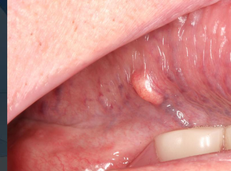
Oral Manifestations Of Systemic Disease
Clinical Significance6
- Early diagnosis prior to systemic manifestations is possible through oral examination.
- The role of the dentist is critical in identifying systemic links.
Common Manifestations
- Dry mouth: Often medication-related or associated with diabetes.
- Lichen Planus
- Pemphigus
Clinical Documentation And Records
Legacy Examination Requirements7
Form 21 requires the following items to be addressed:
- Reason For Attendance
- Extra-Oral Examination
- Intra-Oral Examination
- Radiographs Viewed
- Notes
- Diagnoses
- Further Procedures

Examination Templates And Saliva Assessment8
 |  |
Saliva Assessment Parameters
- Resting Saliva
- Stimulated Saliva
- Hydration: >60 Secs
- Viscosity: Sticky/frothy
- pH: 5.0-5.8
- Quantity: <3.5 ML
- Buffering: 0-5 Points
Exam Saliva Caries Risk
Sample Case Presentation Template9
- P/C (Presenting Complaint): Three “snapped off” teeth and a P/- Cr-Co denture that is “falling out”; patient is using Polydent for retention.
- M.Hx (Medical History): See 011.
- D.Hx (Dental History):
- Last dental visit: Temporary filling for broken tooth.
- Denture history: Cr-Co P/- made 4 years ago at OHCWA. Loss of tooth 24 (which had the retaining clasp) resulted in no retention. Patient happy with appearance of clasps. Tooth 24 snapped 6 months ago while brushing.
- OH (Oral Hygiene): Brushes 2x/day with electric toothbrush and Colgate charcoal toothpaste. No flossing or other products.
- Diet assessment:
- Breakfast: Banana, oats.
- Lunch: Toasted sandwich.
- Dinner: Weight-watchers meals.
- Snacks: Infrequent sugar-free lollies, potato sticks (one sitting).
- Drinks: Water, 375mL Pepsi Max sipped throughout the day, coffee throughout the day.
- S.Hx (Social History): Works in a bakery.
- EOE (Extra-Oral Exam):
- Skin/hair: Healthy, clear complexion.
- Facial symmetry: Symmetrical.
- Thyroid, TMJ, Lymph nodes, Muscles of mastication, Lips: All NAD (No Abnormalities Detected).
- IOE (Intra-Oral Exam):
- Labial mucosa: Ulcer Mx R.
- Palate: Denture stomatitis; nicotinic stomatitis on posterior hard palate.
- Tongue: Large fungiform papillae at back; otherwise NAD.
- Saliva: Inadequate; patient reports dry mouth. Recommended Oral 7 or olive oil spray.
- Sulci, Frena, Oropharynx, Alveolar ridge, Buccal mucosa, FOM: All NAD.
- Tooth charting: No teeth TTP (Tender To Percussion).
Digital Records And Odontograms
Documentation Tools10
- Utilize 011 and eform for record keeping.
- TOHM odontogram or tooth chart should be used, though note they cannot be ‘locked’.
Periodontal And Hard Tissue Examination
Periodontal Probing And Indices
CPITN/BPE Assessment11
- PPD (Probing Pocket Depth)
- Recession
- BOP (Bleeding on Probing)
- Furcation involvement
- Mobility
- Marginal discrepancy

Examination Of Teeth And Restorations
Systematic Tooth Inspection12
- Follow quadrant order: Q1 → 2 → 3 → 4.
- Dry teeth thoroughly with a triple syringe.
- Check for plaque and staining.
- Utilize mirror and sickle probe for inspection.
- Perform percussion testing.
- Evaluate grooves and fissures.
- Screen for caries.
- Evaluate existing restorations.
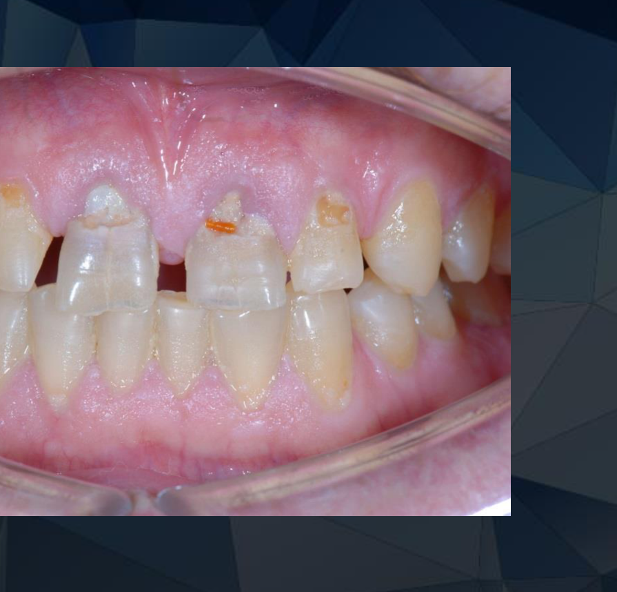
Restoration Evaluation Criteria13
- Type of material
- Integrity of margins
- Presence of overhangs
- Discoloration
- Surface texture
- Anatomical contour
- Interproximal contact quality
- Remaining volume of tooth structure
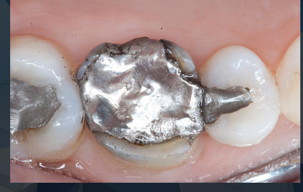
Differentiating Restorative Materials14
- Tooth-coloured GIC vs. Composite:
- Location: Check if restoration is in the gingival 1/3 vs. incisal area.
- Surface texture: GIC typically appears more porous.
- Unknown materials: Classify as TC (Tooth Coloured) restoration if uncertain.
- Clinical Judgment: Decide if the restoration is satisfactory or unsatisfactory.

Evaluation Of Crowns And Bridges
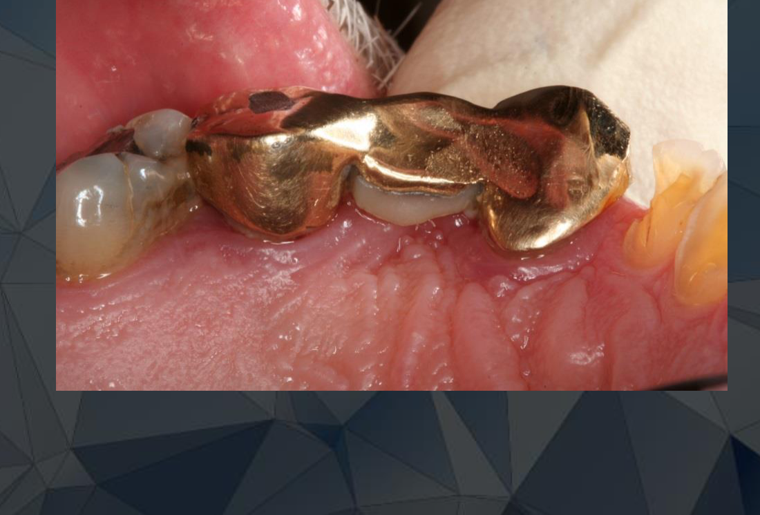
Fixed Prosthodontics Checklist15
- Margins
- Contour
- Colour/shade match
- Proximal contacts
- Associated gingival health

Additional Evaluation Factors16
- Age: Note that patient recall regarding the age of the restoration is often poor.
- Aesthetics: Patient satisfaction with appearance.
- Structural Components: Evaluate pontics, abutments, and connectors.
- Hygiene: Presence of plaque or food impaction.
Removable Prosthodontics And Dentures
Denture Assessment17
- General Details: Type, age, and teeth replaced.
- Mechanical Properties: Retention, support, and stability.
- Aesthetics:
- Appearance of prosthetic teeth.
- Soft tissue and lip support.
- Occlusion: Evaluation of worn teeth and masticatory effectiveness.

Identifying Unsatisfactory Restorations
Criteria for Replacement18
- Structural Failure: Defective margins, leakage, fractures, or excessive wear.
- Caries: Identified via clinical examination or radiography.
- Anatomical Issues: Poor contour.
- Aesthetic Failure: Staining, discoloration, or visible margins.

Clinical Findings And Case Analysis
Case Study Sensitivity Analysis
Case Description: N.H. (61-year-old Female)19
Patient presented with 1.5 years of severe, unrelenting left anterior sensitivity. Examination revealed a vertical root fracture on the left anterior third molar, but no other obvious trauma or lesions.
 |  |
Patient Complaints
- Sensation of teeth falling out and feeling loose.
- Halitosis and numb tongue.
- Morning dry mouth with acidic taste.
- Gingival pain during eating.
- Extreme thermal sensitivity (unable to drink soup).
Reported History
- Dental caries and periodontal disease.
- Bruxism.
- Ryphenoid syndrome (acid reflux).
Contributing Factors
- Genetic Predisposition: Ammonium treatment processing potentially increasing susceptibility to caries/periodontal disease and exacerbating reflux/bruxism.
- Environmental Exposure: Exposure to toxic chemicals (e.g., Mercury, Lead, Arsenic, various metals) linked to dental disease and masticatory apparatus ossification.
- Psychological Stressors: Anxiety and depression leading to self-mutilation coping mechanisms, medication reliance, and social avoidance.
Diagnosis and Management
- Underlying Condition: Fear of the unknown.
- Treatment Approach:
- Behavioral modification to reduce self-mutilation.
- Medication management.
- Psychotherapy for emotional relief and mental health support.
Commonly Observed Dental Anomalies
Structural and Positional Anomalies20
- Spaces and missing teeth
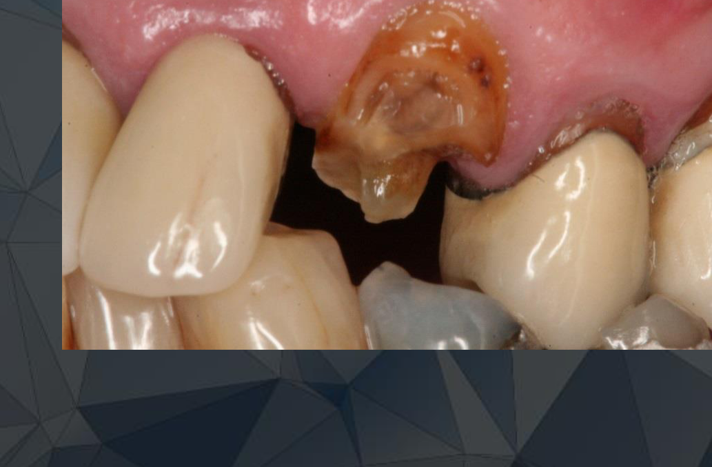
- Broken or fractured teeth
- Tooth wear and cracks
- Rotated or tilted teeth
- Arch malalignment (e.g., supra-eruption or submergence)
Frequently Overlooked Areas21
- Upper Molars: Buccal surfaces, particularly on teeth positioned buccally.
- Lower Molars: Lingual surfaces, where the tongue and saliva often interfere with clear vision.

Professional Communication Standards22
- Use appropriate clinical terms; avoid colloquialisms (e.g., avoid phrases like “p***ing blood” or “hook the tooth”).
- Maintain a non-judgmental attitude.
- Educate the patient on observed issues to facilitate future treatment planning.
Your patient is aware of the discussions you are having with your tutor.
Occlusal Analysis
Static And Dynamic Occlusion
Occlusal Evaluation Steps23
- Identify static occlusal contacts in maximum intercuspation (MIP).
- Identify dynamic occlusal contacts during excursive and protrusive movements.
Articulating Paper And Contact Location24

Analyzing Articulating Paper Marks25
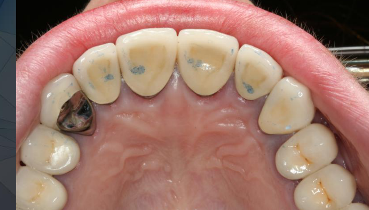
Note the specific location of marks on the tooth surface, as these affect the restoration of:
- Marginal ridges
- Cuspal inclines/slopes
- Transverse ridges
- Grooves and fissures
Bite down for me… no, with your back teeth
Information Collection And Patient Discussion
Data Collection Phase26
- Focus on collecting clinical information; additional diagnostic tests may be required.
- Avoid finalizing the diagnosis and treatment plan at this stage (maintaining suspicion is acceptable).
- Discuss findings briefly and objectively with the patient, offering initial thoughts without losing objectivity.
 |  |
 |
Patient Discussion27
- Discuss findings in a general manner.
- Always frame the discussion with reference to the patient’s perspective.

The Spikes Framework For Communication
The SPIKES Framework28
- Setting
- Perspective & Perception
- Invitation
- Knowledge
- Emotion & empathy
- Strategy and Summary
Source: Curtin and McConnell 2012. While it risks being formulaic, it is a helpful tool for challenging conversations.
SPIKES Component Definitions29
- Setting: Arrange an appropriate venue, time, and duration.
- Perception: Determine the patient’s current understanding of the problem.
- Invitation: Ask the patient how much detail they wish to receive.
- Knowledge: Provide information without using clinical jargon.
- Emotion and empathy: Acknowledge patient emotions and allow time for processing.
- Strategy and summary: Summarize the discussion and outline the next steps for improvement.
Diagnostic Investigations And Guidelines
Clinical Examination And Treatment Planning
Diagnostic Investigations30
- Pulp Vitality: Test 2 sets as part of a new patient exam.
- Specialized Tests: Diet analysis (50g), Extra Hot Test, and testing for cracked teeth where necessary.
- Symptom Assessment: Pain on biting and tooth history.
Radiographic Guidelines
- Take radiographs only when clinically necessary to aid diagnosis.
- Diagnostic cuts may be required for treatment planning, education, or specialist referrals.
Specialist Referrals
Referral is required for procedures beyond the scope of general practice, including:
- Endodontics
- Oral Surgery
- Orthodontics
- Periodontics
- Physiotherapy
- Pharmacology
- Psychological Status (e.g., cognitive impairment)
- Laboratory Tests
- Complex cases involving foreign bodies, trauma, or previous surgical therapy.
Clinical Documentation
- Record the status of teeth to be treated, especially when replacing unsatisfactory restorations.
- All clinical assessment findings should be noted in the radiograph comments section. If a history cannot be obtained, document the reason.
Examination Duration And Workflow
60-Minute Examination Workflow31
- 10-15 minutes: History taking
- 20-30 minutes: Patient examination
- 10-15 minutes: Patient discussion
- 5 minutes: Clinical notes
Note: Focus on the patient rather than the clock.
Additional Diagnostic Steps And Special Tests
Beyond the Clinical Examination32
- Radiographs
- Pulp tests
- Periodontal charting
- Articulated study models
- Dietary analysis
- Plaque scores
- Saliva analysis
- Clinical photographs
Footnotes
-
Original PDF page 1: L3 exam and diagnostic steps, p.1 ↩
-
Original PDF page 22: L3 exam and diagnostic steps, p.22 ↩
-
Original PDF page 2: L3 exam and diagnostic steps, p.2 ↩
-
Original PDF page 3: L3 exam and diagnostic steps, p.3 ↩
-
Original PDF page 4: L3 exam and diagnostic steps, p.4 ↩
-
Original PDF page 5: L3 exam and diagnostic steps, p.5 ↩
-
Original PDF page 6: L3 exam and diagnostic steps, p.6 ↩
-
Original PDF page 7: L3 exam and diagnostic steps, p.7 ↩
-
Original PDF page 8: L3 exam and diagnostic steps, p.8 ↩
-
Original PDF page 9: L3 exam and diagnostic steps, p.9 ↩
-
Original PDF page 10: L3 exam and diagnostic steps, p.10 ↩
-
Original PDF page 11: L3 exam and diagnostic steps, p.11 ↩
-
Original PDF page 12: L3 exam and diagnostic steps, p.12 ↩
-
Original PDF page 13: L3 exam and diagnostic steps, p.13 ↩
-
Original PDF page 14: L3 exam and diagnostic steps, p.14 ↩
-
Original PDF page 15: L3 exam and diagnostic steps, p.15 ↩
-
Original PDF page 16: L3 exam and diagnostic steps, p.16 ↩
-
Original PDF page 17: L3 exam and diagnostic steps, p.17 ↩
-
Original PDF page 18: L3 exam and diagnostic steps, p.18 ↩
-
Original PDF page 19: L3 exam and diagnostic steps, p.19 ↩
-
Original PDF page 20: L3 exam and diagnostic steps, p.20 ↩
-
Original PDF page 21: L3 exam and diagnostic steps, p.21 ↩
-
Original PDF page 23: L3 exam and diagnostic steps, p.23 ↩
-
Original PDF page 25: L3 exam and diagnostic steps, p.25 ↩
-
Original PDF page 24: L3 exam and diagnostic steps, p.24 ↩
-
Original PDF page 26: L3 exam and diagnostic steps, p.26 ↩
-
Original PDF page 27: L3 exam and diagnostic steps, p.27 ↩
-
Original PDF page 28: L3 exam and diagnostic steps, p.28 ↩
-
Original PDF page 29: L3 exam and diagnostic steps, p.29 ↩
-
Original PDF page 30: L3 exam and diagnostic steps, p.30 ↩
-
Original PDF page 31: L3 exam and diagnostic steps, p.31 ↩
-
Original PDF page 32: L3 exam and diagnostic steps, p.32 ↩