Occlusion in Prosthetic Treatment1
Oral Health Centre of Western Australia
The University of Western Australia

Learning Outcomes and Definitions
Learning Outcomes2
- Importance of occlusal analysis for prosthetic treatment (indications)
- Discuss clinical parameters of occlusion: TMJ, muscles of mastication, dental occlusion
- Know how to assess TMJ, masticatory muscles, parafunctional habits, fremitus
- Differentiate Confirmative and Reorganized approaches of prosthetic treatment.
Definition of Occlusion3
occlusion \a-klō′shən\ n (1645):
- The act or process of closure or of being closed or shut off.
- The static relationship between the incising or masticating surfaces of the maxillary or mandibular teeth or tooth analogues.
Clinical Application
Analysis is required for patients suspected of having dysfunctional occlusion or signs of occlusal-related issues.
Related Terms: Articulation, Centric Occlusion, Components of Occlusion, Eccentric Occlusion, Line of Occlusion, Linear Occlusion, Monoplane Occlusion, Pathogenic Occlusion, Spherical Form of Occlusion.
Source: Glossary of Prosthodontic Terms

The Masticatory System4
The masticatory system is composed of the teeth, periodontium, and the articulatory system. These components are interconnected; however, the primary focus of this discussion is the articulatory system and the teeth.
- The articulatory system specifically includes the TMJ and muscles.

Indications for Occlusal Analysis
- Prosthodontic treatment
- TMJ/muscles assessment
- Periodontal assessment
- Mobility assessment
- Functional discomfort
- Mechanically failed restorative treatment
- Bruxism diagnosis
- Orthodontic treatment
System Components:
- Temporomandibular Joint (TMJ)
- Muscles
- Teeth
- Periodontium
Treatment Planning Principles5
- Isolated teeth cannot be treated separately.
- For simple treatments like a single crown where the occlusal scheme is stable and functional, a detailed assessment session may not be necessary.
- Complex treatments for oral rehabilitation require assessment to determine complication risks; failing to address existing challenges like repeatedly failing restorations sets the treatment up for failure.
- Systematic and organized treatment planning is necessary.
- Systematic planning involves gathering relevant information to reach an evidence-based conclusion before carrying out treatment based on those findings.
Importance of Occlusal Records6
Obtaining a good record of the patient’s occlusion is essential for:
- Establishing a baseline record
- Monitoring occlusal changes
- Monitoring disease development
- Assessment of treatment implications
Monitoring Treatment Success
Occlusal records allow for the evaluation of treatment success over time, serving a similar function to a periodontal chart in monitoring health and disease.
Clinical Examination Parameters
Examination Overview7
Clinical examination of occlusion is performed in conjunction with routine dental examination and includes:
- TMJ
- Masticatory muscles
- Dental occlusion
Assessment Categories
Extra-oral assessment:
- Masticatory muscles (masseter, temporalis, medial pterygoid, cervical muscles, suprahyoid muscles)
- TMJ
Intra-oral assessment:
- Masticatory muscles (lateral pterygoid, medial pterygoid)
- Dental occlusion
Temporomandibular Joint Examination
Determining Masticatory Disorders8
Examination aims to identify:
- Pain: Chronic or acute
- Sound: Clicking or crepitus
- Limited movement: Locking or trismus
- Restricted function may require referral to an oral medicine specialist or physiotherapist to increase opening for posterior access.
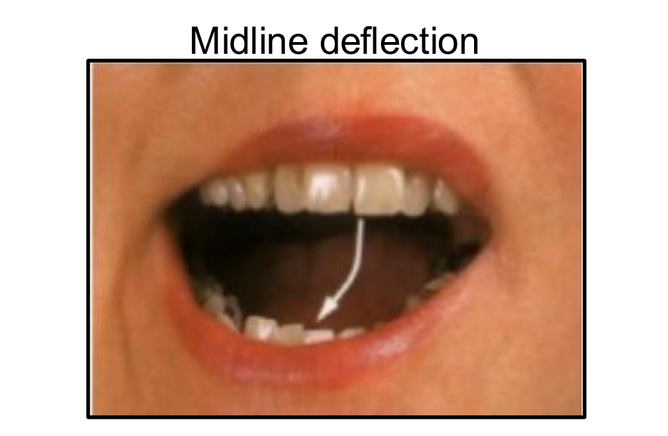
- Midline deviation and Midline deflection
*Note: Average opening is measured as part of the functional assessment.
Quick Functional Test
A quick clinical test for normal opening range is the "three-finger width" rule.
 |  |
Clinical Indicators of Disorder9
- Pain (chronic, acute)
- Sound (clicking, crepitus)
- Limited movement (locking, trismus)
- Midline deviation and Midline deflection
 |  |
Clinical Findings and Evidence Based Practice
Evidence-Based TMD Management
- Occlusion alteration was commonly applied to treat TMD and facial arthromyalgia, but this is not supported by evidence.
- Any TMD-related occlusion treatment should be conservative and reversible (e.g., occlusal splint and removable prosthesis).
- TMD should be stabilized prior to extensive prosthodontic treatment.
- Patients should be informed that prosthodontic treatment is not aimed at restoring TMJ health.
Palpation Techniques
- TMJ Capsules: Palpating the joint area.
- Posterior Palpation: Index fingers in the ears as the patient opens and closes.
- Lateral Palpation: External assessment of the joint
- Palpate anterior to the auricular tragi to assess for symmetrical condylar movement; asynchronous movements suggest disc displacement..
Evaluation Criteria:
- Pain
- Joint sound
- Disc movement
Mandibular Movement and Midline Analysis
Mandibular Movement Evaluation
- Assessment of movement smoothness
- Assessment of mandibular translation
Midline Deflection
- Continuous displacement of the mandibular midline.
- A sign of Anterior Disc Displacement (ADD) without reduction.
Midline Deviation
- The mandible returns to the centered position.
- Indicative of interference during condyle movement.
- A prominent sign of Anterior Disc Displacement (ADD) with reduction.
- Midline deviation often presents as a jagged transition during opening/closing where the mandible returns to the center.
Internal Derangement Classifications
| Condition | Closed Position | Open Position |
|---|---|---|
| Normal | ||
| Disc Displacement with Reduction | ||
| Disc Displacement without Reduction |
Masticatory Muscles Examination
Masseter and Temporalis Muscles
Masseter Muscle Assessment
- Palpation of the masseter muscle at rest and during clenching.
- Tenderness during palpation may suggest occlusal interferences or nighttime bruxing, while hypertrophy indicates strong clenching.
Anatomy of the Masseter:
- Origin: Zygomatic arch and maxillary process of zygomatic bone.
- Insertion: Angle surface of ramus of mandible, coronoid process.
- Artery: Masseteric artery.
- Nerve: Mandibular nerve (V3).
- Actions: Elevation (closing of the mouth) and protrusion of mandible.
Temporalis Muscle Assessment
- Palpation of the temporalis muscle at rest and during clenching.
- Pain in the temporalis is often reported by patients as headaches or pain behind the eye.
Anatomy of the Temporalis:
- Origin: Temporal lines on the parietal bone and superior temporal surface of the sphenoid bone.
- Insertion: Coronoid process of the mandible and retromolar fossa.
- Artery: Deep temporal arteries.
- Nerve: Deep temporal nerves (branches of V3).
- Actions: Elevation and retraction of mandible.
Pterygoid Muscles
Medial Pterygoid Muscle
- Palpation: Medial to the mandibular angle.
- Intra-oral palpation is performed behind the molars along the buccal surface of the ramus; tenderness here is a dependable landmark for occluso-muscular imbalance.
Anatomy:
- Origin:
- Deep head: Medial side of lateral pterygoid plate.
- Superficial head: Pyramidal process of palatine bone and maxillary tuberosity.
- Insertion: Medial angle of the mandible.
- Artery: Pterygoid branches of maxillary artery.
- Nerve: Mandibular nerve via nerve to medial pterygoid.
- Actions: Elevates mandible, closes jaw, assists lateral pterygoids in side-to-side movement.
Lateral Pterygoid Muscle
Anatomy:
- Origin:
- Superior head: Infratemporal surface of sphenoid bone.
- Inferior head: Lateral pterygoid plate.
- Insertion:
- Superior head: Anterior side of the mandibular condyle.
- Inferior head: Pterygoid fovea.
- Artery: Pterygoid branches of maxillary artery.
- Nerve: Lateral pterygoid nerve from mandibular nerve.
- Actions: Depresses and protrudes mandible; facilitates side-to-side movement.
Lateral Pterygoid Assessment
This muscle is difficult to palpate directly. Assessment is done via resistance testing: ask the patient to protrude their chin against the resistance of the clinician's fist. Pain during this movement suggests lateral pterygoid involvement.
Suprahyoid Muscles
Suprahyoid Muscle Palpation
Clinical examination includes the palpation of the suprahyoid muscle group to assess function and tenderness.
Digastric Muscle
- Attachments:
- Anterior belly: Arises from the digastric fossa of the mandible.
- Posterior belly: Arises from the mastoid process of the temporal bone.
- Connection: The two bellies connect via an intermediate tendon attached to the hyoid bone.
- Actions: Depresses the mandible and elevates the hyoid bone.
Stylohyoid Muscle
- Attachments: Arises from the styloid process of the temporal bone and attaches to the lateral aspect of the hyoid bone.
- Actions: Initiates swallowing by pulling the hyoid bone in a posterior and superior direction.
Geniohyoid Muscle
- Attachments: Arises from the inferior mental spine of the mandible; travels inferiorly and posteriorly to attach to the hyoid bone.
- Actions: Depresses the mandible and elevates the hyoid bone.
Mylohyoid Muscle
- Attachments: Originates from the mylohyoid line of the mandible and attaches onto the hyoid bone.
- Actions: Elevates the hyoid bone and the floor of the mouth.
Suprahyoid Muscles Overview
Comprehensive palpation of the suprahyoid muscle group is a standard component of the masticatory muscle examination.
- These muscles are often involved when the mandible is postured forward to avoid deflective occlusal interferences.
Functional Range of Motion
Clinical Range of Motion Parameters
- Degree of opening: An inter-incisal opening of less than 40 mm may hinder efficient prosthodontic treatment.
- Dynamic movements: Assessment of mandibular movements against resistance.
Normal Range of Motion Values
- Opening: 40–50 mm
- Lateral movements: 7–15 mm
- Protrusive: 7–15 mm
Dental Occlusion Assessment
Static and Dynamic Parameters10
Static Parameters
- Centric occlusion and MIP (long centric or eccentric)
- Freedom in centric
- Extent of posterior tooth support
- Angle’s classification
- Overbite and overjet
- Cross bite
Dynamic Parameters
- Protrusion
- Lateral movements:
- Canine guidance
- Group function
- Balanced occlusion
- Interferences
- Centric Interferences: Premature contact during closure to optimal position.
- Working/Non-working Interferences: Contacts on the side of or opposite to the direction of mandibular movement (non-working are particularly destructive).
- Protrusive Interferences: Premature posterior contacts during forward movement.
Evaluate the following during examination:
- Bruxism/clenching
- Mobility
- Periodontitis
- Tooth stability
- Mechanical failure
 |  |
 |
Bruxism and Periodontal Health
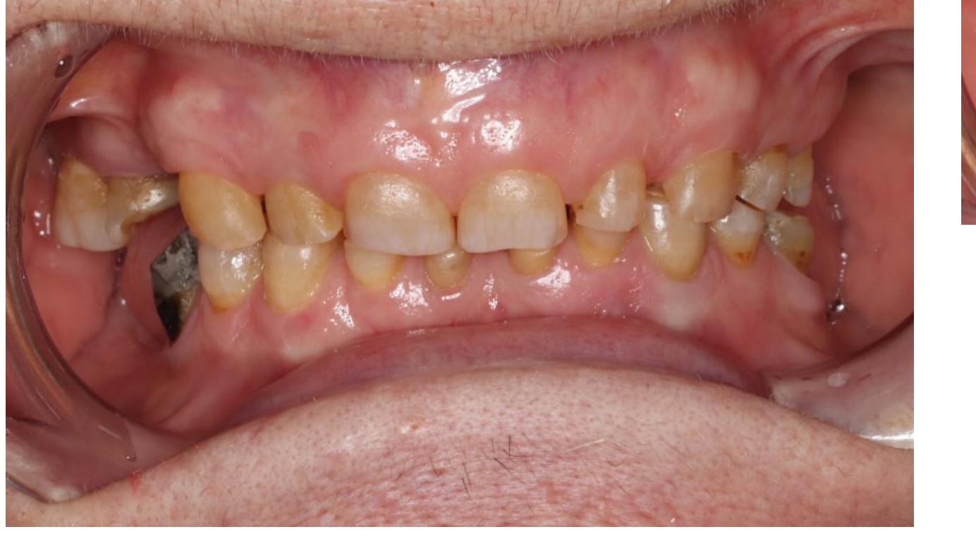
Clinical Signs of Bruxism
- Worn teeth
- Muscle tenderness
- Muscle hypertrophy
- Cracked teeth
- TMJ pain, locking, or clicking
Periodontal Considerations
- Periodontitis: Altering occlusion specifically to treat periodontitis is not recommended due to lack of evidence.
- Mobility: Increasing mobility that concerns the patient may require occlusal management, often in conjunction with splinting.
- Occlusal trauma may appear as a thickened PDL or blunted root tips on radiographs.
Tooth Mobility and Stability
Fremitus Assessment
- Definition: Vibration or movement of teeth during light tapping.
- Method: Observation of tooth movement against a finger placed across the tooth while the patient taps teeth together.
- Significance: Indicates the presence of deflective contacts.
- Fremitus indicates excessive loading or premature contacts.
Miller Classification of Tooth Mobility
- Degree 0: “Physiological” mobility (0.1–0.2 mm horizontal).
- Degree 1: ≤ 1 mm horizontal mobility.
- Degree 2: > 1 mm horizontal mobility.
- Degree 3: Both horizontal and vertical mobility.
Tooth Stability and Migration
- Posterior deflective contacts or loss of posterior support can result in the drifting of maxillary incisors.
- This migration can lead to open contacts and food impaction.
Positional Changes
- Over-eruption of unopposed teeth.
- Drifting or tilting of teeth.
Vertical Dimensions and Mechanical Failure11121314
Causes of Mechanical Failure
Mechanical failure in restorations can be attributed to:
- Poor restoration design
- Lack of occlusal stability
- Repeatedly failing restorations (cracking, debonding) often stem from unresolved occlusal issues like lack of posterior support.
Vertical Dimension Definitions
- Rest Vertical Dimension (RVD)
- Occlusal Vertical Dimension (OVD)
- Freeway Space (FWS)
- Rest Vertical Dimension (RVD) can be measured after the patient says the word "Emma".
- Freeway Space is usually approximately 3mm.
 |  |  |
 |  |  |
 |  |  |
 |  |
Clinical Case Planning and Mock Ups
Case Planning
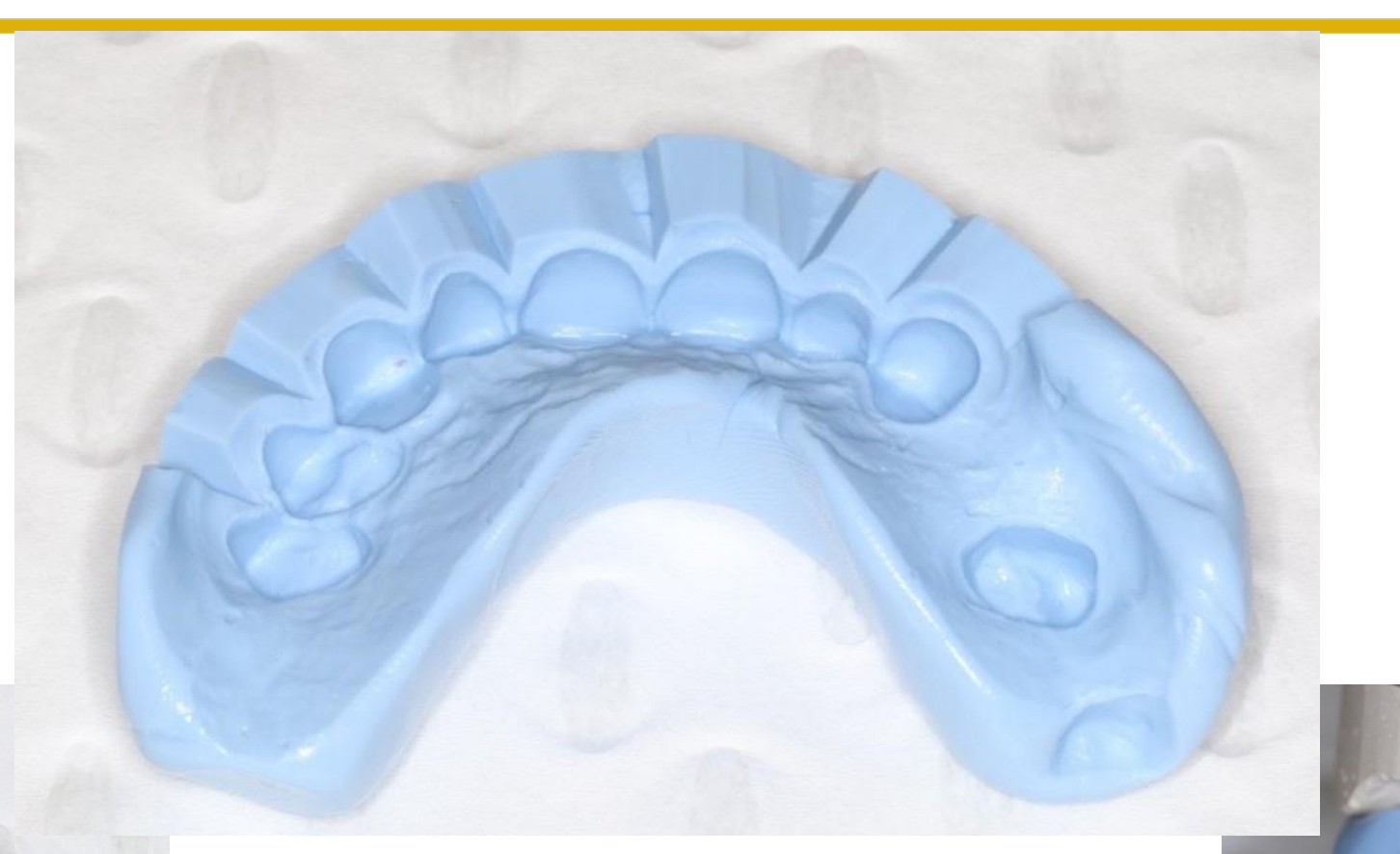
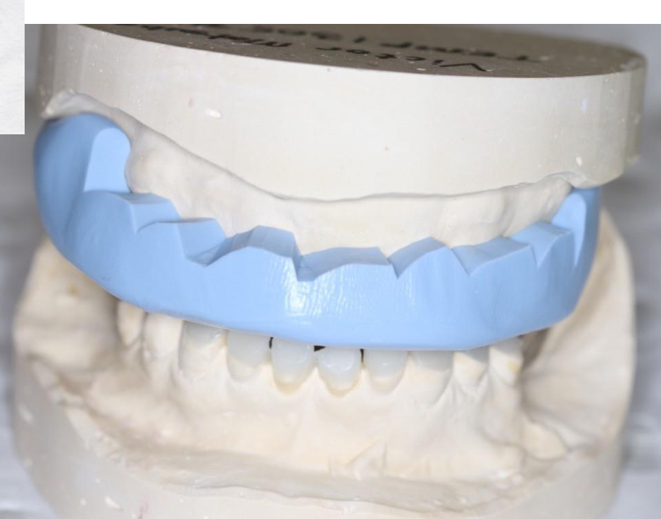
Use of materials like Luxatemp for creating mock-ups during the planning phase of prosthetic treatment.
Mock-up Procedures
Planning your case using Luxatemp mock-ups to visualize and test occlusal changes.
Principles of Ideal Occlusion
Criteria for Ideal Occlusion15
- Mandibular stability
- Axial occlusal load
- No interference on the working side during lateral excursions
- Disocclusion on the non-working side during lateral excursions
- Disocclusion of posterior teeth during protrusion
Prosthetic Treatment Approaches
Treatment Planning Considerations16
Occlusal treatment is typically provided alongside prosthodontic work. The approach must be decided during the planning stage based on:
- Address the root cause (e.g., lack of posterior support) before repairing anterior teeth.
- Consider orthodontic treatment prior to prosthodontics to improve the path of insertion or upright tilted molars.
- Assess prosthetic space (height and width) before proposing crowns or bridges.
- Complexity of treatment
- Required modifications
- Condition of existing dentition/occlusion
- Presence of occlusal abnormalities
Prosthetic Approaches17
There are two primary approaches to prosthetic treatment:
- Conformative
- Reorganized
Conformative Approach
Principles of the Conformative Approach18
- Provision of restorations in harmony with existing jaw relations
Financial Commitment
The choice of prosthetic approach is influenced by the patient's financial commitment in addition to clinical complexity and existing abnormalities. hips.
- Restorations are made according to the Maximal Intercuspal Position (MIP).
- Restorations must fit into the existing occlusal scheme.
- Occlusal contacts on unrestored teeth remain unaltered following treatment.
Advantages of the Conformative Approach19
- Most cost-effective restorative method.
- Requires the least restorative intervention.
- Most common method in restorative dentistry.
- Applicable for single or multiple restorations.
- Easiest and safest method; less likely to introduce new problems to the teeth, periodontium, muscles, or TMJ.

Reorganized Approach
Principles of the Reorganized Approach20
- Altering the existing occlusal scheme to establish an ideal (or near-ideal) occlusion.
- Restorations are made according to Centric Relation.
- Centric Relation (CR) is used because it is a reproducible position independent of tooth contact.
- Requires additional stages to design and establish the new occlusion (using provisional restorations) before the definitive prosthesis.
- Once the new occlusion is established, subsequent treatment conforms to that new scheme.
- Establishing a reorganized occlusion requires thorough planning, often involving temporary mock-ups (e.g., Luxatemp) before final restorations.
 |  |
 |
Indications for the Reorganized Approach21
- An increase in occlusal vertical dimension (OVD) is required.
- Teeth are significantly malpositioned (over-erupted, tilted, or rotated).
- History of repeated restoration failures within the existing occlusal scheme.
- Absence of posterior occlusal contacts at the desired vertical dimension.
- The current occlusal scheme is dysfunctional or parafunctional.

Summary and Clinical Recommendations22

Clinical Recommendations23
- Thorough occlusal analysis is mandatory prior to any prosthetic treatment.
- The patient’s occlusion should be recorded before starting prosthodontic treatment.
- The conformative approach is easier and more predictable; it should be considered the first treatment option.
- If a patient has confusing symptoms (e.g., pain of unknown origin), err on the side of caution with reversible occlusal adjustments rather than invasive endodontic treatment.
- Refer complex reorganized cases to specialists if necessary.
Thank you
Audio Appendix
Additional Audio Content
The following sections from the lecture audio did not correspond to any heading in the main document.
Case Study: Complex Oral Rehabilitation (Dr. Matsubara)
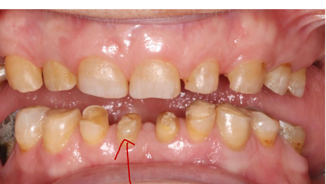
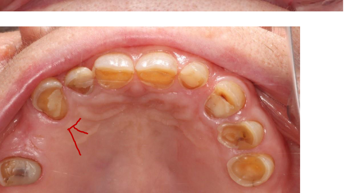
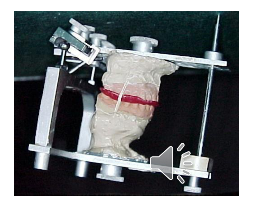
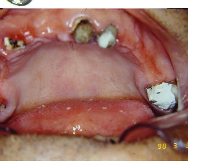
- Patient Presentation: Loss of posterior support, severe wear on lower central incisors and palatal surfaces of upper teeth.
- Procedure:
- Maxillomandibular Relationship (MMR) recorded via bite rims.
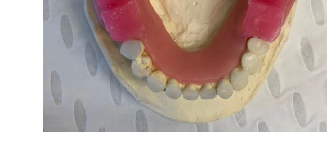
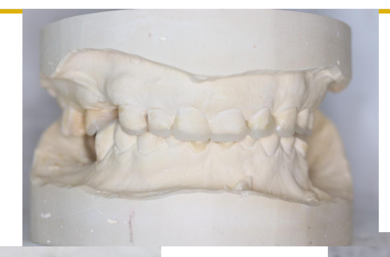
- Diagnostic wax-up on models.
- Transfer to the patient using a putty key and Luxatemp as a mock-up.
- Purpose: To test the patient’s tolerance for a change in vertical dimension and aesthetic results before committing to permanent crowns.
Footnotes
-
Original PDF page 1: L5. Clinical Exam of Occlusion, p.1 ↩
-
Original PDF page 2: L5. Clinical Exam of Occlusion, p.2 ↩
-
Original PDF page 3: L5. Clinical Exam of Occlusion, p.3 ↩
-
Original PDF page 4: L5. Clinical Exam of Occlusion, p.4 ↩
-
Original PDF page 6: L5. Clinical Exam of Occlusion, p.6 ↩
-
Original PDF page 7: L5. Clinical Exam of Occlusion, p.7 ↩
-
Original PDF page 8: L5. Clinical Exam of Occlusion, p.8 ↩
-
Original PDF page 9: L5. Clinical Exam of Occlusion, p.9 ↩
-
Original PDF page 10: L5. Clinical Exam of Occlusion, p.10 ↩
-
Original PDF page 28: L5. Clinical Exam of Occlusion, p.28 ↩
-
Original PDF page 38: L5. Clinical Exam of Occlusion, p.38 ↩
-
Original PDF page 39: L5. Clinical Exam of Occlusion, p.39 ↩
-
Original PDF page 40: L5. Clinical Exam of Occlusion, p.40 ↩
-
Original PDF page 41: L5. Clinical Exam of Occlusion, p.41 ↩
-
Original PDF page 44: L5. Clinical Exam of Occlusion, p.44 ↩
-
Original PDF page 45: L5. Clinical Exam of Occlusion, p.45 ↩
-
Original PDF page 46: L5. Clinical Exam of Occlusion, p.46 ↩
-
Original PDF page 47: L5. Clinical Exam of Occlusion, p.47 ↩
-
Original PDF page 48: L5. Clinical Exam of Occlusion, p.48 ↩
-
Original PDF page 49: L5. Clinical Exam of Occlusion, p.49 ↩
-
Original PDF page 50: L5. Clinical Exam of Occlusion, p.50 ↩
-
Original PDF page 52: L5. Clinical Exam of Occlusion, p.52 ↩
-
Original PDF page 51: L5. Clinical Exam of Occlusion, p.51 ↩