Historical Development of CT[^18]
Sixth Generation CT: Helical (1990s)[^17]
- Slip-ring technology: Developed to allow the gantry to rotate continuously without the restriction of wires.
- Helical CT scanners: These systems acquire data while the table is moving.
- Introduced helical scanning capabilities, improving speed and volume coverage.
Seventh Generation CT (Late 1990s – 2000)
- Multiple Detector Array: Introduction of systems capable of capturing multiple slices simultaneously.
Tomographic Reconstruction[^19][^20]
- An algorithm is used to produce the CT image, which serves as an attenuation coefficient map.
- Filtered back projection is the most widely used reconstruction method in clinical CT scanners.
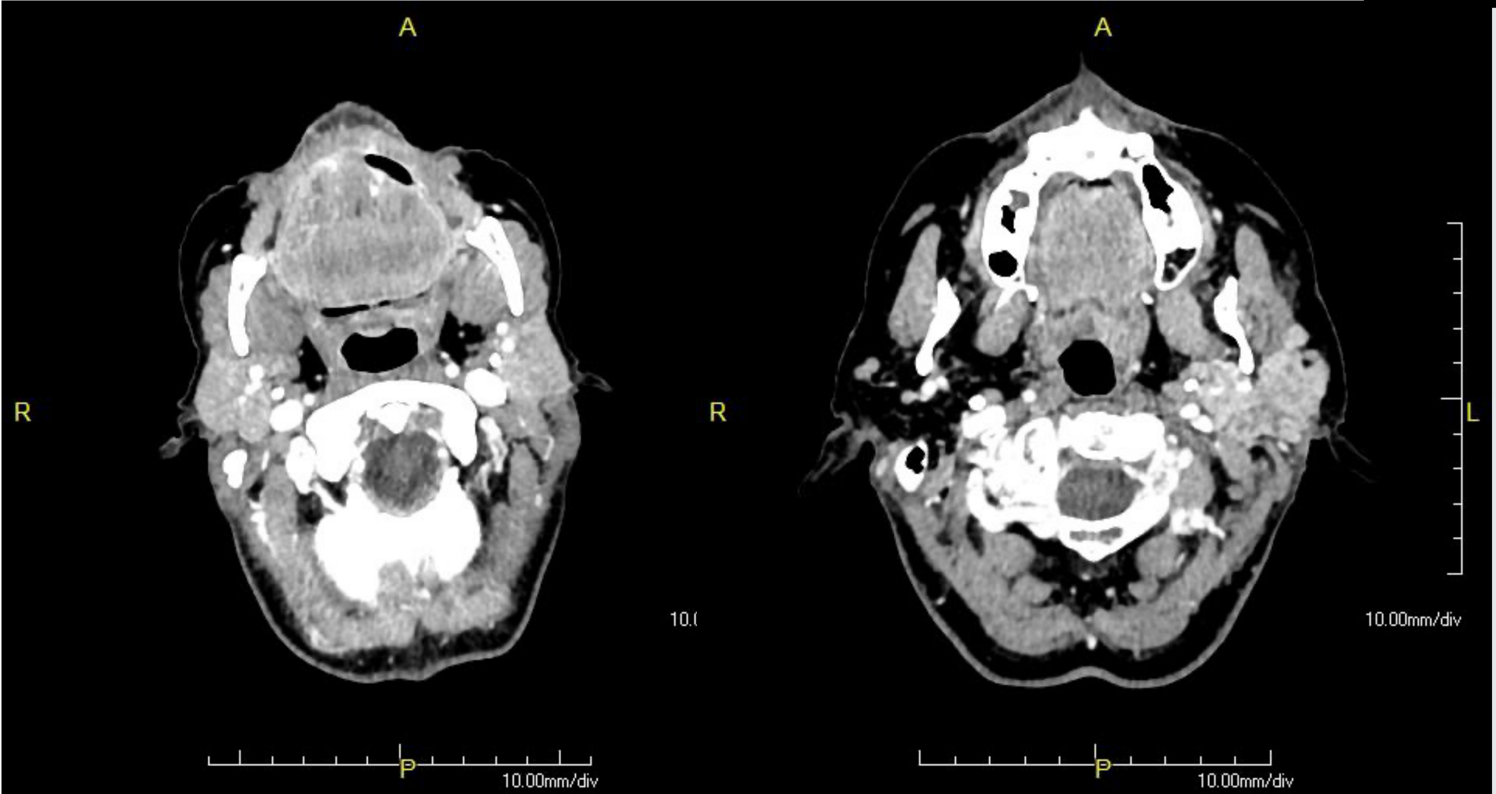
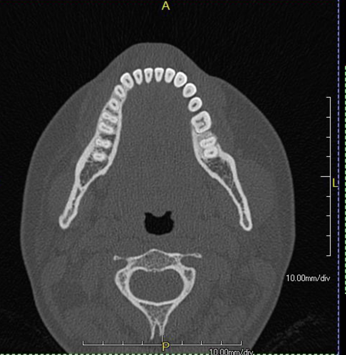
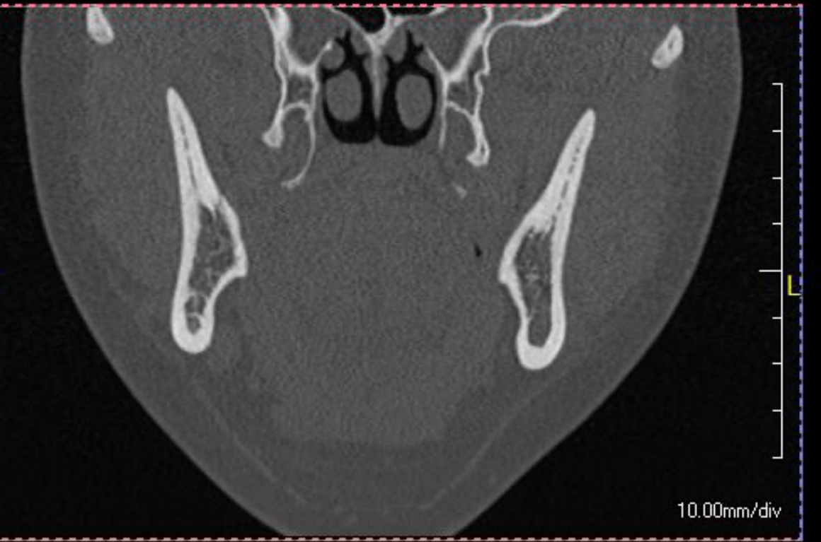
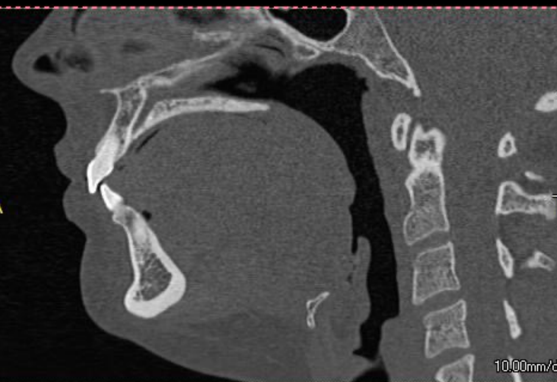
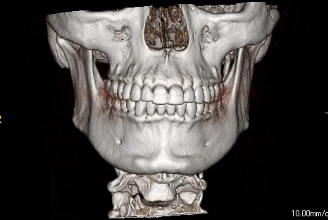
- Bony pathology is very accurately seen with Multislice CT (MSCT).

Hounsfield Unit[^21]
- The Hounsfield unit (HU) is a quantity commonly used in CT scanning to express CT numbers in a standardized and convenient form.
- Hounsfield units, created by and named after Sir Godfrey Hounsfield, are obtained from a linear transformation of the measured attenuation coefficient.
- This transformation is based on the arbitrary definitions of air and water.
- Quantitative measurement of tissue density is possible, unlike in CBCT.

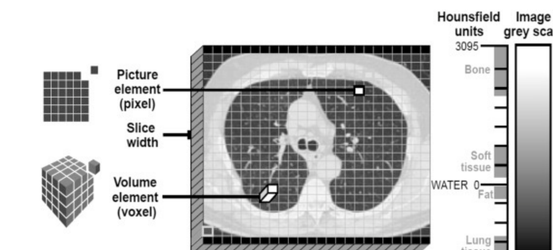
CT Numbers and Hounsfield Units for Various Substances[^22]
| Substance | HU |
|---|---|
| Air | -1000 |
| Lung | -500 |
| Fat | -100 to -50 |
| Water | 0 |
| CSF | 15 |
| Kidney | 30 |
| Blood | +30 to +45 |
| Muscle | +10 to +40 |
| Grey matter | +37 to +45 |
| White matter | +20 to +30 |
| Liver | +40 to +60 |
| Soft Tissue, Contrast | +100 to +300 |
| Bone | +700 (cancellous) to +3000 (dense) |
Digital Image Display Window Level

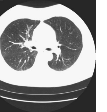

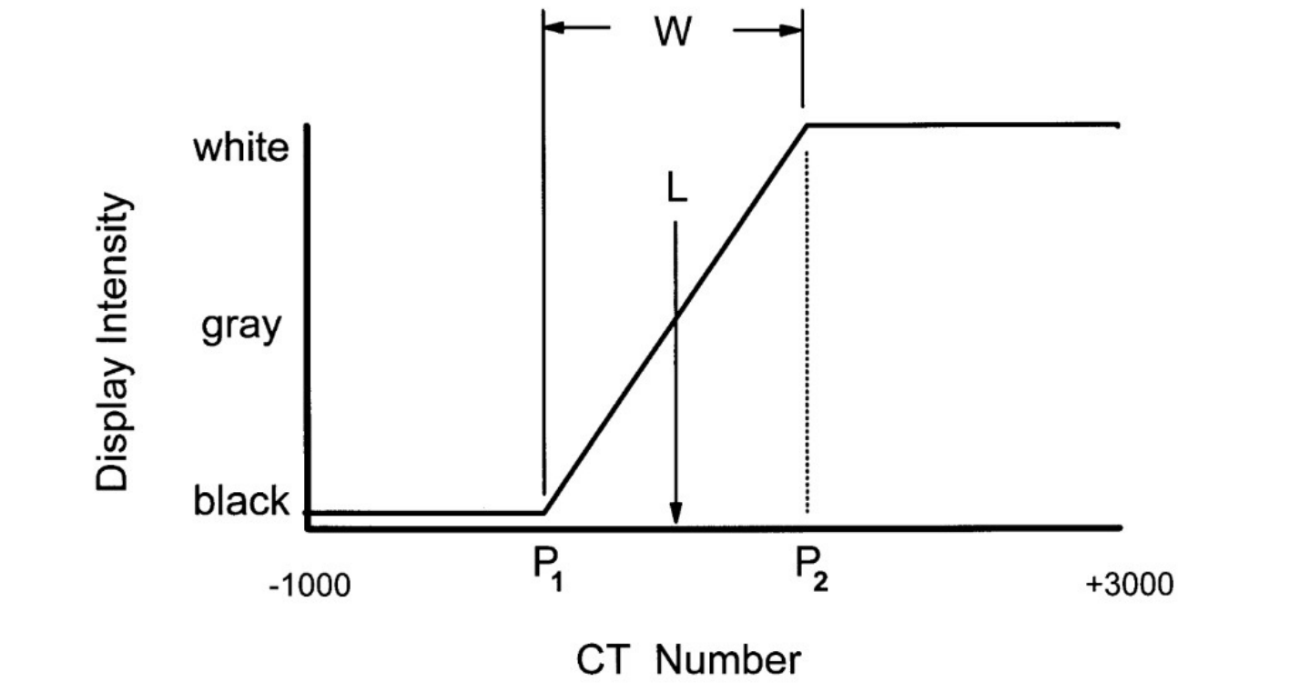

Display Intensity Mapping
| CT Number | Display Intensity |
|---|---|
| -1000 to P1 | Black |
| P1 to P2 | Gray (linearly) |
| P2 to +3000 | White |
Calculation Formulas:
- P1 = L – ½ W
- P2 = L + ½ W
Example Settings:
- W = 4095, L = 1048
- W = 600, L = -100
- W = 700, L = -650
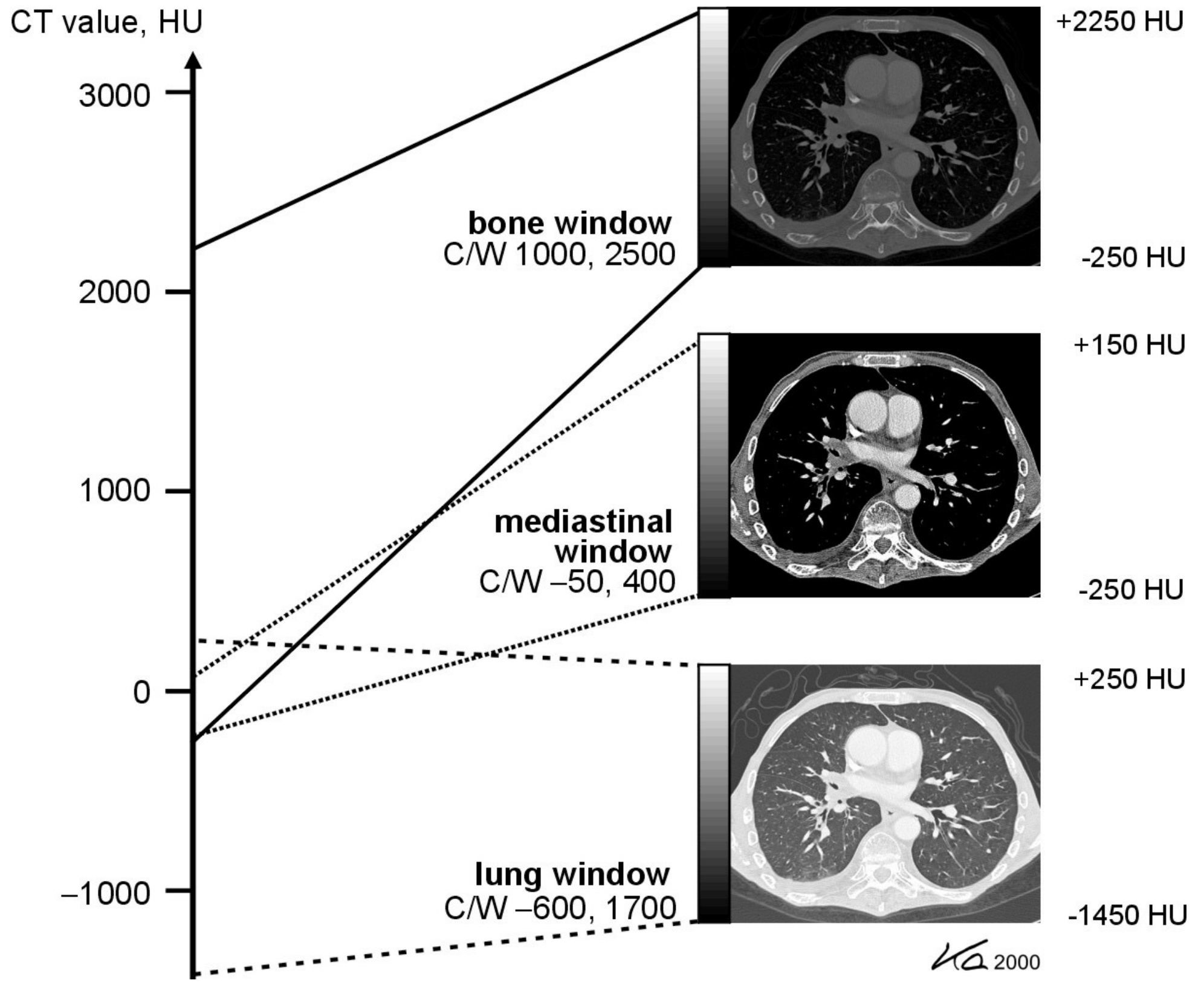
Common Clinical Window Settings[^24]
-
Bone Window
- Center/Width (C/W): 1000, 2500
- Range: +2250 HU to -250 HU
-
Mediastinal Window
- Center/Width (C/W): -50, 400
- Range: +150 HU to -250 HU
-
Lung Window
- Center/Width (C/W): -600, 1700
- Range: +250 HU to -1450 HU
Comparative Windowing Examples[^25]
- Soft Tissue Focus: W:300, L:50
- Bone/Dense Tissue Focus: W:3000, L:500
Contrast Agents[^26][^27]
- Contrast agents frequently utilize Iodine.
- These agents help to better visualize internal structures.
- Tumors: Highly vascular tumors show enhancement, revealing the full extent and vascularity.
- Differentiation: Helps differentiate tumors from cysts.
- Speed: CT is the fastest imaging available, ideal for patients who are kyphotic, frail, or cannot stand still.