TMJ Imaging Anatomy
TMJ Disc Displacement Case Study
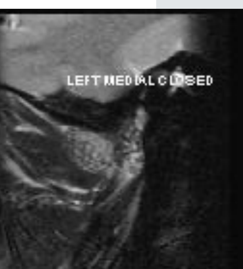
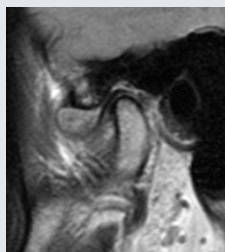
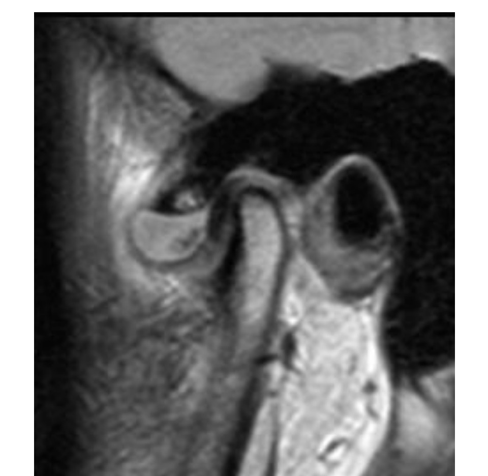
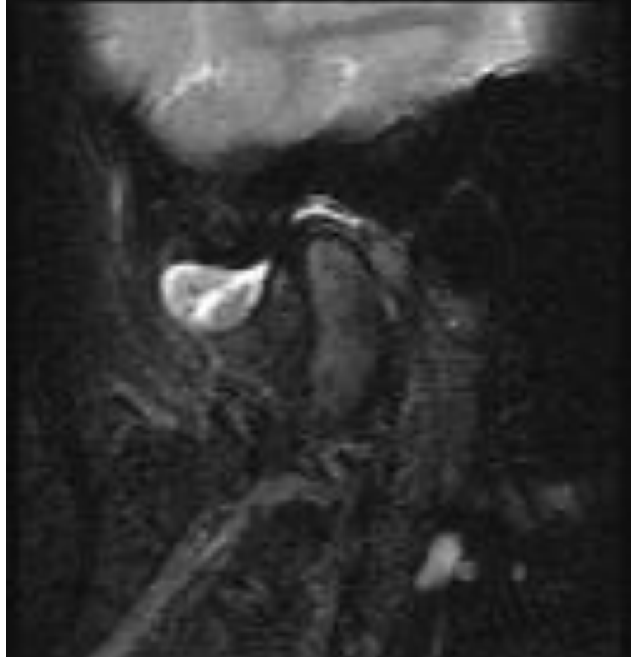
Case 1: TMJ Internal Derangement
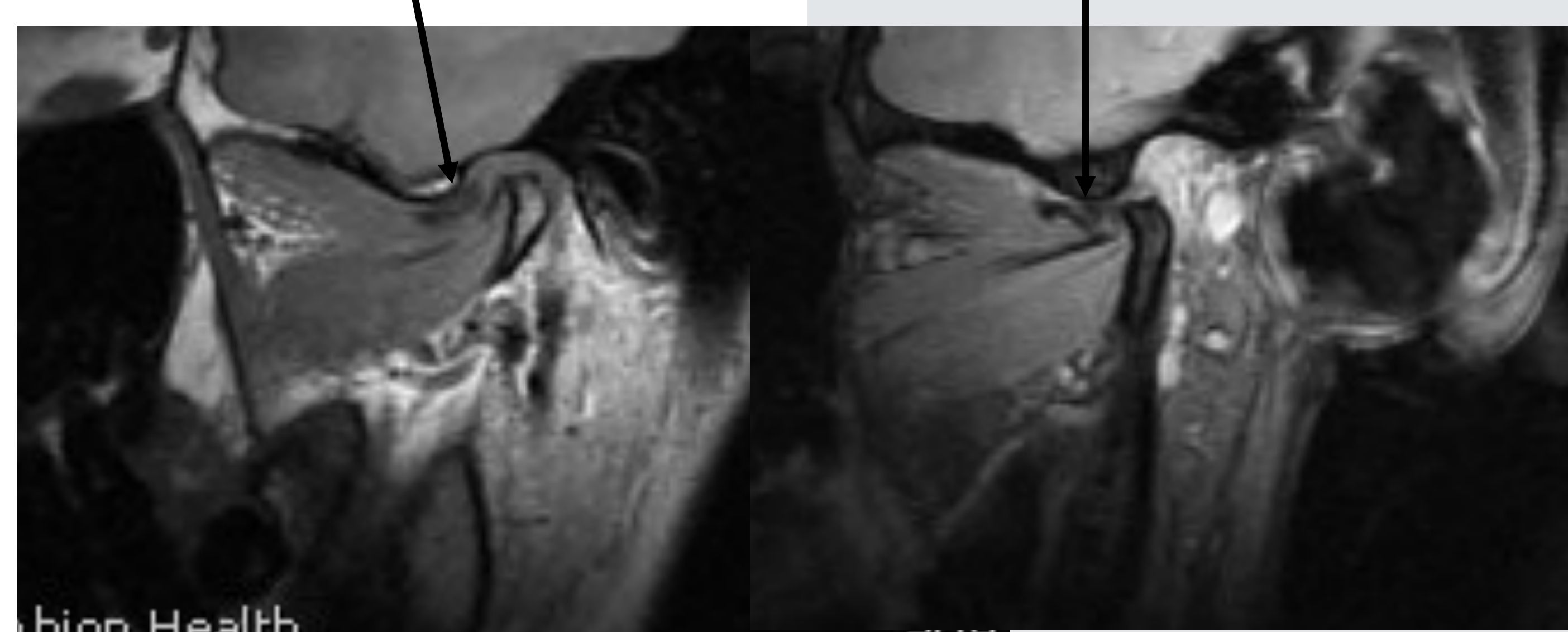
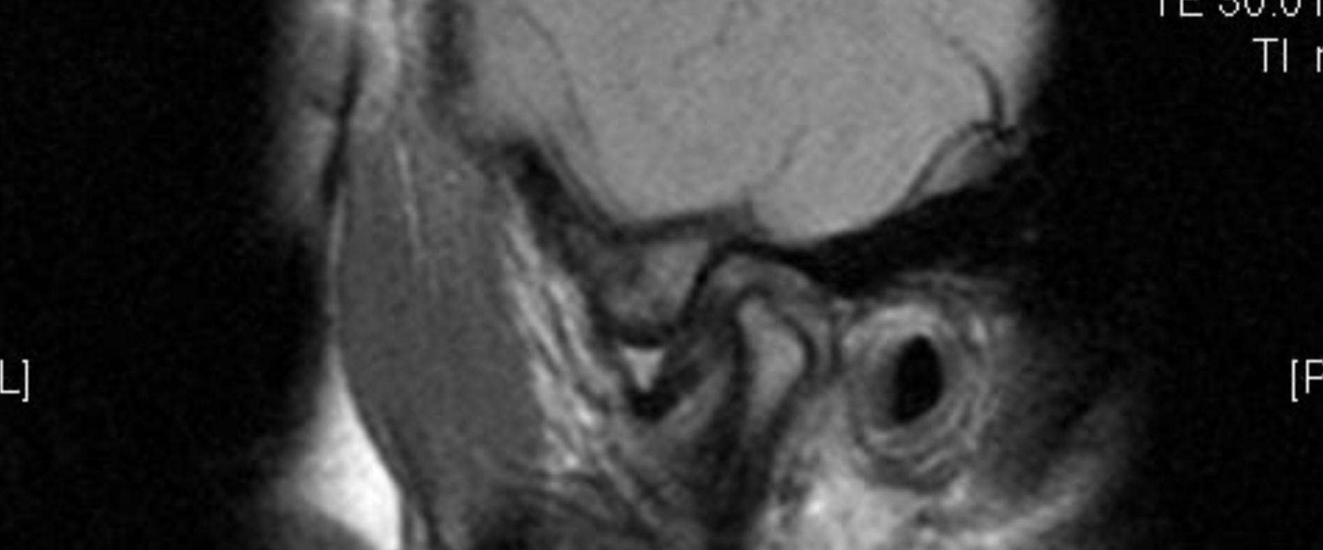
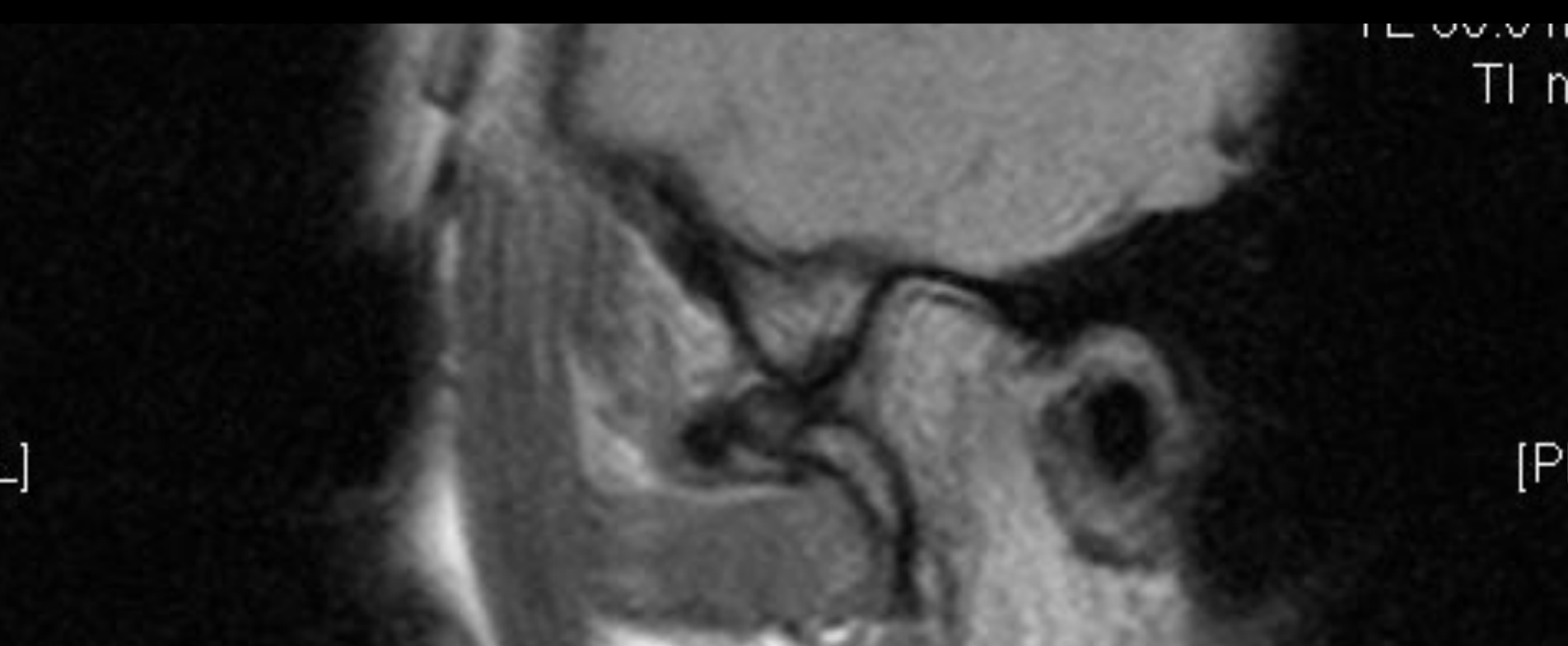
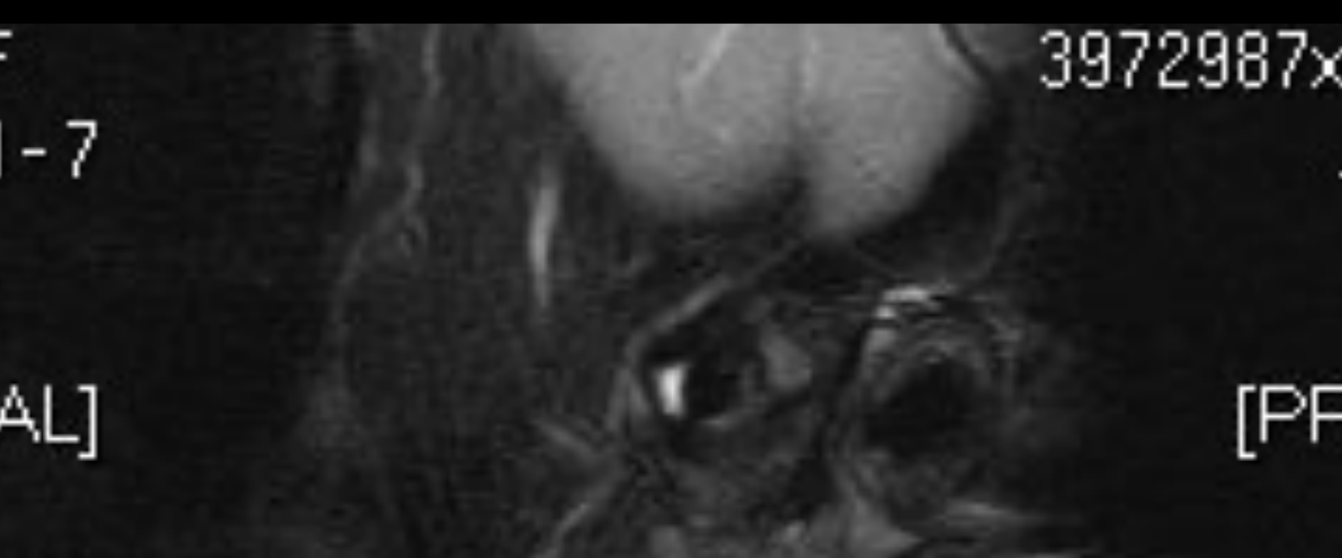
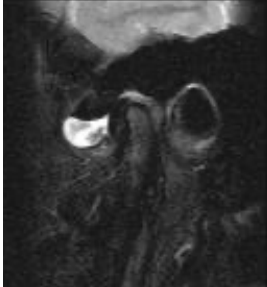
- ==Imaging: Open and closed mouth MRI==
- ==Findings: Disc was anteriorly positioned relative to the condylar head in closed mouth position. On opening, disc remained anteriorly displaced.==
- ==Diagnosis: Non-reducing anterior disc displacement==
- ==Clinical Correlation: Patient experiences popping/clicking sounds because the disc is not recaptured.==
Understanding MRI Signals
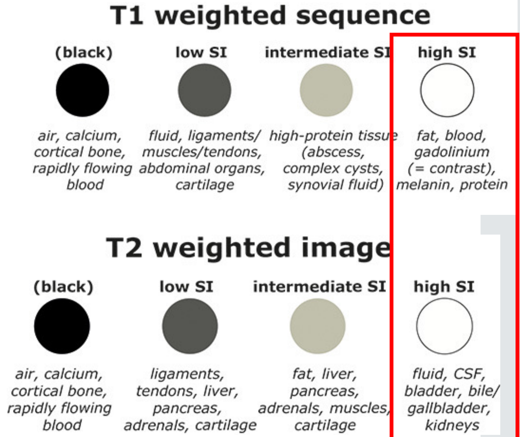
T1 Weighted Sequence (T1WI)
- Low Signal Intensity (Black): Air, calcium, cortical bone, rapidly flowing blood.
- Intermediate Signal Intensity: Fluid, ligaments, muscles, tendons, abdominal organs, cartilage.
- High-Protein Tissue: Abscess, complex cysts, synovial fluid.
- High Signal Intensity (Bright): Fat, blood, gadolinium (contrast), melanin, protein.
T2 Weighted Image (T2WI)
- Low Signal Intensity (Black): Air, calcium, cortical bone, rapidly flowing blood.
- Intermediate Signal Intensity: Ligaments, tendons, liver, pancreas, adrenals, cartilage.
- Variable Intermediate/High: Fat, liver, pancreas, adrenals, muscles, cartilage.
- High Signal Intensity (Bright): Fluid, Cerebrospinal Fluid (CSF), bladder, bile/gallbladder, kidneys.
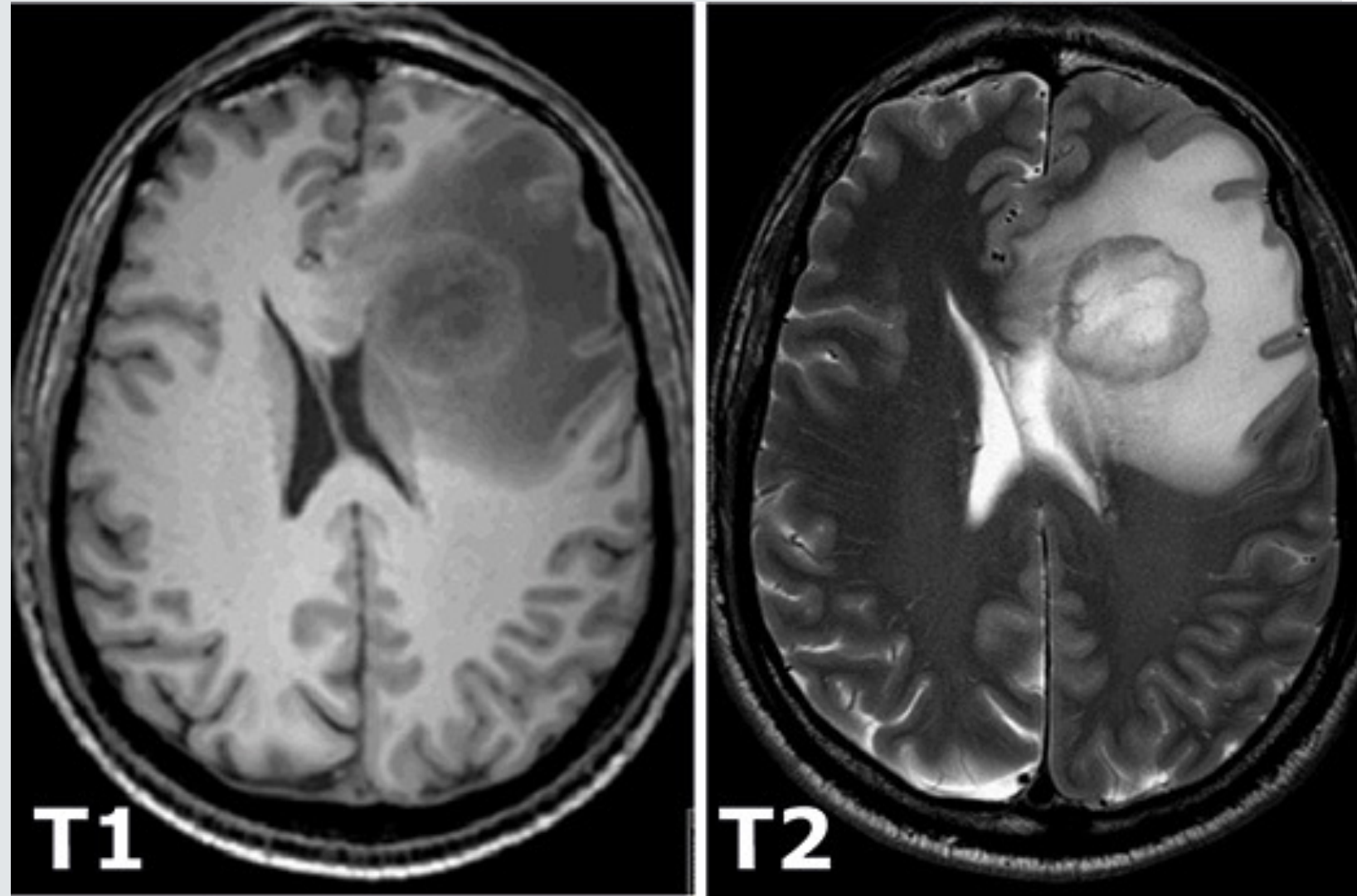
T1WI vs T2WI Comparison[^46][^47][^48][^49][^50][^51][^52][^53]
Primary Signal Differentiators
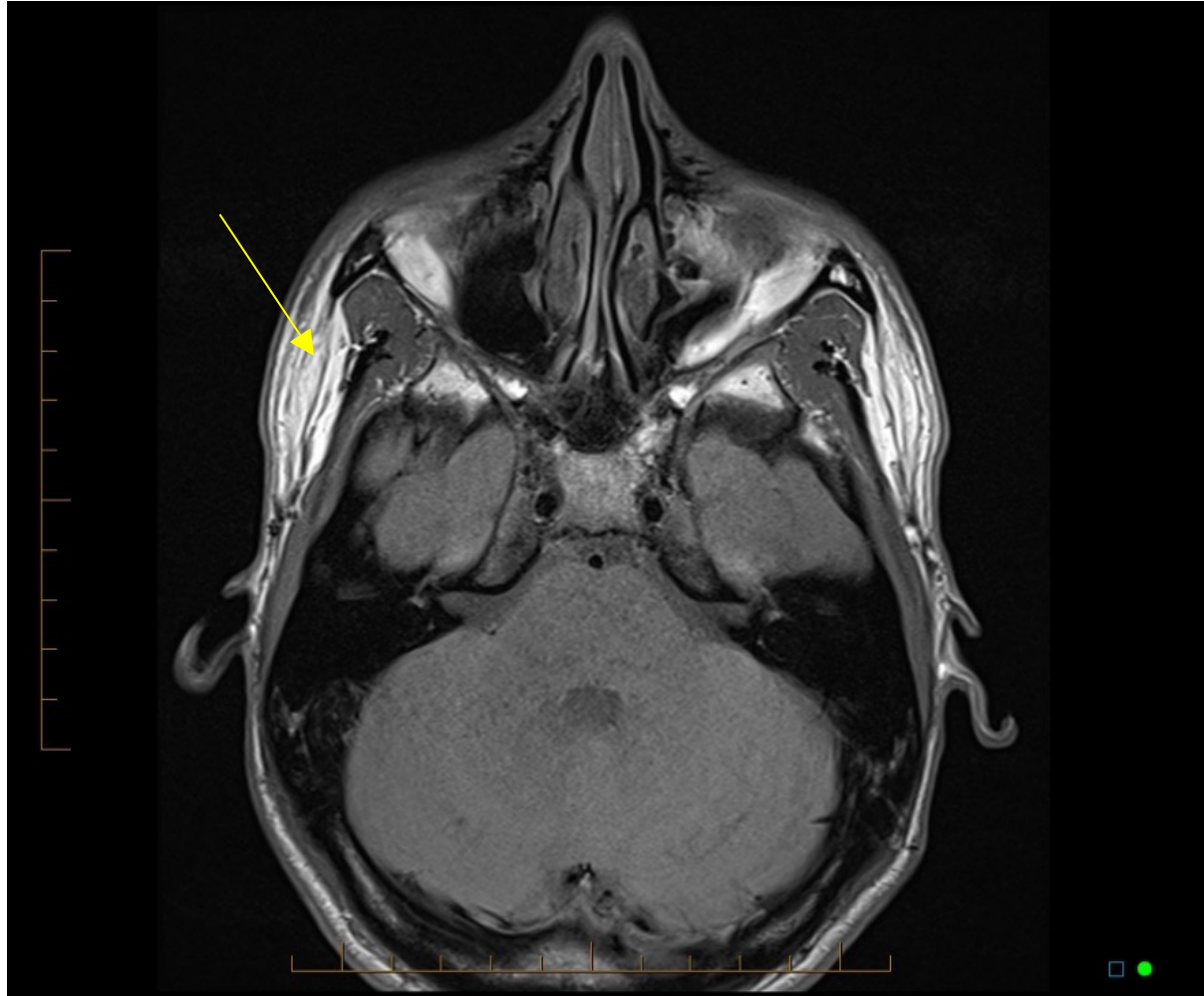
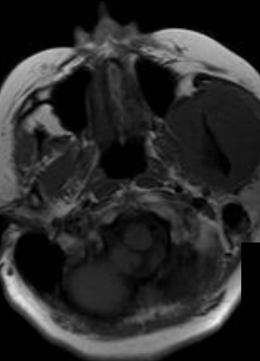
- T1WI: Fat appears bright.
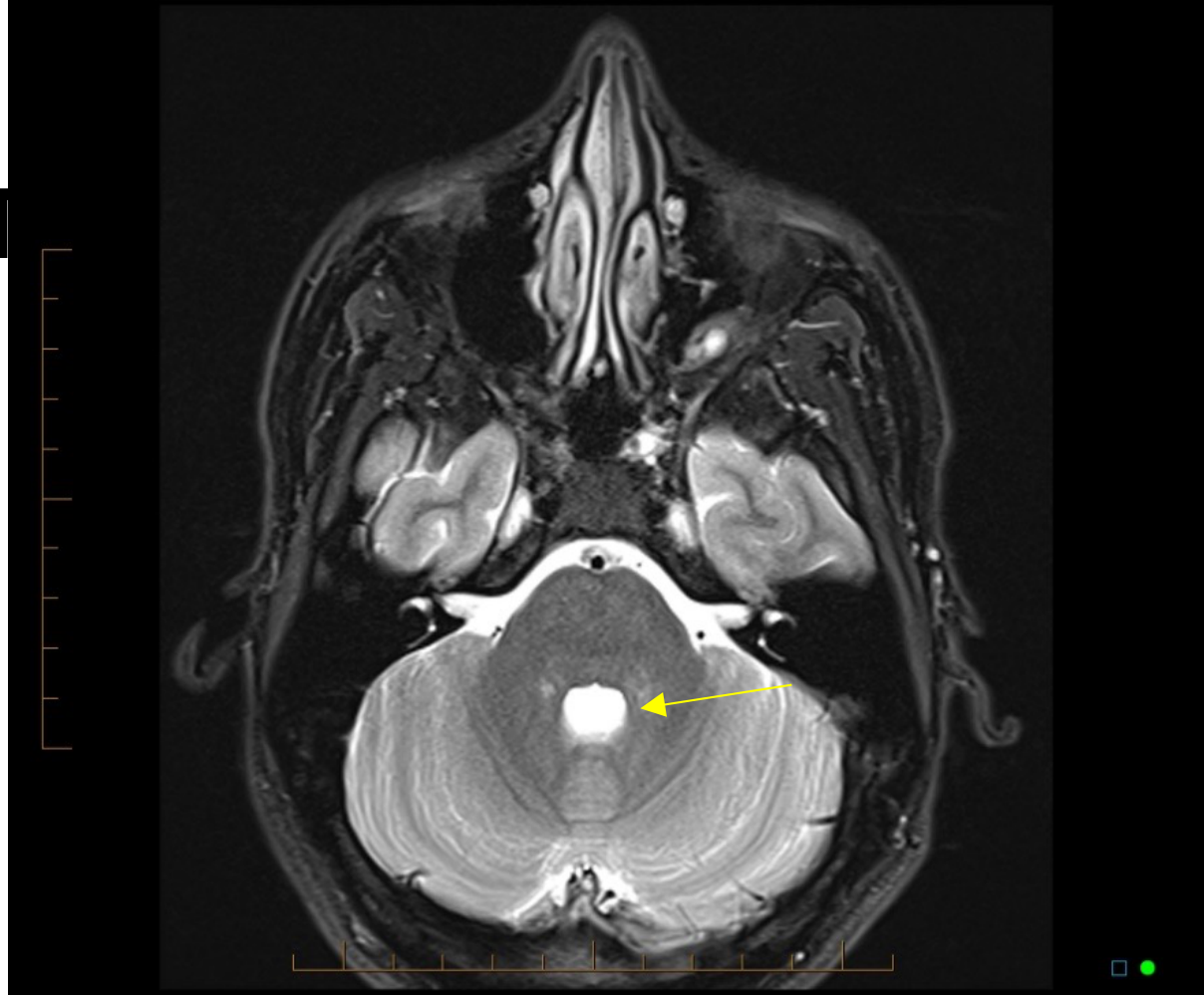
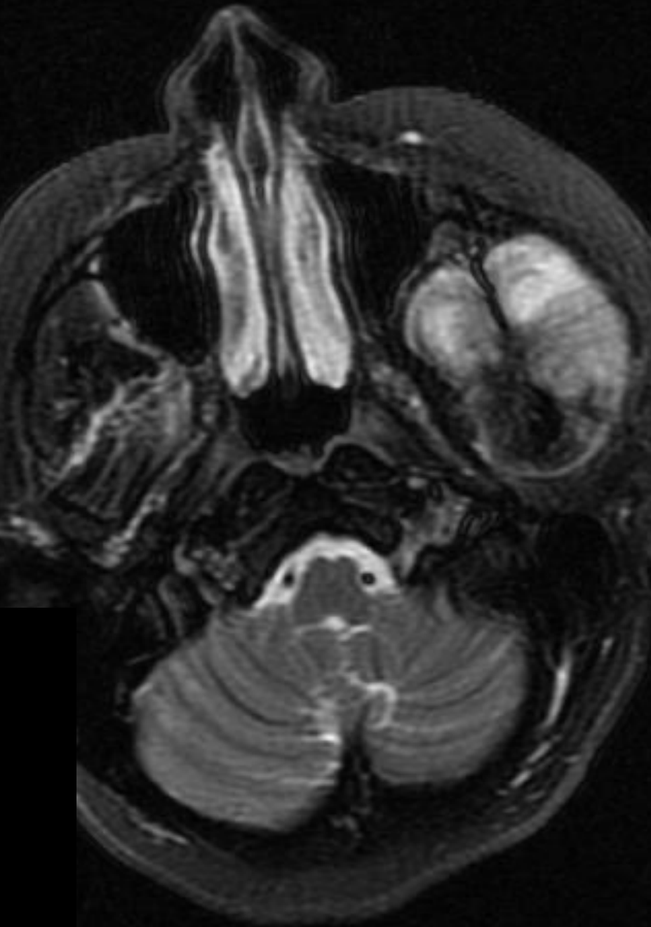
- T2WI: Fluid appears bright.
TMJ Disc Visualization
- Closed Position: Evaluation of the disc relationship to the condyle and glenoid fossa.
- Open Position: Evaluation of the disc translation and morphology during mandibular excursion.
This page intentionally left blank in the source material.
MRI Principles
Image Generation Process[^41]
- Magnetic field and radiofrequency pulses: The patient is placed in a relatively strong magnetic field.
- Hydrogen alignment: Nuclei of many atoms, mainly hydrogen, align with the magnetic field.
- Radiofrequency signal: Several pulses are applied, causing protons to rotate away from the direction induced by the imaging magnet.
- Relaxation and detection: At the end of the pulse sequence, relaxation occurs, and the signal is detected by a receiver coil.
- Signal intensity: A larger number of nuclei of loosely bound hydrogen atoms results in a larger intensity of the recovered signal.
- Reconstruction: Fourier transform and reconstruction techniques are used to produce the final images.
- Signals are generated based on tissue properties (fluid, fat, etc.) rather than density