Oral Burning Sensation Case Study1
DISCUSSION CASE
 |  |  |
 |  |  |
Case Description and Patient History
Patient Profile and Chief Complaint2
A 58-year-old female presents with a burning sensation in the oral cavity that has persisted for two months.
 |  |
Medical and Social History
- Medical History: Significant for hypertension and diabetes. The patient's diabetic status is relevant for treatment planning due to the risk of oral candidiasis when using steroids.
- Medications: Currently managed with hydrochlorothiazide (for hypertension) and metformin (for diabetes).
- Smoking History: Ex-smoker with a 30-year history (one pack per day); quit 10 years ago.
- Alcohol Consumption: None.
- Dental History: Last professional dental visit was 12 months ago.
Clinical Activity and Differential Diagnosis
Clinical Assessment Tasks12
- Identify the pathosis and describe the clinical features.
- Determine the differential diagnosis.
- Identify the relevant clinical investigations required.
- Establish the definitive diagnosis.
- Outline the management plan for this patient.
1. Idnetify the pathosis and describe the clinical features
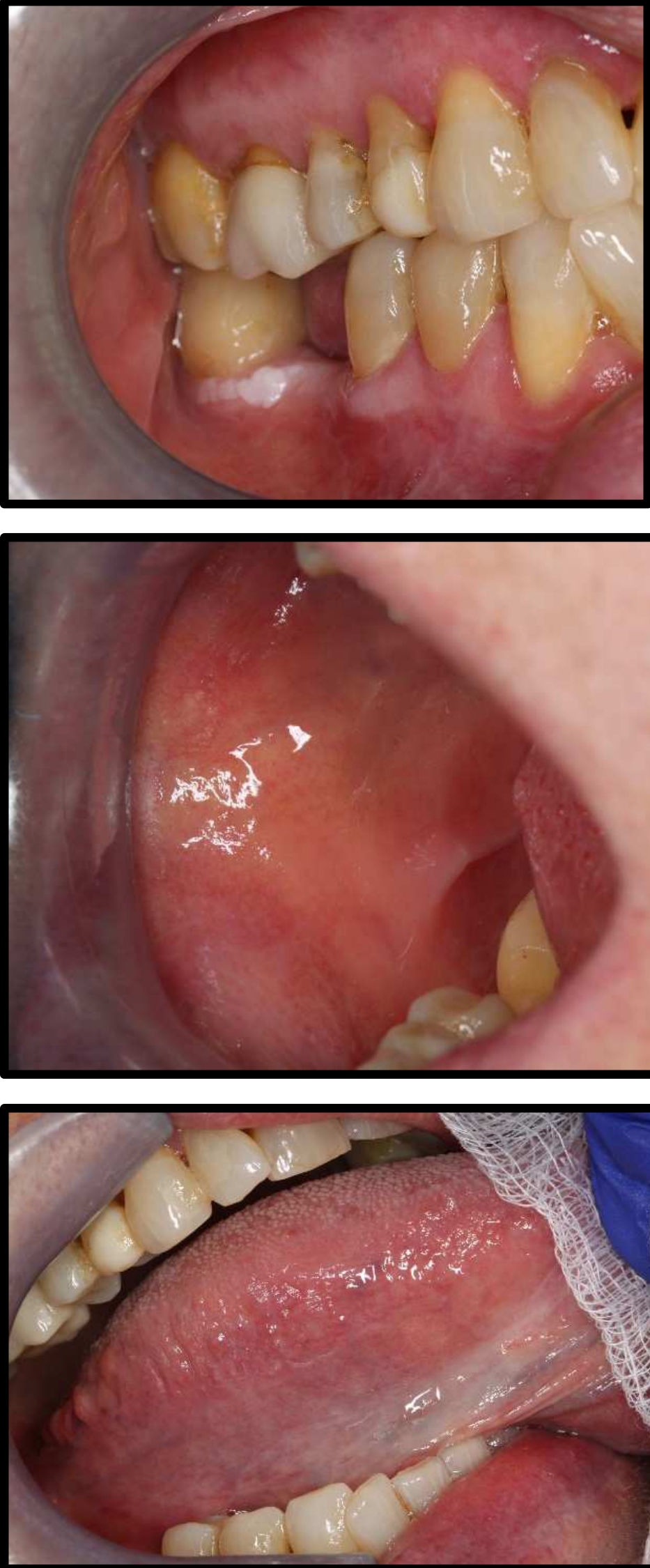
- Has plaque like lesion on 46 could be leukoplakia ⇒ do blood tests
- An obvious white lesion is present around a molar.
- This specific plaque-like lesion may or may not be part of the same process as the reticular lesions.
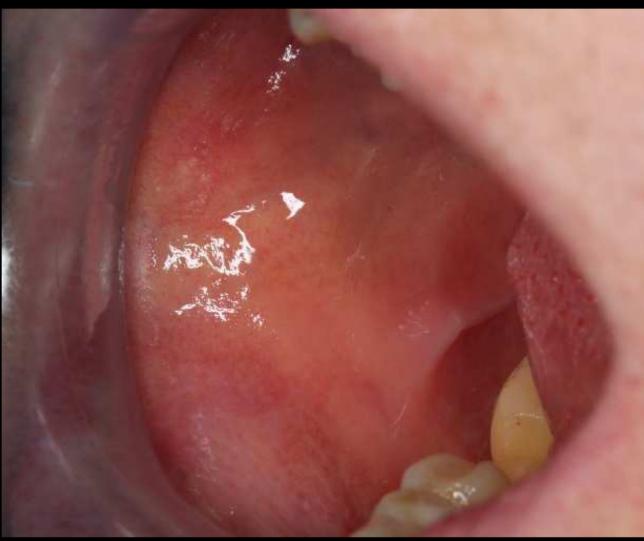
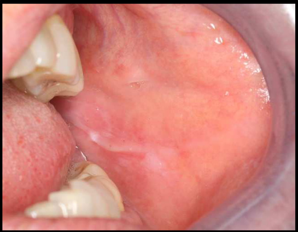
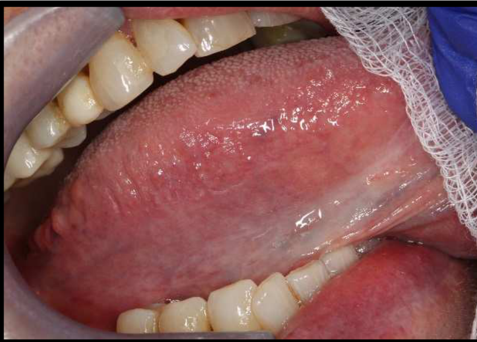
- Reticular erythmatous lesions
- Subtle reticular lesions are visible on the maxillary gingiva and bilaterally on the buccal mucosa.
- The lesions appear bilateral and more or less symmetric.
- ==Frictional Hyperkeratosis: Some areas on the buccal mucosa are attributed to frictional hyperkeratosis from occlusal trauma.==
- ==Symptoms: The patient reports a burning sensation.==
3. Diagnostics
- she has diabetes so do Hba1c
- HBA1C is required to check diabetes control, which impacts the risk of developing candidiasis during treatment.
- Biopsies
- take multiple biopsies
- Multiple biopsies are necessary because there are different types of lesions (reticular vs. plaque-like).
- Recommended sites: The plaque-like lesion and one or two reticular sites.
- ==Biopsy is essential to check for dysplasia (e.g., keratosis with moderate dysplasia).==
Differential Diagnosis
- ==Lichen Planus: The primary consideration due to the bilateral, symmetric reticular lesions.==
- ==Leukoplakia: Specifically considered for the plaque-like lesion, which may exist as a separate entity.==
- ==Proliferative Verrucous Leukoplakia (PVL): A possibility when white lesions show dysplasia or transform; it can initially mimic lichen planus.==
- ==Malignant Transformation: Lichen planus has a quoted malignant transformation risk of approximately 1%.==
Clinical Monitoring
- Use of clinical photography to track changes over time.
- Close follow-up (no more than three months between examinations) is required, especially if dysplasia is present.
- The highest risk of transformation is within the first five years.
Management and Treatment
- ==Symptom Control: The goal is to manage burning and ulceration while using the minimum amount of medication necessary.==
- ==Topical Steroids:==
- Used to control symptoms, but use should be minimized if dysplasia is present.
- ==Dexamethasone mouthwash: Effective but carries a higher risk of oral candidiasis.==
- ==Targeted approach: Using a medicinal spray or targeting specific symptomatic sites is preferred over a general mouthwash to limit exposure to non-affected areas.==
- ==Alternative/Initial Therapy: Starting with “D-flab” (Diflam) to avoid steroids initially if there is no ulceration.==
- ==Systemic vs. Topical: Topical is preferred over systemic immunomodulating drugs to minimize side effects.==
- ==The “Inflammation Balance”: Treating ulcerated lichen planus is a “fine balance.” While inflammation can be a defense mechanism against dysplasia, chronic inflammation and tissue damage also increase malignancy risks.==
: Original PDF page 1: 16. Allergies and Immune mediated disease case 2, p.1 : Original PDF page 2: 16. Allergies and Immune mediated disease case 2, p.2