Bone Diseases Tutorial
Course Information1
- Subject: Oral Medicine
- Course Code: DENT5310
- Instructor: A/Prof Omar Kujan
Case One: Maxillary Swelling In An Elderly Patient
Case Summary2
A 73-year-old female with dementia presented to a general dentist with swelling and purulence drainage involving the right posterior maxillary teeth (14, 15, 16, and 17), which were subsequently extracted.
Clinical Presentation And History
Past Medical History
The patient’s medical history is significant for:
- Dementia
- Hypertension
- Liver disease
Clinical Examination And Activity34
Clinical exam
 |  |
Clinical and Radiological Objectives5
- Discuss the clinical and radiological presentations
- Discuss the treatment and differential diagnosis
Clinical And Imaging Descriptions
Intraoral Findings6
- The oral cavity exhibits a well-defined, smooth, dome-shaped lesion.
- The lesion appears translucent and pinkish in color with an ulcerated surface.
- There are no evident signs of inflammation, ulceration, or bleeding in the tissue surrounding the lesion.
- The lesion is round to oval, smooth-surfaced, and protrudes significantly from the surrounding tissue.
- The approximate size of the lesion is 1 cm.
Radiographic Findings7
- A panoramic view of the CBCT imaging taken at the first clinical presentation demonstrates a large right maxillary sinus that appears cloudy.
- In comparison, the contralateral left maxillary sinus is clear.
- The alveolar ridge shows slight erosion, but there is no evidence of an intra-osseous lesion.
Differential Diagnosis And Biopsy891011
 |  |
Potential Diagnoses12
- Reactive Benign Lesion: Pyogenic granuloma
- Malignant Tumors:
- Squamous Cell Carcinoma (SCC)
- Melanoma
- Lymphoma
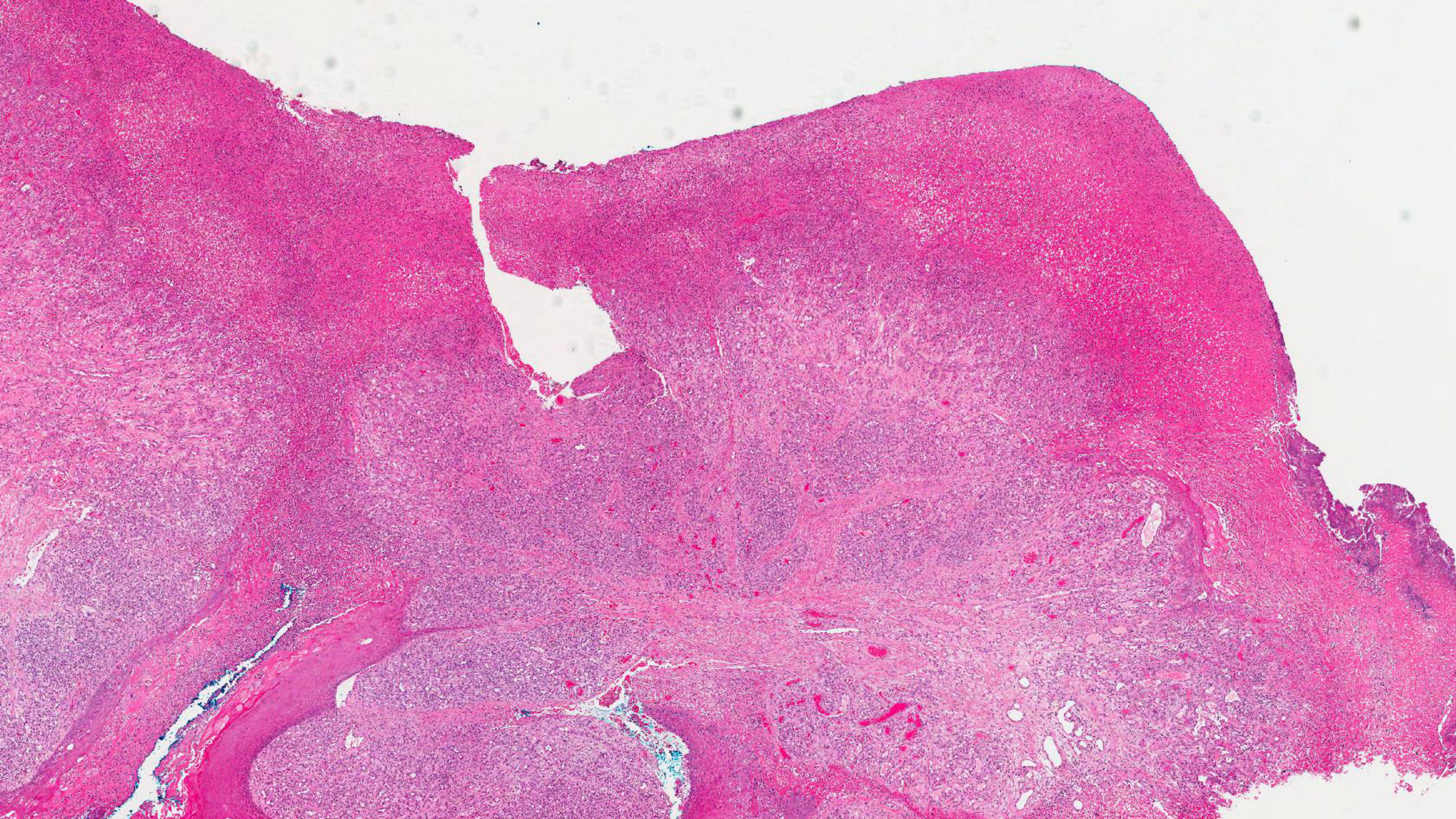
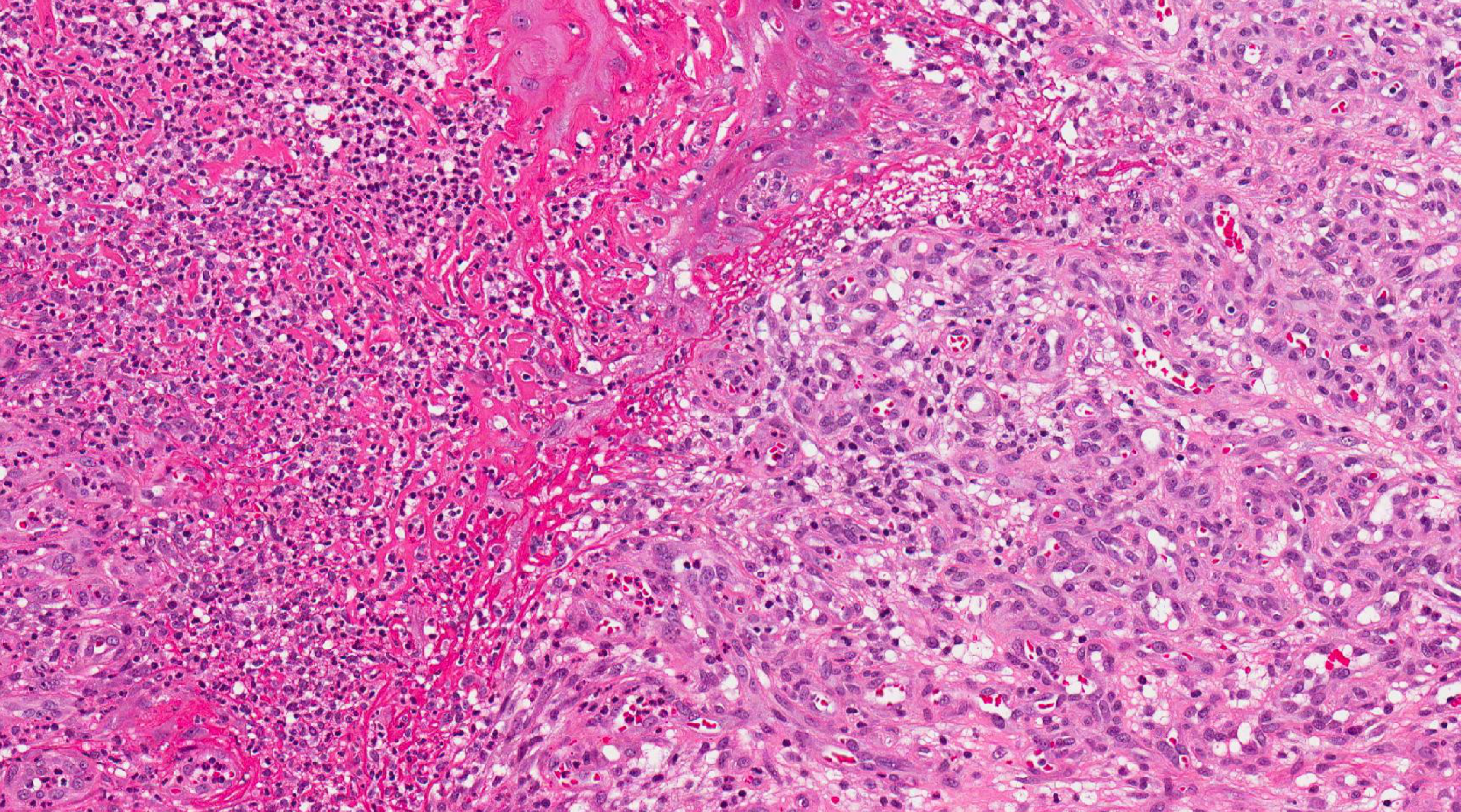
Incisional biopsy results
Microscopic Description And Diagnosis13
Histopathological Features14
- Vascular Patterns: Lobular pattern of vascular proliferation with inflammation and edema resembling granulation tissue.
- Surface Characteristics:
- Thin epidermis at the top with variable ulceration.
- Acanthosis and hyperkeratosis at the lateral aspects.
- Vascular Structure: Central branching vessels (capillary or vascular lobules) surrounded by endothelial cells, containing no or rare red blood cells.
- Deep Tissue Presentation: Deep lesions often lack the edema and inflammation seen in superficial areas.
Pyogenic granuloma
Treatment Plan
Recommended Intervention15
- Surgical removal of the lesion.
Case Two: Mandibular Asymmetry In A Juvenile Patient
Patient Presentation16
A 17-year-old male presented with a chief complaint of right facial swelling and asymmetry that has persisted for six months.
History of Present Illness
The patient has maintained regular dental and medical care. Clinical evaluation revealed:
- No evidence of pain
- No neurological abnormalities
- No malocclusion
- No involvement of the condyle or coronoid processes
Case Summary And History17
Past Medical History
The patient’s medical history is significant for Type 1 diabetes.
Clinical Activity And Imaging
Clinical Discussion Points18
- Discuss the clinical and radiological presentations
- Discuss treatment options and differential diagnosis
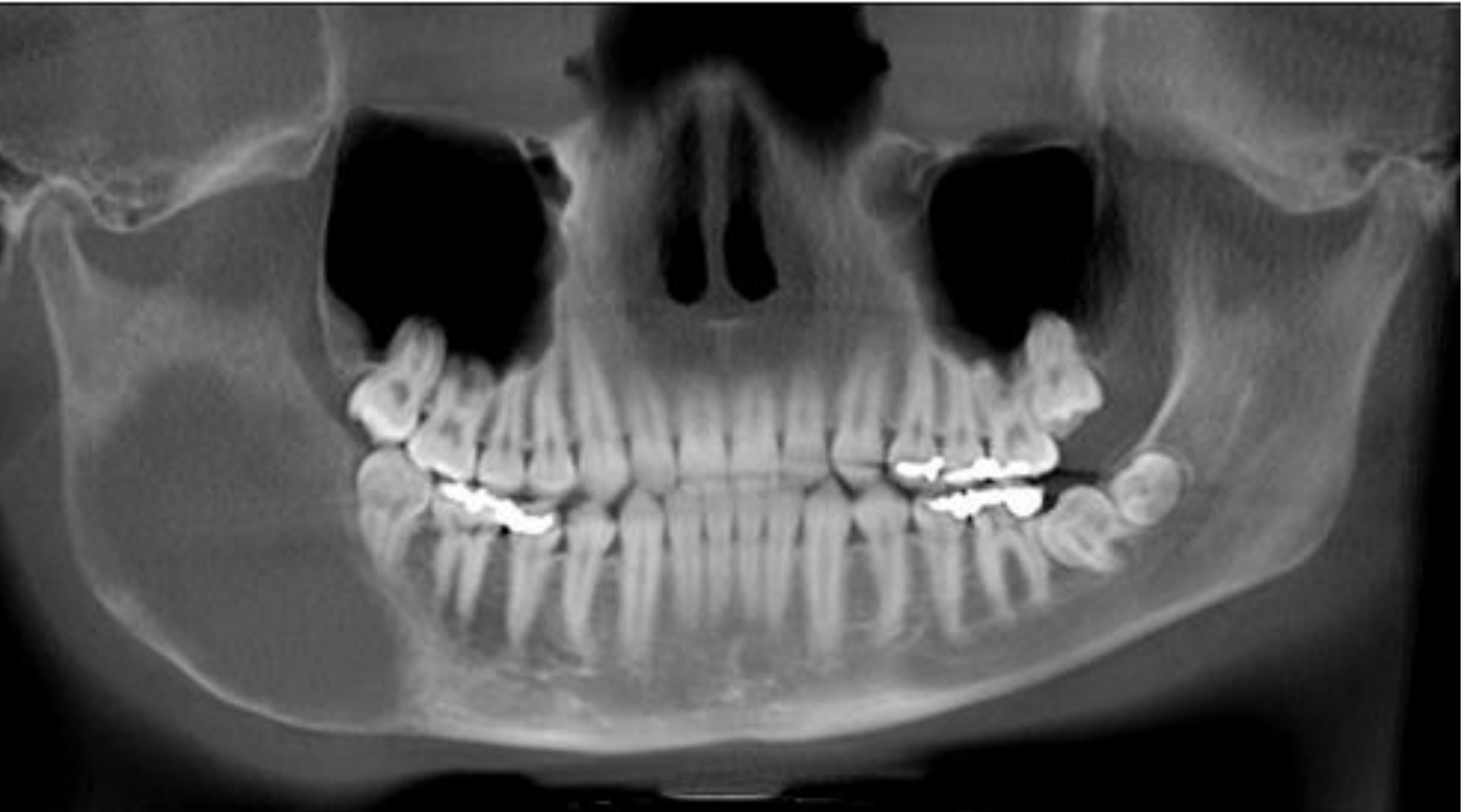
Radiographic Findings19
Cone Beam CT (CBCT) at the first presentation demonstrates:
- A large, unilocular, and expansile radiolucency
- Generalized fine radiopacity within the lesion
- Involvement of most of the right posterior mandible and part of the ramus
- Sparing of the condyle and the coronoid processes
Differential Diagnosis202122
 |  |
 |
Clinical Considerations23
- Aneurysmal bone cyst
- Fibrous dysplasia
- Brown tumour of hyperparathyroidism
- Juvenile ossifying fibroma
Microscopic Examination And Diagnosis
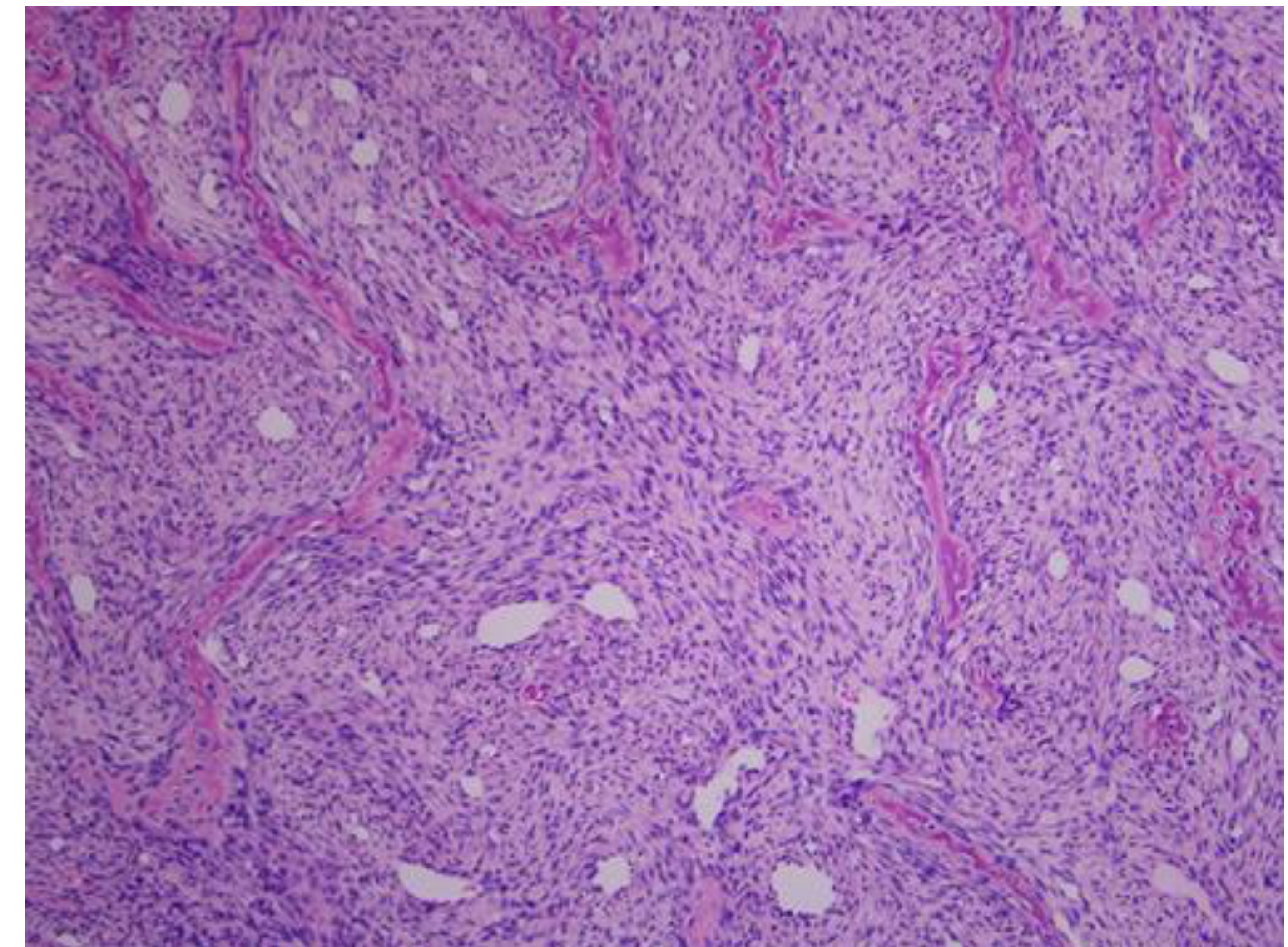
Histopathological Features24
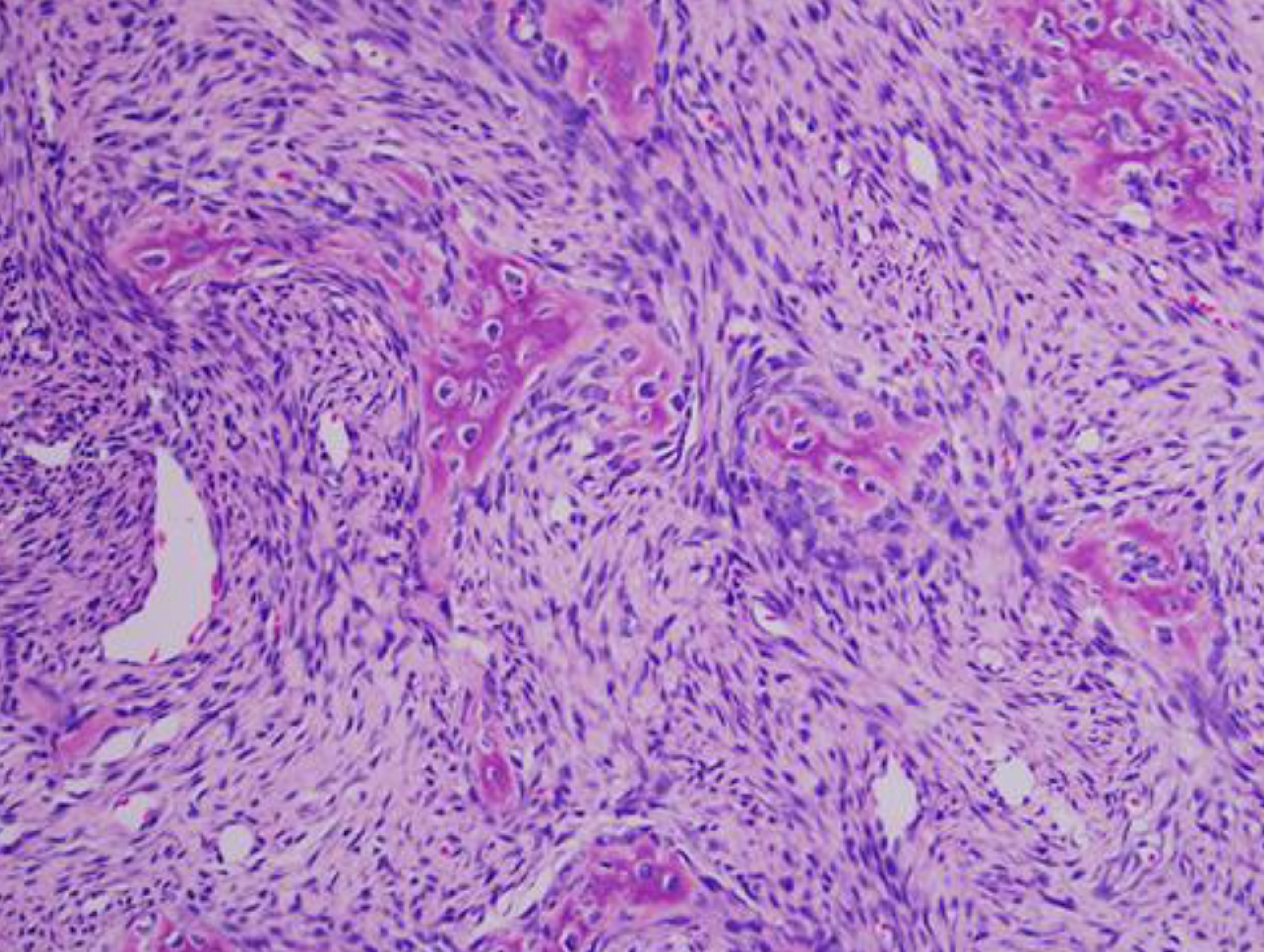
Multiple fragments of decalcified hard and soft tissue reveal a benign fibro-osseous lesion characterized by:
- Irregularly shaped and woven-type bony trabeculae
- Cellular fibrous connective tissue stroma surrounding the trabeculae
- Bony trabeculae of variable shapes and sizes, with some exhibiting significant osteoblastic rimming
- Focal areas within the connective tissue stroma containing clusters of multinucleated giant cells
Final Diagnosis25
- Juvenile ossifying fibroma
Treatment Plan For Case Two
Footnotes
-
Original PDF page 1: 19.Bone diseases Tutorial 2026, p.1 ↩
-
Original PDF page 2: 19.Bone diseases Tutorial 2026, p.2 ↩
-
Original PDF page 3: 19.Bone diseases Tutorial 2026, p.3 ↩
-
Original PDF page 4: 19.Bone diseases Tutorial 2026, p.4 ↩
-
Original PDF page 5: 19.Bone diseases Tutorial 2026, p.5 ↩
-
Original PDF page 6: 19.Bone diseases Tutorial 2026, p.6 ↩
-
Original PDF page 7: 19.Bone diseases Tutorial 2026, p.7 ↩
-
Original PDF page 9: 19.Bone diseases Tutorial 2026, p.9 ↩
-
Original PDF page 10: 19.Bone diseases Tutorial 2026, p.10 ↩
-
Original PDF page 11: 19.Bone diseases Tutorial 2026, p.11 ↩
-
Original PDF page 12: 19.Bone diseases Tutorial 2026, p.12 ↩
-
Original PDF page 8: 19.Bone diseases Tutorial 2026, p.8 ↩
-
Original PDF page 14: 19.Bone diseases Tutorial 2026, p.14 ↩
-
Original PDF page 13: 19.Bone diseases Tutorial 2026, p.13 ↩
-
Original PDF page 15: 19.Bone diseases Tutorial 2026, p.15 ↩
-
Original PDF page 16: 19.Bone diseases Tutorial 2026, p.16 ↩
-
Original PDF page 17: 19.Bone diseases Tutorial 2026, p.17 ↩
-
Original PDF page 18: 19.Bone diseases Tutorial 2026, p.18 ↩
-
Original PDF page 19: 19.Bone diseases Tutorial 2026, p.19 ↩
-
Original PDF page 21: 19.Bone diseases Tutorial 2026, p.21 ↩
-
Original PDF page 22: 19.Bone diseases Tutorial 2026, p.22 ↩
-
Original PDF page 23: 19.Bone diseases Tutorial 2026, p.23 ↩
-
Original PDF page 20: 19.Bone diseases Tutorial 2026, p.20 ↩
-
Original PDF page 24: 19.Bone diseases Tutorial 2026, p.24 ↩
-
Original PDF page 25: 19.Bone diseases Tutorial 2026, p.25 ↩