Case Discussion Of Oral Pathosis
Patient presentation
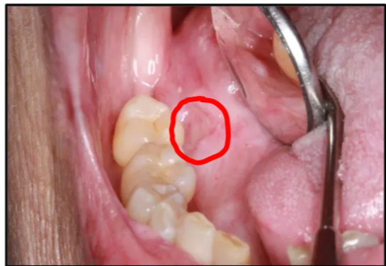
Snapshot
- Patient: 73-year-old female
- Chief complaint: Painful, non-healing tongue lesion/ulcer for 6 weeks
- Risk context: Non-smoker, non-drinker; considered “high risk” due to medical history
- Key background: Previous oral cancer at the same site (treated 2 years ago)
History of presenting complaint
- Painful, non-healing lesion/ulcer of six weeks duration
- Site: right lateral surface of tongue
- The patient does not smoke and does not drink alcohol, and is considered "high risk" due to her medical history.
Medical history
- Osteoarthritis
- Hypertension
- Hypercholesterolemia
==Current Status: Presents with a non-healing lesion/ulcer in the mouth.==
Relevant dental & oncological history
Prior malignancy and treatment
==* Previous Malignancy: History of oral cancer at the same site as the current lesion.
- Treatment History: Completed cancer treatment two years prior, which included:
- Surgery: Resulted in altered anatomy and scarring.
- Radiation Therapy: Leads to long-term side effects such as xerostomia (dry mouth) and reduced blood supply to the tissues.
- Chemotherapy: Contributes to the overall complexity of the case.==
Additional relevant oral history
- Biopsy-proven Oral Lichen Planus (OLP).
- Squamous Cell Carcinoma (SCC) of the right lateral tongue, treated with surgery and chemo-radiation therapy (treatment completed two years ago).
- The patient is a non-smoker and non-drinker.
Clinical examination / lesion description
Current lesion characteristics
==* Location: The lesion is located at the site of the previous surgery, in close proximity to a tooth.
- Type: Ulcerated lesion (not a lump).
- Size: Approximately 1 cm in diameter.
- Appearance:
- The lesion looks “punched out.”
- Margins are raised and appear white (indication of a chronic process).
- No evidence of keratosis.
- The surrounding anatomy is disfigured due to previous surgical intervention.==
Local factors affecting symptoms/healing
Why this site is vulnerable
==* Sensory Changes: The patient has altered sensation in the area (paresthesia/anesthesia) due to previous surgery, meaning she may not feel trauma occurring at the site.
- Tissue Quality: The area has significant scarring and poor blood supply (hypovascularity) due to radiation, making the mucosa more susceptible to breakdown and slower to heal.==
Question: Identify the pathosis and describe the clinical features.
Marks
3 marks for location
3 marks for description
Identification
- Pathosis: Chronic ulcerated lesion
- ==Margins: Well-defined but with raised, white margins==
Clinical features
- Location: Left lateral tongue
- Ulcer characteristics:
- NOT RED, therefore no inflammation, indicating it is a chronic ulcer
- Associated with dry mouth (lack of lubrication causes mucosa to rub against teeth)
- The dorsal surface of the tongue may appear disfigured or "crazy" due to previous surgical reconstruction
Question: Is it a primary or recurrent lesion?
Definitions used in this case
- Second Primary: A new lesion appearing at the site after a 5-year disease-free interval (AFTER 5 years)
- Recurrence: lesion appearing at the same site within 5 years of treatment
==Current Classification: This is the primary concern because the lesion is at the same site as the previous cancer. Since this patient is only 2 years post-treatment, it would be classified as a recurrence (defined as a lesion appearing at the same site within 5 years of treatment).==
Question: What is the differential diagnosis?
Differential diagnosis (ranked/priority implied by notes)
- Recurrent OSCC (Squamous Cell Carcinoma): Must be the #1 differential given the history.
- Traumatic Ulcer (Chronic): Likely due to the combination of dry mouth, altered sensation, and scarring
- Its very close tooth (the tissue rubs against the adjacent tooth)
- Shes particularly sensitive because
- radiotherapy: low salivary flow (dry mouth)
- susceptible to trauma (altered sensation)
- ==Secondary Candidal Infection: A secondary infection of a pre-existing benign ulcer.==
Question: What are the relevant clinical investigations?
Clinical assessment
- Check for sharp teeth, rough fillings, or calculus that could be causing trauma.
Biopsy
Biopsy timing in a high-risk patient
- When should biopsy be undertaken: ==Unlike a standard case where you might wait 2 weeks for a traumatic ulcer to heal, a biopsy should be performed immediately (first visit) because the patient is high-risk for recurrence.== She has super sus symptoms and has lots of points to OSCC so take biopsy right away.
- Type: Incisional biopsy (scalp biopsy).
- Repeat biopsy: If the lesion does not respond to treatment as expected, a repeat biopsy is necessary to rule out a "false negative" (non-representative sample) or to check for secondary changes.
Blood tests
Hematinic blood tests
- Iron, B12, folate, etc. Check levels to rule out nutritional deficiencies that prevent healing.
Full blood counts
- Should be performed as a routine investigation for non-healing ulcers.
Diabetes screening
- Fasting Glucose: Recommended as a minimum to rule out undiagnosed diabetes in a patient with a non-healing lesion. For an unknown diabetic the best test would be Fasting Glucose.
- HbA1c: Don’t necessarily have to do HbA1c. Can be used for diagnosis but may have Medicare billing restrictions for general dentists; fasting glucose is often more accessible.
Question: What is the diagnosis?
Results / working diagnosis from notes
- Biopsy was taken and came back as traumatic ulcer
- ==Subsequent finding: Secondary candidal infection (identified when the ulcer failed to heal after initial management)==
Question: How would you manage this patient?
Benign traumatic ulcer
Remove contributing trauma + support healing
do all the obvious like check occlusion and interferences—smooth down any sharp tooth surfaces, remove calculus, and ensure no rough fillings are irritating the site
- ==Hygiene: Use Chlorhexidine (e.g., Curasept) to keep the area clean==
Meds
- Chlorhexidine (highest concentration) to keep the area clean
- ==Antifungals: Required once a secondary infection is suspected==
Recall / follow-up
- ==Initial follow-up: 2 weeks==
- longer recall period because the biopsy gave a diagnosis , so 6 weeks
- In post-radiation patients, healing is extremely slow due to poor blood supply
- because we aren’t expecting healing to be quick
- ==Total healing time: It took 12 months for the ulcer to fully epithelialize==
If not improving (theoretical course in notes)
- patient comes in after 6 weeks and is not better even after medications
- a rebiopsy is not recommended because it will just make a better wound!
- Tell the patient to continue with the prescribed treatment and recall again in 6 weeks
- it got a bit better , so its a sign to kep doing it
- In the end this was done for 3 months, at the end of the 3 month period the ulcer wasn’t getting worse or better
- At this point the diagnosis should be questioned ex: if a benign ulcer does not reduce by at least 50% within 3 months despite compliance, you must question if the biopsy was representative or if there is a secondary factor (like infection or undiagnosed diabetes)
- could the pathology be wrong
- Yes
- Therefor biopsy should be repeated
- When biopsy was retaken
- A secondary candida infection was found
- could the pathology be wrong
- At this point the diagnosis should be questioned ex: if a benign ulcer does not reduce by at least 50% within 3 months despite compliance, you must question if the biopsy was representative or if there is a secondary factor (like infection or undiagnosed diabetes)
Antifungal tablet (agent selection considerations)
- Amphotericin isn’t a good candidate because p/t has a dry mouth (lozenges are difficult for patients with severe dry mouth as they cannot dissolve the tablet). Theoretically, a compounding pharmacy could create a custom gel version of Amphotericin, though it is expensive
- Nystatin drops are bad because they are sugary and can promote caries —avoided in this case due to high sugar content and the patient's high caries risk from xerostomia
- Miconazole oral gel is a good alternative
- For 6 weeks 4 times a day
- Tell GP about miconazole due to interactions with drugs (e.g., Statins, Warfarin). If the patient is on a Statin, consult the GP to monitor or temporarily suspend the medication