Case Discussion Of Oral Lesion
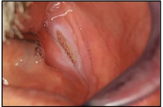
Case 1 — Chronic non-healing ulcer (mandibular lingual sulcus)
Chief complaint / HPI
- A 52-year-old male presented with a 6-week history of a painful oral lesion.
- Self-treatment: Kenalog, with no clinical response.
- Location: Mandibular lingual sulcus (Case 1).
Key history point (Case 1)
The patient was wearing dentures constantly (including at night), which contributed to the lack of healing.
Medical + social history
- Medical conditions: Diabetes, hypertension, hypercholesterolemia.
- Social history:
- Long-term smoker (30 years), currently 15 cigarettes/day.
- Alcohol: ~2 drinks/day.
- Dental status: Edentulous; current dentures are 5 years old.
Clinical examination (physical findings)
- Location: Mandibular lingual sulcus (Case 1).
- Duration: 6 weeks.
- Appearance: Poorly defined ulcer with white, raised/thickened margins.
- Palpation: Induration (palpable thickness/firmness under the lesion, like scar tissue).
Additional case notes (contributing factors)
- The lesion was being inappropriately treated with Kenalog (a glucocorticoid) obtained from a pharmacy.
Assessment
Identify the pathosis + clinical features
- Pathosis: Chronic traumatic ulcer
- Clinical features:
- Ulcer definition: Break in continuity of epithelium
- Margins: Raised and thickened
- Color: White areas (hyperkeratosis) around margins
- Texture: Indurated/firm on palpation (scarring + hyperkeratosis)
Chronic vs acute ulcer (features)
| Feature | Acute ulcer | Chronic ulcer |
|---|---|---|
| Surrounding inflammation | Redness + active inflammation | Lacks intense redness |
| Keratosis | — | Hyperkeratosis → white, raised margins |
| Base/margins | Softer | Induration from scarring |
Differential diagnosis
- Traumatic ulceration (likely denture-related, e.g., dental plate)
- Oral squamous cell carcinoma (must always be considered for any non-healing ulcer present for more than 2–3 weeks)
- Leukoplakia (could be an ulcerated leukoplakia, but a bit of a stretch)
- Tuberculosis (infectious)
- Fungal infection (infectious)
- Syphilis (infectious)
- Chemical injury (possible, less likely given raised margins)
Since OSCC is a differential
The differentials for OSCC are fair game too (e.g., TB, fungal infection).
Relevant clinical investigations
Principle
Investigations should rule out systemic factors that complicate healing.
Blood tests
- Full blood count: general health + immune status
Ulcerated lesions — hematinics
ALWAYS DO HEMATINICS FOR ULCERS
B12, folate, iron by default for ulcers.
Deficiencies can cause or complicate oral ulcerations.
- HbA1c: check diabetes control (poorly controlled diabetes hinders wound healing); relevant for diabetic patients with poorly healing ulcers.
Biopsy
- Should be done after other less invasive tests.
- Timing: If suspicious for SCC, biopsy is essential. If a clear local trauma (e.g., denture) is identified, may delay 1–2 weeks while removing the irritant to see if healing occurs.
- Procedure: If ulcer does not resolve or significantly improve after removing suspected cause → biopsy to rule out malignancy.
Misc / targeted checks
- Ask if denture is worn at night (in this case: yes).
- Denture assessment: check fit and stability.
- Microbiology/Imaging: consider if TB or deep fungal infections suspected (lower on differential list).
Diagnosis
- Chronic traumatic ulcer
Management
Meds and rinses
- Chlorhexidine mouthwash (highest concentration)
- Antibacterial — primary recommendation to keep area clean and reduce bacterial load
- 2×/day every day until next appointment
- Difflam (benzydamine): PRN for pain control (anti-inflammatory/analgesic rinse)
- Salt water rinses: not highly recommended vs chlorhexidine (less antibacterial), though can help clear debris
Chlorhexidine gel vs mouthrinse
Applying gels (e.g., Chlorofluor gel) or patches is discouraged because the patient may poke the wound with a finger (dirty/irritating), interfering with delicate new tissue.
Kenalog
- Discontinue immediately.
- Interferes with healing (glucocorticoid) and increases risk of secondary infections like Candida.
- Inappropriate for traumatic ulcers.
Why the ulcer persisted
Part of the reason the ulcer is lasting so long is repeated trauma/irritation from continued Kenalog use.
Dentures
- Adjust/relieve any sharp or overextended areas.
- Wear only if necessary (eating/going out).
- Never wear at night to allow healing.
Recall / follow-up
- Initial follow-up: 2 weeks (range 1–2 weeks)
- At review:
- If smaller (even 2–3 mm) and pain reducing → continue conservative management
- If no improvement → proceed to biopsy
- Long-term: observe until complete epithelialization (can take up to 2 months in chronic cases)
Clinical pearl (scenario)
- If patient returns at 2 weeks and ulcer is slowly improving but not healed:
- A biopsy is not needed (chronic ulcer won’t be healed in 2 weeks)
- Continue same protocol (chlorhexidine BID, Difflam, etc.)
- Review again in 2–3 weeks
- A clinician is not considered negligent for delaying biopsy 1–2 weeks if actively removing suspected traumatic cause and closely monitoring. However, failure to follow up until fully healed is a clinical risk.
- Took two months for lesion to resolve.