Case Discussion Of Oral Pathosis
Patient Presentation and History1
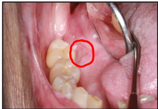 A 73-year-old female presented with a painful lesion of six weeks duration affecting the right lateral surface of her tongue. Her medical history is significant for the following conditions:
- Osteoarthritis
- Hypertension
- Hypercholesterolemia
Relevant Dental and Oncological History
The patient has a complex oral history, including:
- Biopsy-proven Oral Lichen Planus (OLP).
- Squamous Cell Carcinoma (SCC) of the right lateral tongue, treated with surgery and chemo-radiation therapy (treatment completed two years ago).
- Post-Treatment Complications:
- Xerostomia (Dry Mouth): Lack of lubrication makes the mucosa more susceptible to friction and traumatic ulceration.
- Altered Sensation/Nerve Damage: Reduced sensation means the patient may not feel trauma occurring.
- Scarring: Surgical scarring has reduced the elasticity and blood supply of the tissue.
- The patient is a non-smoker and non-drinker.
Clinical Case Description
Lesion
Lesion: 1 cm "punched out" ulcer with raised, white margins; no significant keratosis.
Case Description And Patient History
Comparison of Diagnostic Approaches
- Low-Risk Patient: Remove trauma source and wait two weeks to observe healing before biopsy.
- High-Risk Patient (Oncology History): Perform an incisional biopsy on the first visit to rule out malignancy immediately due to high recurrence risk.
Biopsy Techniques
- Incisional Scalpel Biopsy: Preferred to obtain a representative sample.
- Punch Biopsy: Common tool, though preferences vary.
- Representative Sampling: Critical to avoid a "false sense of security" (false negative) from non-representative sites.
Identify the pathosis and describe the clinical features.
Marks
3 marks for location 3 marks for description
- Location
- Left lateral tongue
- Ulcer
- NOT RED, therefore no inflammation thus its a chronic ulcer
Is it a primary or recurrent lesion?
The patient had OSCC, so when would we call it a secondary primary lesion?
- AFTER 5 years
What is the differential diagnosis?
Differential diagnosis
Differential Diagnosis: Recurrent SCC (primary concern as it is within 5 years of original), Chronic Traumatic Ulcer (due to xerostomia/sensory deficit), or Secondary Fungal Infection.
- Recurrent OSCC
- Traumatic Ulcer
- its very close tooth
- shes particularly sensitive because
- radiotherapy: low salivary flow
- susceptible to trauma
What are the relevant clinical investigations?
Investigations
Investigations: Immediate incisional biopsy (required for high-risk patients), Hematinics (Iron, Folate, B12), FBC, and Fasting Glucose (to rule out Diabetes).
Biopsy
- When should biopsy be undertaken
- she has super sus symptoms and has lots of points to OSCC so take biopsy right away
Blood tests
Hematinic blood tests
- iron, b12, folate etc
Full blood counts
Diabetes
- Don’t necessarily have to do HbA1c
- for an unknown diabetic the best test would be Fasting Glucose
What is the diagnosis?
- Biopsy was taken and came back as traumatic ulcer
How would you manage this patient?
Initial management
Management: Initial treatment involved smoothing sharp teeth and Chlorhexidine. Healing was extremely slow due to hypovascularity.
Benign traumatic ulcer
do all the obvious like check occlusion and interferences
Meds
- Chlorhexidine (highest concentration)
Recall
- longer recall period because the biopsy gave a diagnosis, so 6 weeks
- because we aren’t expecting healing to be quick
Theoretical
- patient comes in after 6 weeks and is not better even after medications
- a rebiopsy is not recommended because it will just make a better wound!
- Tell the patient to continue with the prescribed treatment and recall again in 6 weeks
- it got a bit better, so its a sign to kep doing it
- In the end this was done for 3 months, at the end of the 3 month period the ulcer wasn’t getting worse or better
- At this point the diagnosis should be questioned ex:
- could the pathology be wrong
- Yes
- Therefore biopsy should be repeated
- When biopsy was retaken
- A secondary candida infection was found
- could the pathology be wrong
- At this point the diagnosis should be questioned ex:
Secondary diagnosis + outcome
Secondary Diagnosis: After 3 months of minimal improvement, a repeat biopsy identified a secondary candidal infection.
Outcome: The ulcer took 12 months to fully epithelialize.
Antifungal tablet
Pharmacology
Pharmacology: Miconazole Oral Gel was used. Nystatin was avoided due to high sugar/caries risk; Amphotericin B was avoided as lozenges won't dissolve in a dry mouth.
- Amphotericin isn’t a good candidate because p/t has a dry mouth
- Nystatin drops are bad because they are sugary and can promote caries
- Miconazole oral gel is a good alternative
- For 6 weeks 4 times a day
- Tell GP about miconazole due to interactions with drugs
Footnotes
-
Original PDF page 1: Injuries Case 1, p.1 ↩