Radiology Workshop Part 1: Lucencies and Opacities
Workshop Overview1
This session focuses on the identification and analysis of radiographic findings in dental imaging, specifically categorizing them into two primary classifications:
- Lucencies: Areas of decreased density that appear dark on a radiograph.
- Opacities: Areas of increased density that appear light or white on a radiograph.
Presented by Dr. Dayea Oh for the DMD3 curriculum.
Orthopantomogram Interpretation Fundamentals
To ensure accurate interpretation of an Orthopantomogram (OPG), the following core principles should be applied:
Key Points of Orthopantomogram Interpretation2
- Review the initial clinical indication for the OPG.
- Identify any present OPG artefacts.
- Maintain a comprehensive knowledge of the normal radiographic anatomy of the maxillofacial region.
- Study the radiographic features of various jaw pathologies, specifically determining if they are lucent or opaque.
- Utilize a systematic approach by establishing a consistent routine for review.
Initial Clinical Indication and screening
Medico-legally this counts more as a comprehensive exam with no previous opgs instead
Orthopantomogram Artefacts Revision
Common Radiographic Artefacts3
- Foreign materials: Examples include earrings, necklaces, or dentures left in situ.
- Ghost images
- Incorrect patient positioning:
- Chin down
- Chin up
- Head tilting
- Head rotation
- Movement:
- Horizontal (following the beam or opposite to the beam direction)
- Vertical
- Swallowing
- Soft tissue position:
- Tongue not pressed to the palate
- Lips not closed
- Focal trough position:
- Head in front of the focal trough
- Head behind the focal trough
- Neck extension
- Collision with shoulder
Instructional Methodology
A structured review of the radiograph should follow these steps:
Systematic Approach to Radiographic Analysis4
- Address Clinical Concerns
- Dentoalveolar Analysis
- Count teeth and identify ectopic and/or impacted teeth.
- Identify dental anomalies (position, shape, etc.).
- Assess periodontal bone loss.
- Identify dental pathology (non-carious tooth loss, caries, fractures, periapical pathologies, etc.).
- Maxilla and Mandible
- Identify abnormal (radio)lucencies.
- Identify abnormal (radio)opacities.
- Note altered trabecular patterns.
- Identify fractures.
- Assess jaw asymmetry.
- Maxillary Sinuses: Evaluate for mucosal changes.
- TM Joints: Evaluate for morphological changes of the condyles.
- Soft Tissues: Identify swelling or soft tissue calcifications (e.g., tonsilloliths, salivary gland stones, etc.).
- Other Sites: Evaluate the spine, orbits, and other visible structures for structural changes.
Descriptive Features of Lucencies and Opacities5
When analyzing lucencies and opacities, consider the following descriptive features:
- Location: Where is the lesion located in relation to anatomical or dental structures?
- Number: How many lesions are present?
- Symmetry: Are they unilateral or bilateral?
- Definition: Are the margins well-defined?
- Shape: For example, round, ovoid, teardrop, lobulated, or irregular.
- Size: Use anatomical or dental landmarks for measurement (e.g., the 38-34 region).
- Homogeneity: Is the lesion uniform in density?
- Internal Architecture: Does the lesion have internal structures or patterns?
- Effects on Adjacent Structures: What is the impact on surrounding anatomy? Examples include:
- Displacement of teeth, the sinus floor, or mandibular canals.
- Resorption of teeth.
- Scalloping around adjacent teeth.
- Sclerosis of the surrounding bone.
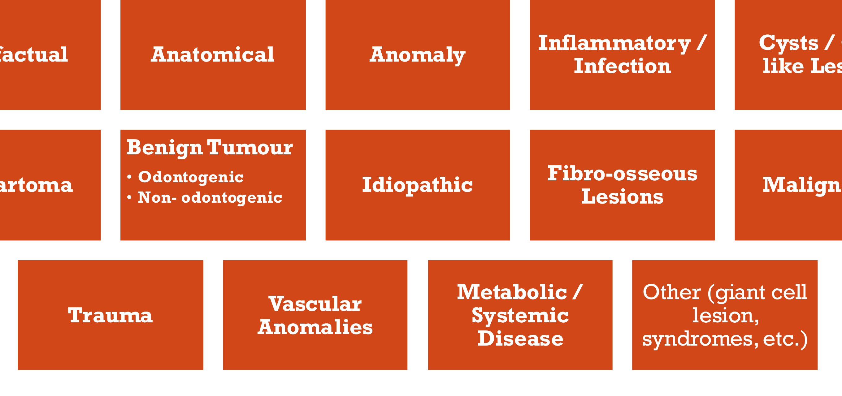
Surgical Sieve for Lesion Identification6
The surgical sieve is used to identify the nature of a lesion by categorizing it into one of the following groups:
- Artefactual
- Anatomical
- Anomaly
- Inflammatory / Infection
- Cysts / Cyst-like Lesions
- Hamartoma
- Benign Tumour
- Odontogenic
- Non-odontogenic
- Idiopathic
- Fibro-osseous Lesions
- Malignancy
- Trauma
- Vascular Anomalies
- Metabolic / Systemic Disease
- Other: Including giant cell lesions, syndromes, etc.
Indications for Further Imaging7
If the initial OPG is insufficient for a definitive diagnosis, consider the following advanced imaging modalities:
- CBCT (Cone Beam Computed Tomography)
- MSCT (Multi-Slice Computed Tomography)
- MRI (Magnetic Resonance Imaging)
If you are unsure of the appropriate next step, contact a radiologist for consultation.
Footnotes
-
Original PDF page 1: W4.1 Radiology Workshop, p.1 ↩
-
Original PDF page 2: W4.1 Radiology Workshop, p.2 ↩
-
Original PDF page 3: W4.1 Radiology Workshop, p.3 ↩
-
Original PDF page 4: W4.1 Radiology Workshop, p.4 ↩
-
Original PDF page 5: W4.1 Radiology Workshop, p.5 ↩
-
Original PDF page 6: W4.1 Radiology Workshop, p.6 ↩
-
Original PDF page 7: W4.1 Radiology Workshop, p.7 ↩