Case 3: Routine Orthopantomogram Image Analysis
Anomaly Identification[^10]
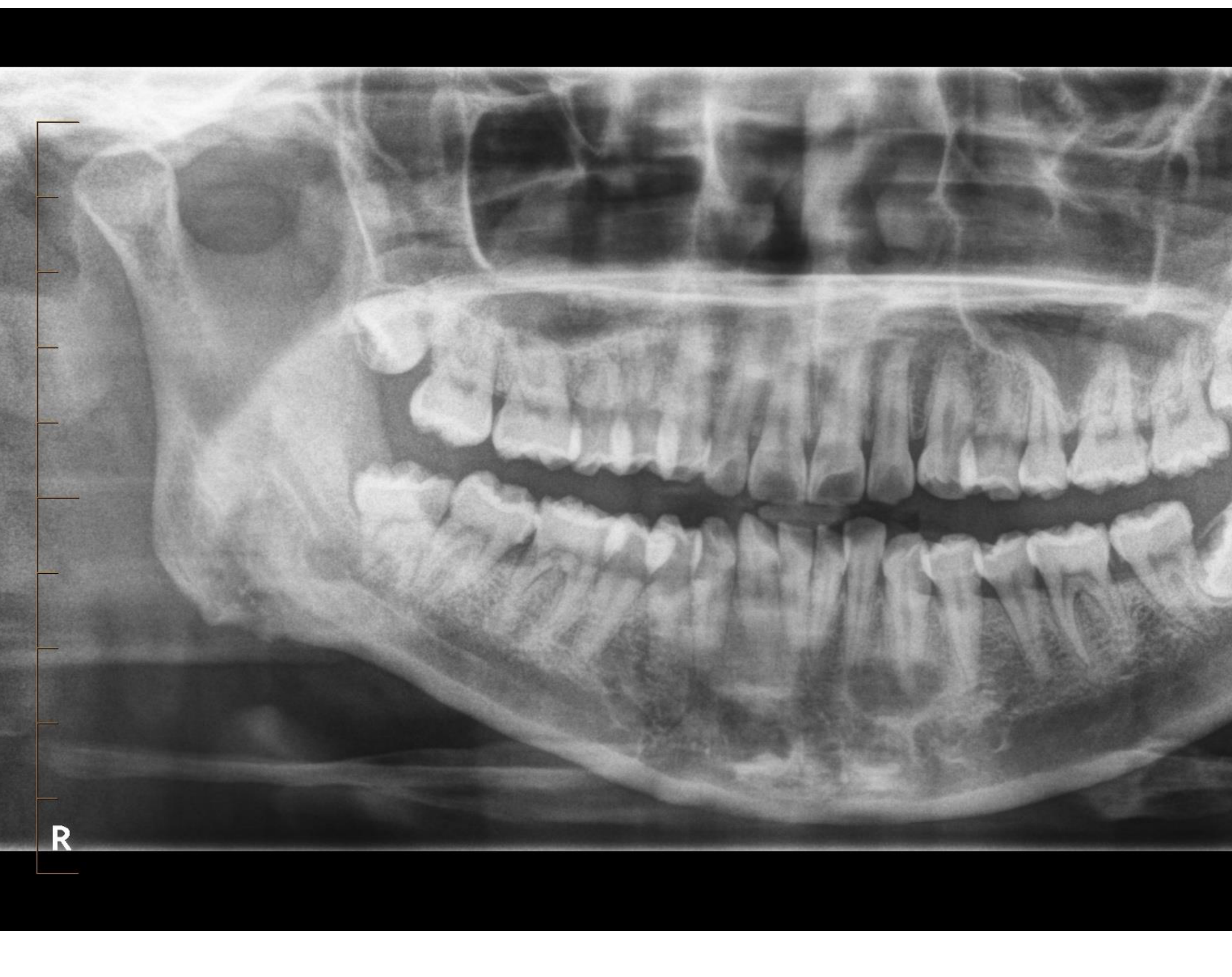
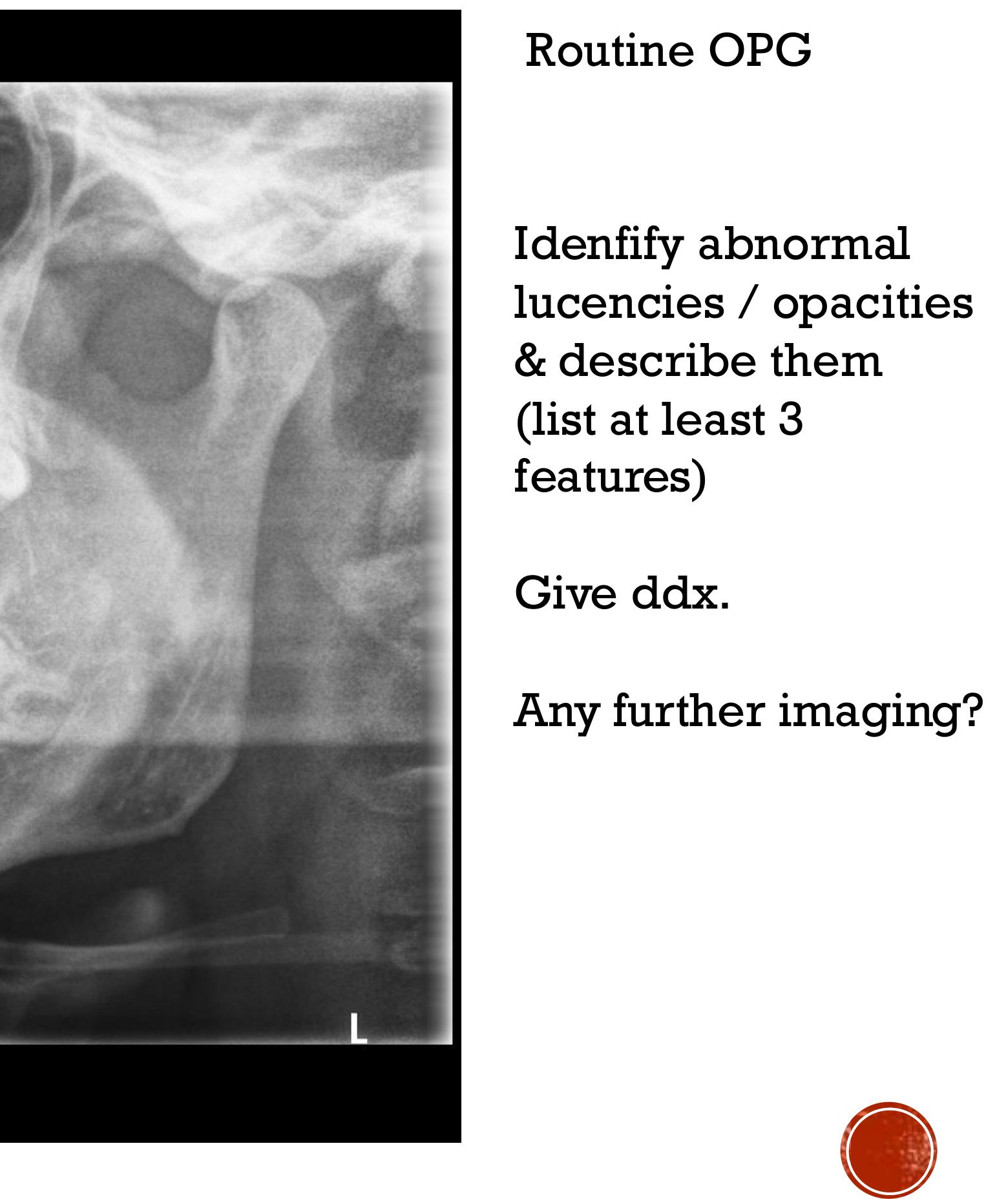
- radiolucency in the 33 in the left side of the mandible
- Well defined
- some lucency at the distal aspect of the 33 root
- but the lamina dura and pdl is preserved, thus it is only a loss of density
- there is no apperent resorption
 |  |
Differential Diagnosis
Further Imaging
Case 4: Orthodontic Assessment
Radiographic Analysis[^11]
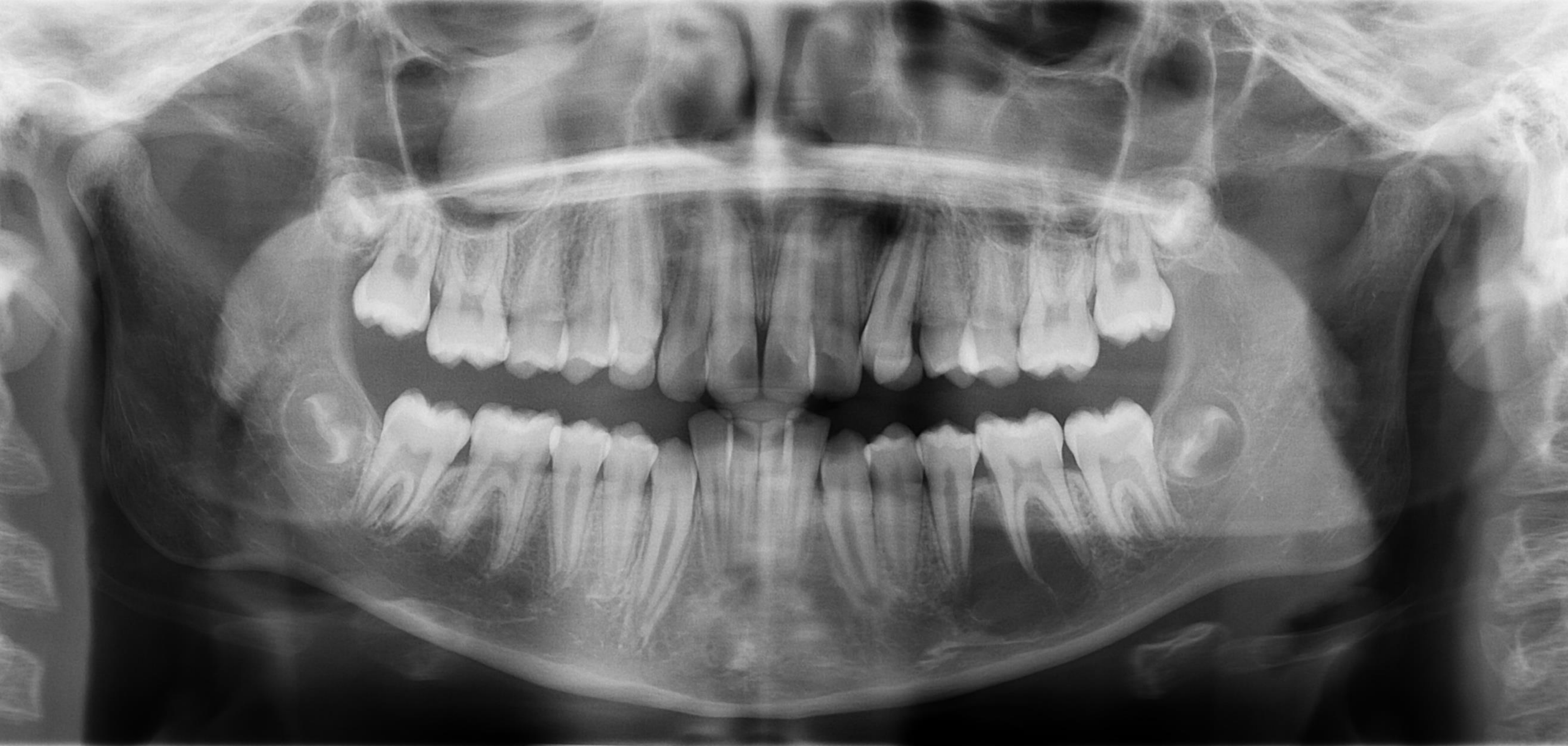
Identify and describe the abnormal lucency and opacity observed in the orthodontic assessment.
Further Investigation
- Is further imaging required?
Case 5: Temporomandibular Joint Assessment
Clinical Symptoms[^12]
- Discomfort in the jaws
- Stiffness in the Temporomandibular Joints (TMJs)
- Limited mouth opening
- Joint crepitus
Radiographic Evaluation[^13]
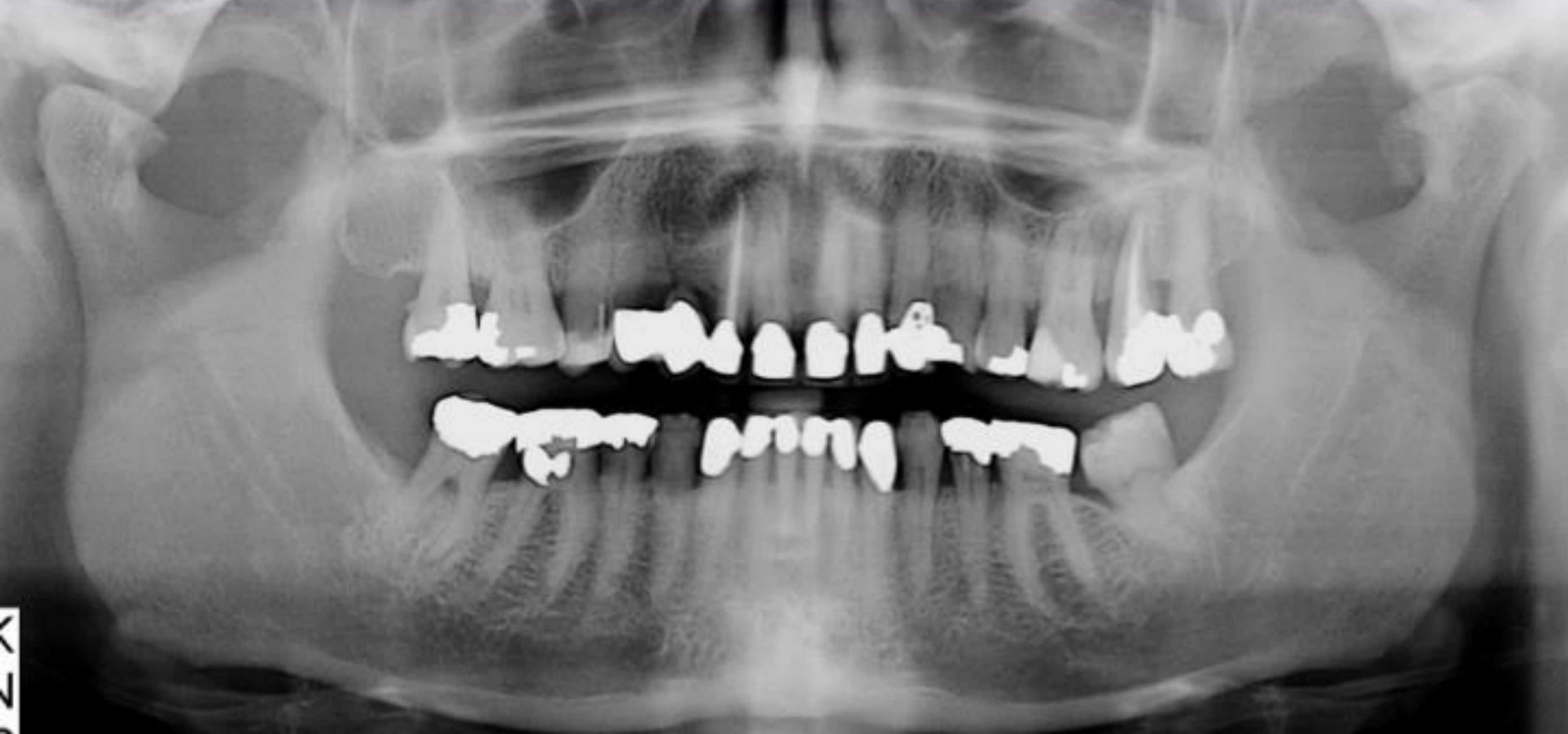
- Describe the appearance and condition of the TMJs.
Advanced Imaging
- Would you consider further imaging?
- Specify which modality should be used and provide the clinical reasoning.