Case 4: Orthodontic Assessment
Radiographic Analysis[^11]
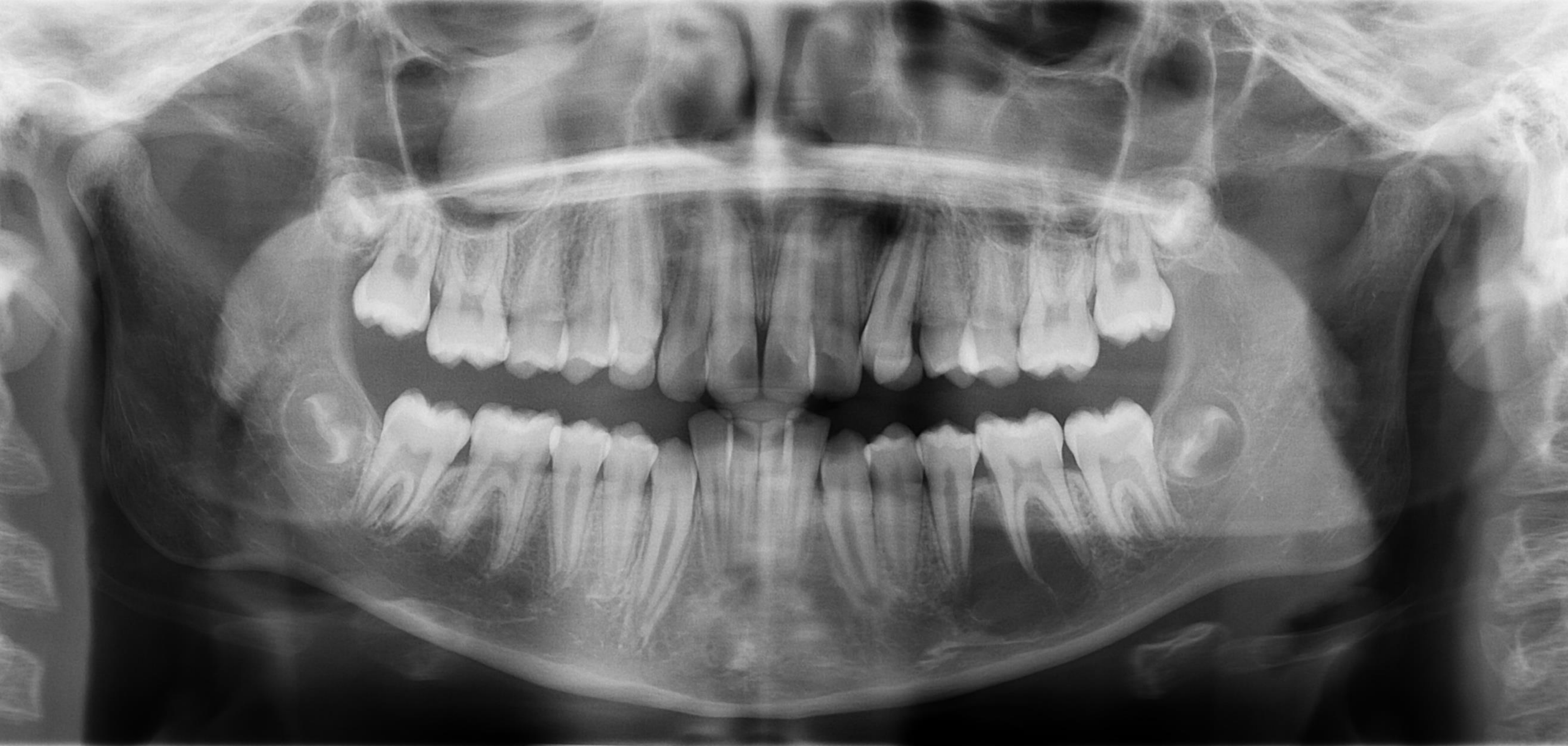
Identify and describe the abnormal lucency and opacity observed in the orthodontic assessment. The assessment begins with a systematic count of the teeth and an evaluation of dental health. In this case, all teeth are present, though several developmental and incidental findings are noted.
Dental and Bone Findings
- Retained Primary Teeth: Opacities are noted over the 23 and 13 regions, identified as retained primary teeth. Teeth 65, 63, and 53 are currently exfoliating.
- General Health: Crowns appear normal with no evidence of caries.
- Bone Levels: There is no periodontal bone loss; areas that appear to have low bone height are attributed to partially erupted teeth rather than pathology.
- Impacted vs partially erupted teeth (OPG reporting point): Differentiate completely embedded/impacted teeth (may have normal surrounding bone density) from partially erupted teeth, where apparent “reduced bone height/density” adjacent to the crown can reflect eruption status rather than pathology.
- Third Molars: Currently in the crown formation stage.
- Routine OPG third‑molar evaluation (document even if incidental): Record the tooth/side (e.g., 18/28/38/48), location (maxilla/mandible), angulation, stage of development/eruption, and relationship to important structures—especially the mandibular canal for lower thirds.
Left Mandibular Lesion
- Description: A well-defined, circular/round lucency located in the left mandible between the 35 and 36.
- Morphology: The lesion is large and extends vertically (cranial-caudally). It is "sharp" at the superior margin where it respects the anatomy of the adjacent roots.
- Effect on Adjacent Structures: ==Despite its size, there is no root resorption. The lamina dura of the 35 and 36 remains faintly visible, indicating the lesion is going around the teeth rather than destroying them.==
- Differential Diagnosis:
- Simple Bone Cyst (Pseudocyst): This is the primary differential. It is not a "true cyst" (it lacks an epithelial lining and hydraulic expansion) but rather a bone cavity. They are common incidental findings in young orthodontic patients, often occur in the mandible, and are typically self-limiting.
- Focal Cemento-Osseous Dysplasia (COD): Ruled out because CODs are typically smaller, centered at the apex, and tend to grow horizontally (sideways), whereas this lesion is larger and grows vertically.
Maxillary Findings
- Lateral Fossa (Canine Fossa): A bilateral (though often asymmetric) anatomical lucency/depression located near the 22 and 23. It should not be mistaken for a periapical lesion.
- Right Maxillary Sinus Lesion:
- Description: A well-defined, homogeneous, teardrop/oval-shaped opaque mass.
- Diagnosis: ==Mucus Retention Cyst. This is a benign, non-epithelial lined cavity filled with mucus. It is not a true cyst and is usually asymptomatic.==
TMJ Assessment (Preliminary)
- Initial observation of the OPG suggests asymmetry between the left and right condyles.
Further Investigation
- Is further imaging required? While many of these findings are incidental, proper clinical management and documentation are required.
- 3D Imaging (CBCT): Because an OPG is a 2D image of 3D structures, a CBCT may be requested if the relationship between a lesion (or an impacted tooth) and the mandibular canal/adjacent structures needs clarification.
- Monitoring: Non‑odontogenic lesions such as focal COD or a simple bone cyst often do not require active treatment but should be monitored/appropriately followed up as indicated.
Clinical Protocol for Simple Bone Cysts
- Referral: General dentists should refer to an oral surgeon or radiologist for a second opinion to confirm the diagnosis and ensure medicolegal safety.
- Surgical Intervention: If the lesion is large or interfering with treatment, a surgeon may perform a "bur hole" or puncture of the cortical bone to induce bleeding, which promotes self-healing.
- Documentation: All consultations with specialists and conversations with the patient must be recorded in the clinical notes, as these are legal documents.
Management of Mucus Retention Cysts
- Observation: These often rupture spontaneously or remain static.
- Review: ==A follow-up OPG should be taken in 2 to 5 years. If the size remains unchanged, no further follow-up is necessary.==
- Referral to ENT: Referral to an Ear, Nose, and Throat (ENT) specialist is only necessary if the lesion fills the entire sinus, grows rapidly, or causes clinical symptoms (e.g., disrupted airflow).
- Clinical Correlation: For the TMJ findings, further investigation is needed regarding the patient's reported symptoms of stiffness, limited opening, and crepitus.