OPG Case 6
-
Address Clinical Concerns
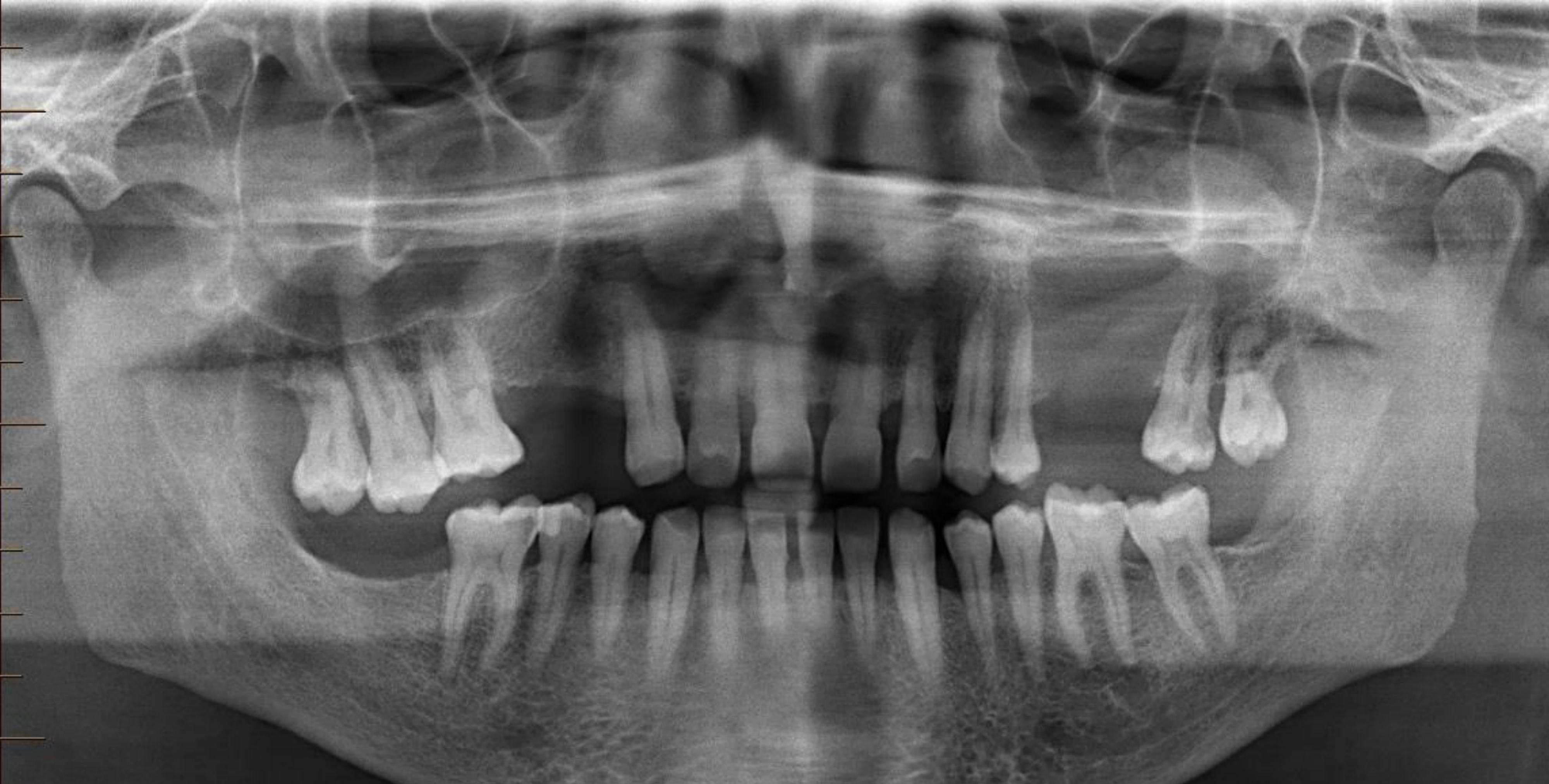
- ==Radiographic Diagnosis: Aggressive tumorous mass destroying the left maxilla and involving the sinus.==
- ==Urgency: This is considered an urgent case due to the aggressive nature of the lesion.==
- ==Management/Referral Pathway:==
- ==The patient needs to see an Oncologist or a Maxillofacial Surgeon.==
- While a GP can be involved, the hospital systems are often busy; direct contact with a Maxillofacial Surgeon is recommended.
- ==Action for Dentist: Call a Maxillofacial Surgeon directly to explain the aggressive nature of the lesion and arrange for the patient to be admitted or seen immediately. Maxillofacial surgeons are familiar with the necessary medical pathways and registrar programs to manage such cases.==
- ==Histopathological Requirement: A definitive diagnosis requires histopathological proof to confirm the specific cell type.==
-
Dentoalveolar Analysis
- Count teeth and identify ectopic and/or impacted teeth.
- Identify dental anomalies (position, shape, etc.).
- Opencontacts
- 44-45
- 13-12
- you get it
- Maxillary midline shifted to the left.
- Interdental spacing in the maxillary incisive region.
- Interdental spacing at the mesial and distal of 33 and distal of 34.
- Tooth 27: Widened PDL space on the mesial aspect. This may be related to mobility from periodontal disease or disease activity from the adjacent soft tissue mass.
- Tooth 24: Poor resolution noted on the distal aspect.
- Apical resorption of 31 and 41.
- Opencontacts
- Assess periodontal bone loss.
- 37: bone resorption causes floating appearance
- generalized periodontal bone loss : Moderate (15-33%)
- Identify dental pathology (non-carious tooth loss, caries, fractures, periapical pathologies, etc.).
- 46 : large triangle radiolucency on mesial cusp
- Possible caries on the distal of 27 and mesial of 46.
- Large diffuse radiolucency in the anterior mandibular region.
-
Maxilla and Mandible
- Identify abnormal (radio)lucencies.
- Identify abnormal (radio)opacities.
- Note altered trabecular patterns.
- Identify fractures.
- Assess jaw asymmetry.
- Maxilla
- chondral dysplasia
- assymetrical condyles
- LHS Posterior region communicated with LHS floor of sinus / possible tissue invasion
- ==Lesion Characteristics:==
- The lesion is a destructive tumorous mass rather than a cyst or a simple Oroantral Communication (OAC).
- While an OAC (communication between the oral cavity and antrum) exists, it is secondary to the destruction of the sinus wall by the mass.
- There is no extraction socket present to explain the lucency.
- ==Growth Pattern: The lesion is fast-growing. It destroys trabecular bone quickly and does not displace or resorb teeth (unlike benign tumors or cysts which grow slowly and have time to move hard structures).==
- ==Quadrant 2 / Left Maxilla Specifics: Lack of corticated edentulous ridge, loss of trabecular bone, and loss of the maxillary sinus floor.==
- ==Differential Diagnosis:==
- ==Squamous Cell Carcinoma (SCC): Suggested by the typical appearance of a lytic, destructive mass in the maxilla and sinus. Given the lytic nature and the epithelial environment, carcinoma is more likely than sarcoma.==
- ==Lymphoma: Another possibility for a lytic lesion in this region.==
- ==Note on Sarcoma: Derived from endothelial or mesenchymal cells; often bone-forming (e.g., osteosarcoma) with a “sunray” appearance, which is not seen here.==
- chondral dysplasia
- Maxillary Sinuses: Evaluate for mucosal changes.
- Large defect between the 24 and 26 region within the sinus.
- ==Soft Tissue: Presence of a large, globular soft tissue mass/density in the sinus.==
- TM Joints: Evaluate for morphological changes of the condyles.
- Soft Tissues: Identify swelling or soft tissue calcifications (e.g., tonsilloliths, salivary gland stones, etc.).
- Tongue position was down during the scan.
- Other Sites: Evaluate the spine, orbits, and other visible structures for structural changes.