OPG Case 7
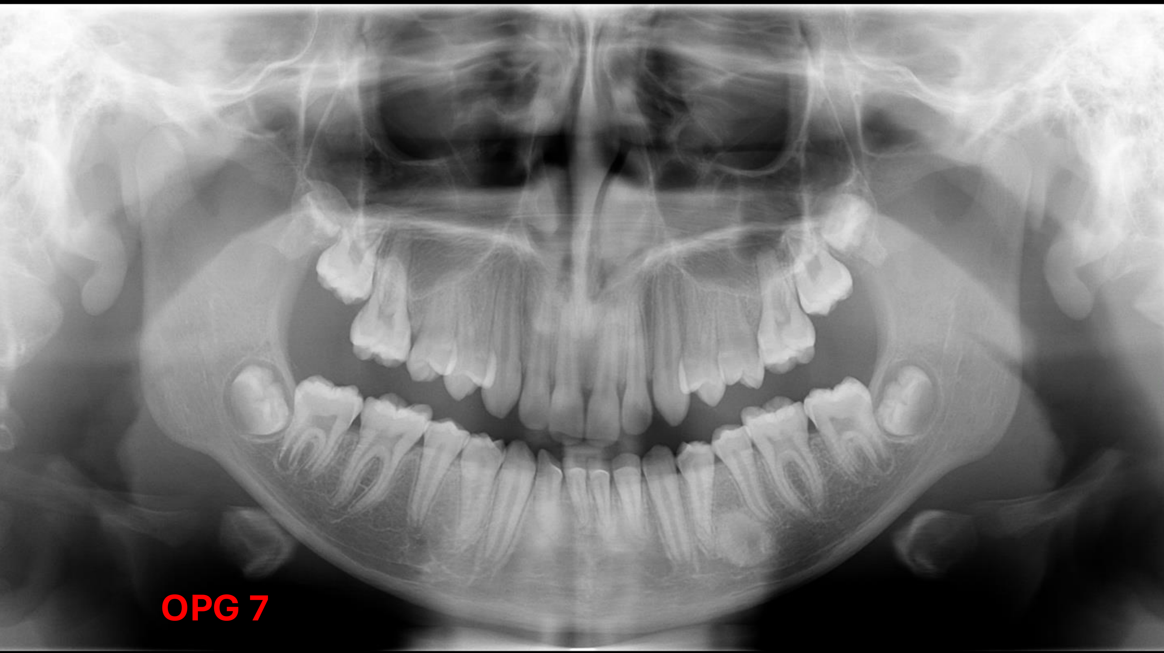
- Address Clinical Concerns
- ==Patient Age: Approximately 13 years old.==
- ==Developmental Status: Roots of the second molars (sevens) have not completely traveled/formed.==
- ==General Observations: Mandible, sinus floor, septum, and orbitals appear normal. Teeth show no signs of caries.==
-
Dentoalveolar Analysis
- Count teeth and identify ectopic and/or impacted teeth.
- 28: hard to tell might be distoangularly impacted
- All teeth are present; all third molars (eights) are unerupted.
- Identify dental anomalies (position, shape, etc.).
- **42: Rotated **
- ==34 and 35 Roots: The roots are not “displaced” in the sense of being pushed by a tumor; rather, they are dilacerated or diverging to avoid the opacity. The roots grew around the lesion because it was present during root formation and was too dense for the roots to grow into.==
- Assess periodontal bone loss.
- Identify dental pathology (non-carious tooth loss, caries, fractures, periapical pathologies, etc.).
- ==PDL and Lamina Dura: Intact for both the 34 and 35.==
- ==Root Resorption: No resorption is present.==
- Count teeth and identify ectopic and/or impacted teeth.
-
Maxilla and Mandible
- Identify abnormal (radio)lucencies.
- Identify abnormal (radio)opacities.
- Between 34-25
- bone island
- Solitary bone cyst
- ==Location: Between the 34 and 35 (lower left first and second premolars).==
- ==Appearance: Well-defined, circular, homogeneous, and very radio-opaque.==
- ==Differential Diagnosis:==
- ==Bony Island (Dense Bone Island): The primary diagnosis. It is identified by its homogeneity and lack of inflammatory or cystic characteristics.==
- ==Exclusions:==
- ==Cyst: Ruled out because the lesion is opaque and the PDL/lamina dura of adjacent teeth are intact.==
- ==Tumor/Inflammatory Lesion: Ruled out due to the lack of root resorption or active displacement.==
- Between 34-25
- Note altered trabecular patterns.
- Identify fractures.
- Assess jaw asymmetry.
- OPG error: Chin tipped too low
- ==The OPG displays a “joker smile,” indicating a chin down positioning error.==
-
- Maxillary Sinuses: Evaluate for mucosal changes. - Asymmetry in the Nasal concha
- TM Joints: Evaluate for morphological changes of the condyles.
- Soft Tissues: Identify swelling or soft tissue calcifications (e.g., tonsilloliths, salivary gland stones, etc.).
- ==Soft Tissue Artifacts: A hook-like radio-opacity is visible behind the ramus of the mandible. This is identified as the earlobes (soft tissue density), which can appear prominent or radio-opaque in some patients (e.g., cauliflower ears).==
- Other Sites: Evaluate the spine, orbits, and other visible structures for structural changes.