OPG Case 8
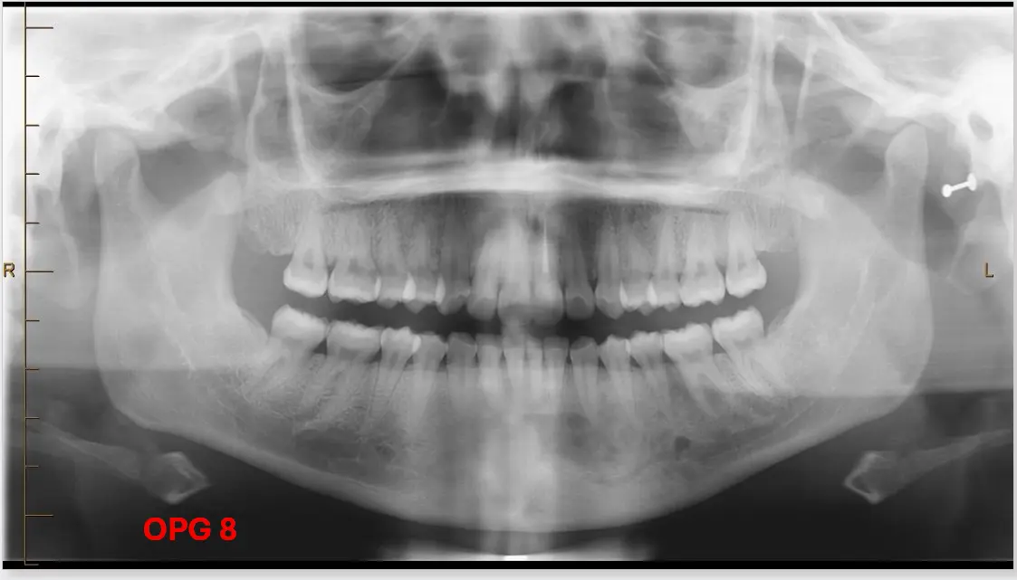
- Address Clinical Concerns
- Dentoalveolar Analysis
- Count teeth and identify ectopic and/or impacted teeth.
- Identify dental anomalies (position, shape, etc.).
- Assess periodontal bone loss.
- Identify dental pathology (non-carious tooth loss, caries, fractures, periapical pathologies, etc.).
- 21: Root canal treated
- ==12: Root appears short at the apex with a hyperdense appearance; may have a history of trauma (“knocked in the past”).==
- ==22: Endodontically treated; possible history of trauma to anterior teeth.==
- ==PDL Widening: Widening observed at 33, 35, and 37 is attributed to occlusal loading rather than the primary lesion.==
- Maxilla and Mandible
-
Identify abnormal (radio)lucencies.
-
Identify abnormal (radio)opacities.
-
Note altered trabecular patterns.
- Mandible : between teeth LHS
- Diffuse sclerosing pattern , mixed radiolucencies
- ==Location: The lesion extends from the 41 to approximately the 33 region, potentially entering the canal. There is an altered normal trabecular bone pattern of the left mandible, possibly involving the anterior mandible up to the 35/36 region.==
- ==Appearance: Non-homogeneous with a mix of radiolucency and radiopacity. It has a sclerotic, fibrous, and “ground glass” appearance.==
- ==Definition: Initially described as well-defined, but upon broader view, the lesion is not well-defined due to the altered trabecular pattern.==
- ==Components: The “ground glass” bone represents the bony component, while small lucencies throughout represent the fibrous component.==
- ==Effect on Surrounding Structures: No displacement of teeth and no resorption noted in the primary lesion area. The lesion involves the periodontal ligament space and the incisive canal.==
- ==Mandibular Canal: This is an atypical presentation as it is not located inferior to the mandibular canal (which would typically displace the canal upwards).==
- ==Clinical Note: The lesion has changed the composition of the left mandible but is “not really doing anything to the teeth.”==
Differential Diagnosis
- ==Fibro-osseous Lesions: The lesion belongs to this family.==
- ==Cyst: Considered because it involves the PDL and lamina dura, suggesting it could be odontogenic.==
- ==Tumors/Inflammation: Ruled out due to the lack of tooth resorption or displacement.==
Diagnosis
- ==Primary Diagnosis: Fibrous Dysplasia.==
- ==Note: It is described as the “only ground glass bone lesion.” This specific case is noted as an “atypical fibrous dysplasia” because of its location relative to the mandibular canal.==
Investigations
- ==CT Scan: A “premium CT” is suggested for further investigation due to the atypical presentation.==
- ==Periapical (PA) Radiograph: Recommended for tooth 12 to check for an apical lesion or apical resorption, and to check the “implant condition.”==
- Mandible : between teeth LHS
-
Identify fractures.
-
Assess jaw asymmetry.
-
- Maxillary Sinuses: Evaluate for mucosal changes.
- TM Joints: Evaluate for morphological changes of the condyles.
- Soft Tissues: Identify swelling or soft tissue calcifications (e.g., tonsilloliths, salivary gland stones, etc.).
- Other Sites: Evaluate the spine, orbits, and other visible structures for structural changes.