W2 Cases (Pediatric Dentistry)
Index
Case 1
Images
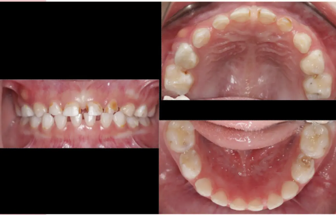
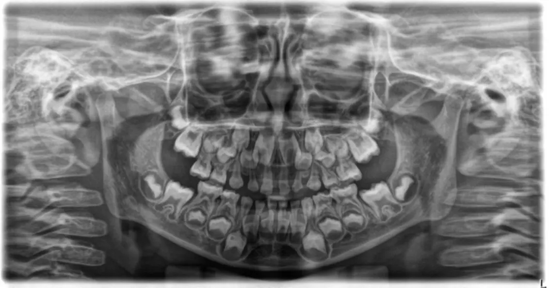
Age / dentition stage
- Age: 3–5
- Second primary molars erupt ~20–30 months
- Lower limit: Fully erupted dentition suggests >36 months (~3 years)
- Upper limit: Absence of first permanent molars indicates <5.5 years
- Corroborating factors: No mobile teeth or history of premature loss
Key findings
Soft tissue
- 53–54 buccal abscess
- Note: abscesses can appear as lumps that come and go as they drain/refill
- Asymptomatic presentation: Common with draining abscesses as infection drains, preventing pressure buildup and pain
- Draining sinus tract indicates chronic infection from necrotic pulp
Radiographic (OPG)
- 84 DO
- 74 DO cavity
- Caries 61, 62
- 54 DO
- 55: Large cavity with marginal ridge breakdown (indicates probable irreversible pulpitis or necrosis)
- Talon cusp vs. Shovel-shaped incisors: Marginal ridges on palatal surface likely represent shovel-shaped incisors (normal trait in East Asian populations)
- Caries are often underestimated radiographically compared to clinical extent
Questions (from class)
- What is the cause of the abscess?
My notes / working answer
-
Possibly 54 or 55 (insufficient information to determine)
-
Pathophysiology: The abscess most likely originates from 54 (existing filling acts like a cork in a bottle, forcing pus through bone)
-
Management of 54: Likely necrotic; requires extraction (pulpectomy rarely indicated for primary incisors)
-
Management of 55: Requires pulpotomy + Stainless Steel Crown (SSC) due to marginal ridge breakdown indicating irreversible pulpitis
-
Clinical Tip: Always examine soft tissues first to look for redness, inflammation, or stretched gingiva before being distracted by caries
Case 2
Images
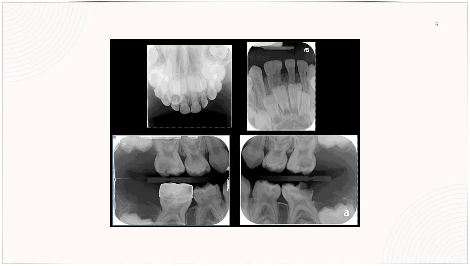
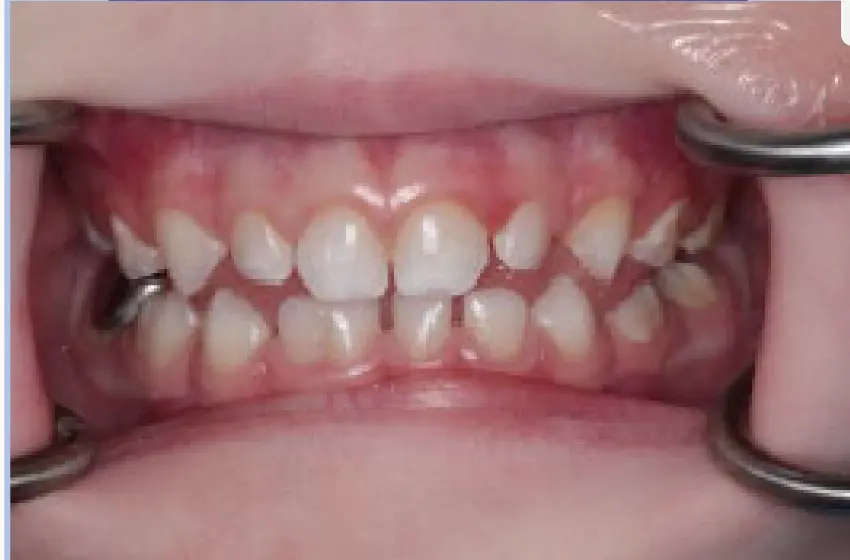
Case Study: Child with Double Tooth (Fusion)
- Family relocated from overseas; medically fit and healthy.
- Dental history: Hall crown placed on 85 ten months ago; other treatment planned but not completed due to move.
- Behavior: Frankel ++.
- Radiographs show a nasal tube (radiopaque wire) indicating a previous General Anesthetic (GA) procedure.
- Caries into inner third/half of dentine on 55, 65, 75, 74.
Age / dentition stage
- Estimate: 4–4.5 years
- First permanent molars visible in bone on BW but not in occlusion
- ~1 year from “in bone” → “in occlusion”
- Estimation is precise because 6s are "just through bone," placing the child at ~4-4.5 years.
Key findings
Teeth / clinical
- Fusion of 81/82
- White spot lesions buccal of 64
- Diagnosis of 81/82: Fusion (not gemination) because there are 19 teeth in the primary count instead of 20.
- 81/82 Anatomy: Two separate roots and two pulp chambers with a distinct notch at the crown junction.
- 85 Hall Crown: Poorly seated (not past contact points) likely due to insufficient crown size or inadequate separator use; open bite should have settled in weeks, not 10 months.
- Crown on 85 not seated properly
- Supported by bitewings and clinically open occlusion
Questions (from class)
- Describe key clinical and radiological findings
- Main caries risk factors and likely etiology?
- Treatment priorities: first visit?
- Treatment priorities: second visit?
- Ongoing management / what to do on extraction visit?
- Why extract 81/82 prophylactically under GA?
- Caries Risk Assessment (CRA) questions: Origin/Fluoride history, open-ended diet questions (e.g., tea with sugar, bedtime bottles), and confirming 1000ppm fluoride toothpaste vs 500ppm.
Management (draft)
Visit 1 (priorities)
- Reinforce OHI
- SSC + pulpotomy: 55, 65
- 75 likely unrestorable
- Avoid immediate exo on first visit unless active/severe infection (e.g., draining sinus, swelling)
- Prevention: Brush twice daily with 1000ppm fluoride toothpaste (note: commercial "3-5 year" pastes are often only 500ppm).
- 85 Crown: Lower priority than active deep caries as it is currently sealing the decay.
- Avoid immediate exo on first visit unless active/severe infection (e.g., draining sinus, swelling)
Visit 2
- (To confirm/fill in)
- Extract 75 AND 74: Same quadrant management to use a single local anesthetic episode and preserve cooperation.
- Space Maintainer: Contraindicated as 6 is not erupted enough for band and loop; must accept space loss risk.
Ongoing / extraction-visit notes
- Space maintainer: usually not yet unless considering distal shoe
- If extracting 75: anesthetize and treat 74 at same appointment
- 81/82 fusion: permanent successors appear normal → monitor
Visit 3 (under GA)
- Extract 81/82: High risk of interfering with permanent successor eruption and high risk of tooth fracture during extraction.
- Informed Consent: Explicitly warn parents that the double tooth may break and require removal in pieces.
Case 3 (Bitewings)
Images
- (add bitewings here)
Questions (from class)
- What factors would influence your treatment decision for these patients?
Important
What age do impacted 6’s self-correct?
(Flag for EXAM/QUIZ)
Case 4
Clinical question
- What is the “double tooth”?
Case Study: Suspected Supernumerary vs. Gemination (Remote Consultation)
A ~4-year-old healthy child living 2 hours from Perth. Parents sent a photo of two tooth-like structures in the upper anterior (62 region). A PA radiograph confirmed a supernumerary tooth (mesiodens) alongside the normal lateral incisor.
Notes / interpretation
Ectopic Eruption of First Permanent Molars
- Mechanism: 6s erupt mesially, impacting distal of E's causing resorption.
- Physiological Resorption: Normal process in the wrong location; related to 6's position.
- Inflammatory Resorption: Indicated by symptoms, abscess, or irregular patterns if bacteria reach subgingival space.
- Space Maintenance: If E is extracted and 6 is unerupted, a distal shoe is difficult to clean and painful; often no maintainer is possible at age 4-5.
-
Clinical photo alone: insufficient to determine
-
Clinical test: wedge flat plastic between the two teeth; twisting/pushing may show crowns moving in opposite directions
-
Best next steps: clinical exam + periapical/occlusal radiograph
-
From occlusograph: supernumerary suggested
-
Gemination usually presents with a single root and normal tooth count, whereas a supernumerary has separate roots and increased tooth count.
-
To identify the supernumerary, compare anatomy (often conical/tuberculate with shorter roots) and check the contralateral side.
-
Risks include resorption or ectopic eruption of the permanent lateral incisor; however, surgical removal at age 4 carries risks of damaging unerupted permanent teeth.
Information needed for management plan
- Check other areas of primary dentition for anomalies
- Family history of supernumeraries
- Maxillary Occlusal Radiographs are superior to OPGs for the anterior region as they avoid the narrow focal trough and are more comfortable for children.
- CBCT is indicated if multiple supernumeraries are suspected, but ideally deferred for 12 months to allow permanent incisor positions to become clearer.
If multiple supernumeraries are permanent
-
Discuss/flag with parents: potential future issues
-
Child may be too young for removal of permanent supernumerary (timing depends on position/root development/risks)
-
If asymptomatic, monitor every 6 months until exfoliation (~age 6).
-
Surgical removal is ideally performed after age 5 to reduce risk to permanent successors.
-
CBCT is mandatory for multiple supernumeraries to map relationships and plan a single GA episode.
Parking lot / to confirm
- Case 2: caries risk factors + etiology
- Case 2: Visit 2 plan
- Case 3: treatment decision factors (list)
- Impacted 6’s: typical self-correction age range
- Specific duration for physiological resorption of primary roots when impacted by permanent successors.
- OPG vs. targeted imaging: whether OPG is needed to screen for additional supernumeraries when one is found.
Audio Appendix
Additional Audio Content
The following sections from the lecture audio did not correspond to any heading in the main document.
Introduction and Professional Expectations
- LMS Engagement: Lecturers can track lecture completion, “in progress” status, and whether content has been opened. Downloading lectures for offline viewing still registers as accessed.
- Pre-clinic Requirements: Students must watch recorded lectures and clinic orientations (including the 5-minute orientation) before entering pediatric clinics. Falling significantly behind in content consumption puts students at risk of failing and being unable to defend their performance at semester end.
- Case-Based Learning: Interactive case discussions using QR codes and polling software to assess clinical reasoning and holistic thinking.
Additional Topics Covered
Pediatric Radiology Techniques
- OPG Positioning: Use foam blocks as holders instead of RIN holders for pediatric patients (also applicable to anxious adults); ensures better anterior region visualization
- Anterior Focal Trough: OPG poor for incisor detail due to narrow focal trough; maxillary occlusal preferred for anterior assessment in young children
Treatment Prioritization Principles
- Pain and infection (abscesses, symptomatic pulpitis)
- Active deep caries (near pulp, risk of exposure)
- Sealed but problematic restorations (asymptomatic but poor fit - lower priority than active disease)
- Efficiency: Combine same-quadrant treatments to minimize visits and anesthesia episodes (e.g., extract 74 when extracting 75)
Informed Consent Specifics
- Double Tooth Extraction: Explicitly warn parents that fused teeth may fracture during extraction due to weak connection between fused portions
- GA Planning: Goal is to complete all necessary treatment in one episode to avoid repeat anesthesia; may justify more aggressive treatment (e.g., prophylactic extraction of 81/82)
Cultural Competency in History Taking
- Avoid assumptions about bottle feeding vs. extended breastfeeding (child-led weaning)
- Ask open-ended diet questions (“What do they drink?”) rather than leading questions (“Do they drink juice?”) to capture culturally specific practices (tea with sugar/honey, undiluted cordial)