Pattern of Caries Spread1
Research and statistical analysis regarding the distribution and progression of dental caries.
 |
Caries Risk Assessment2
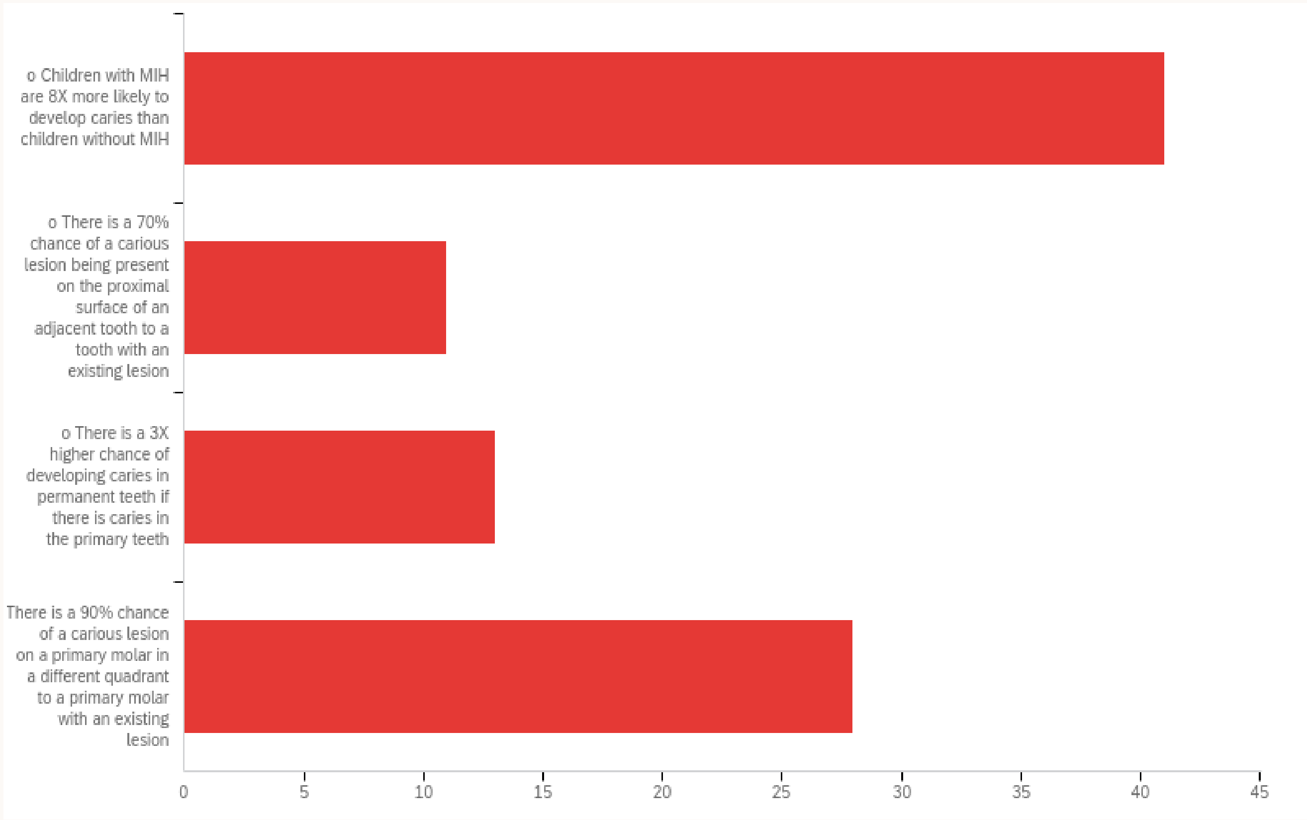
Q39 - Which of these statements is false?
- Statement 1: Children with MIH are 5.89 times more likely to have a DMFT greater than zero than children without MIH.
- Statement 2: 69% of primary teeth with proximal caries developed caries on the adjacent proximal surface.
- Statement 3: 89% of patients with a proximal carious lesion on a primary tooth in one quadrant developed another primary molar proximal lesion in another quadrant.
- Statement 4: Children with MIH are 8X more likely to develop caries than children without MIH.
Proximal Caries Progression
Adjacent Surface Involvement3
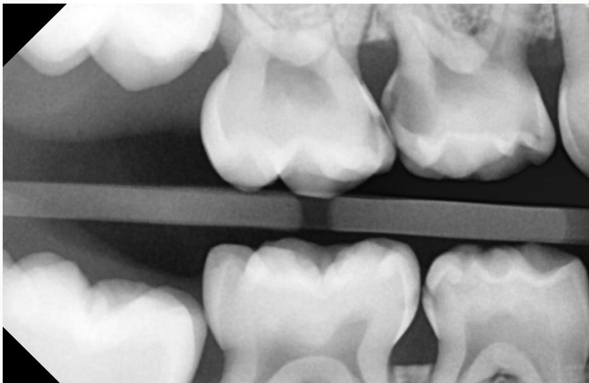
According to research by Dean et al. (1997), 69% of primary teeth with proximal caries developed caries on the adjacent proximal surface. readings
- Clinical Assumption: If a lesion is identified on one tooth, clinicians should assume at least a "white spot" lesion exists on the adjacent tooth until proven otherwise.
 |  |
Cross-Quadrant Progression4
Research published in J Clin Pediatr Dent (1997) titled “Progression of interproximal caries in the primary dentition” by Dean et al. indicates:
- Contiguous Spread: 69% of primary teeth with proximal caries spread to the adjacent surface.
- Quadrant Correlation: 89% of patients with a proximal carious lesion on a primary tooth in one quadrant developed another primary molar proximal lesion in a different quadrant.
- Clinical Assumption: When proximal caries is seen in one quadrant, it should be assumed to be happening elsewhere in the mouth.
Predicting Caries in Permanent Teeth
Longitudinal Cohort Study Results5
An eight-year cohort study (Li and Wang, 2002) conducted on 362 children investigated the correlation between primary and permanent dentition caries.
-
Key Findings:
- Children with caries in primary teeth were approximately three times more likely to develop caries in their permanent teeth (Relative Ratio = 2.6, 95% CI = 1.4-4.7).
- Caries on primary molars demonstrated the highest predictive value at 85.4%.
- Caries in primary molars is a more significant predictor of risk than caries in primary incisors.
Molar Incisor Hypomineralization and Caries Risk
Association Between MIH and Caries Experience6
A case-control study by Americano et al. (2016) readings investigated the relationship between Molar Incisor Hypomineralization (MIH) and the Decayed, Missing, or Filled Index (DMF-T) in children aged 7 to 11.
-
Statistical Risk: Children with MIH are 5.89 times more likely to have a DMFT score greater than zero than children without MIH (95% CI = 2.69 to 12.88; P < 0.05).
-
Study Parameters: The groups were balanced for age, gender, and primary dentition caries experience.
-
Conclusion: There is a significant association between the presence of MIH and increased caries experience in permanent teeth.
-
Children with MIH must be treated as high caries risk.
-
Preventative strategies for these patients include high-strength fluoride toothpaste, fissure sealants, and potentially mouth rinses.
Fact Check:
- True: Children with MIH are 5.89x more likely to have DMFT > 0.
- False: Children with MIH are 8x more likely to develop caries.
Clinical Presentation of MIH7
Data from Americano et al. (2016) confirms that children with Molar Incisor Hypomineralization (MIH) face a significantly higher risk of caries.
- Risk Ratio: 5.89 times increased likelihood of having a DMFT > 0.
- Clinical Observations: MIH often presents as hypoplastic patches and color anomalies on the occlusal surfaces of permanent molars and incisors, increasing susceptibility to decay.
- MIH involves porous enamel and post-eruptive breakdown, which creates plaque retention factors.
- These teeth are often highly sensitive, leading to poor brushing habits.
- A unilateral presentation of plaque and calculus often indicates a source of pain; if a child is not cleaning or chewing on one side, it is often because a tooth on that side is painful.

Risk Factors for Second Primary Molars
Impact on First Permanent Molars
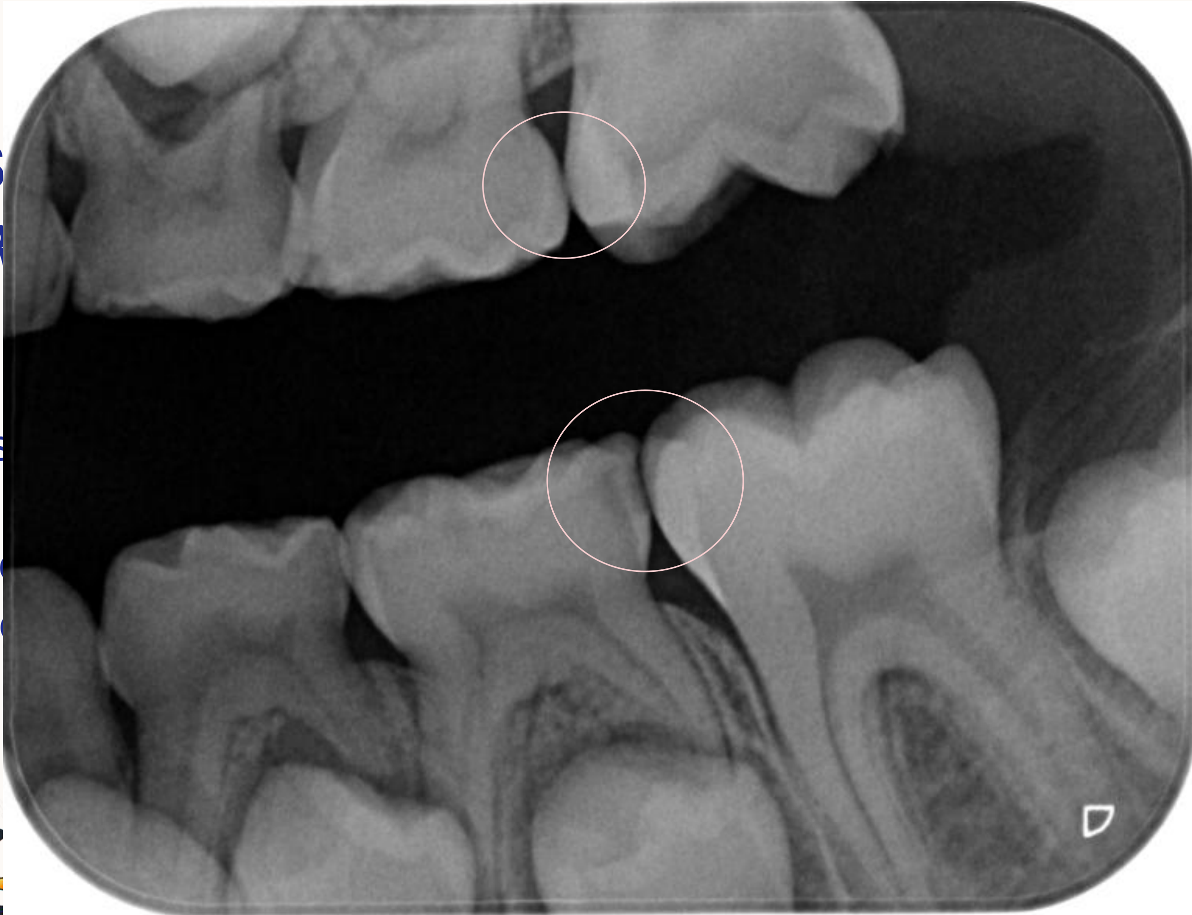
- Risk Factor: Caries on the distal surface of a second primary molar increases the risk for caries on the mesial surface of the first permanent tooth by 15 times.
- If caries is detected on the distal surface of a second primary molar, clinicians should consider active management—such as restoration or Silver Diamine Fluoride (SDF)—to protect the mesial surface of the erupting or erupted first permanent molar.
 |  |
: Original PDF page 1: W2 Case Based Discussion 2, p.1 : Original PDF page 2: W2 Case Based Discussion 2, p.2 : Original PDF page 3: W2 Case Based Discussion 2, p.3 : Original PDF page 4: W2 Case Based Discussion 2, p.4 : Original PDF page 5: W2 Case Based Discussion 2, p.5 : Original PDF page 6: W2 Case Based Discussion 2, p.6 : Original PDF page 7: W2 Case Based Discussion 2, p.7 : Original PDF page 8: W2 Case Based Discussion 2, p.8 : Original PDF page 9: W2 Case Based Discussion 2, p.9 : Original PDF page 10: W2 Case Based Discussion 2, p.10 : Original PDF page 11: W2 Case Based Discussion 2, p.11 : Original PDF page 12: W2 Case Based Discussion 2, p.12 : Original PDF page 13: W2 Case Based Discussion 2, p.13 : Original PDF page 14: W2 Case Based Discussion 2, p.14 : Original PDF page 15: W2 Case Based Discussion 2, p.15 : Original PDF page 16: W2 Case Based Discussion 2, p.16 : Original PDF page 17: W2 Case Based Discussion 2, p.17 : Original PDF page 18: W2 Case Based Discussion 2, p.18 : Original PDF page 19: W2 Case Based Discussion 2, p.19 : Original PDF page 20: W2 Case Based Discussion 2, p.20 : Original PDF page 21: W2 Case Based Discussion 2, p.21 : Original PDF page 22: W2 Case Based Discussion 2, p.22 : Original PDF page 23: W2 Case Based Discussion 2, p.23 : Original PDF page 24: W2 Case Based Discussion 2, p.24 : Original PDF page 25: W2 Case Based Discussion 2, p.25 : Original PDF page 26: W2 Case Based Discussion 2, p.26 : Original PDF page 27: W2 Case Based Discussion 2, p.27 : Original PDF page 28: W2 Case Based Discussion 2, p.28 : Original PDF page 29: W2 Case Based Discussion 2, p.29 : Original PDF page 30: W2 Case Based Discussion 2, p.30 : Original PDF page 31: W2 Case Based Discussion 2, p.31 : Original PDF page 32: W2 Case Based Discussion 2, p.32 : Original PDF page 33: W2 Case Based Discussion 2, p.33 : Original PDF page 34: W2 Case Based Discussion 2, p.34 : Original PDF page 35: W2 Case Based Discussion 2, p.35 : Original PDF page 36: W2 Case Based Discussion 2, p.36 : Original PDF page 37: W2 Case Based Discussion 2, p.37 : Original PDF page 38: W2 Case Based Discussion 2, p.38 : Original PDF page 39: W2 Case Based Discussion 2, p.39