“The development of a reasonable differential diagnosis is of prime importance in determining if biopsy is indicated. Furthermore, the differential diagnosis aids the clinician in selecting the appropriate technique if biopsy is necessary.” (“Contemporary Oral Medicine: A Comprehensive Approach to Clinical Practice”, 2019, p. 343) (pdf)
“A biopsy, even though it may be the gold standard for some diagnosis, is not always indicated for every oral mucosal lesion” (“Contemporary Oral Medicine: A Comprehensive Approach to Clinical Practice”, 2019, p. 343) (pdf)
“There are several main reasons why a clinician may perform a biopsy: 1. To rule out possible cancer or potentially cancerous pathology or to monitor the possible malignant transformation of an oral potentially malignant disorder (OPMD). This is especially important when one encounters white, red, indurated, and/or ulcerated lesions. 2. To help in establishing the diagnosis when the clinician has doubts, regardless of the nature of the pathology. Appropriate treatment can only” (“Contemporary Oral Medicine: A Comprehensive Approach to Clinical Practice”, 2019, p. 343) (pdf)
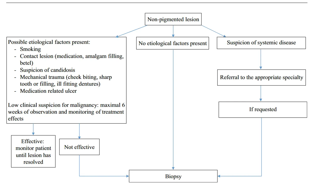
“Fig. 1 Flowchart for non–pigmented mucosal lesions. (Adapted from van der Waal 2010)” (“Contemporary Oral Medicine: A Comprehensive Approach to Clinical Practice”, 2019, p. 344) (pdf)
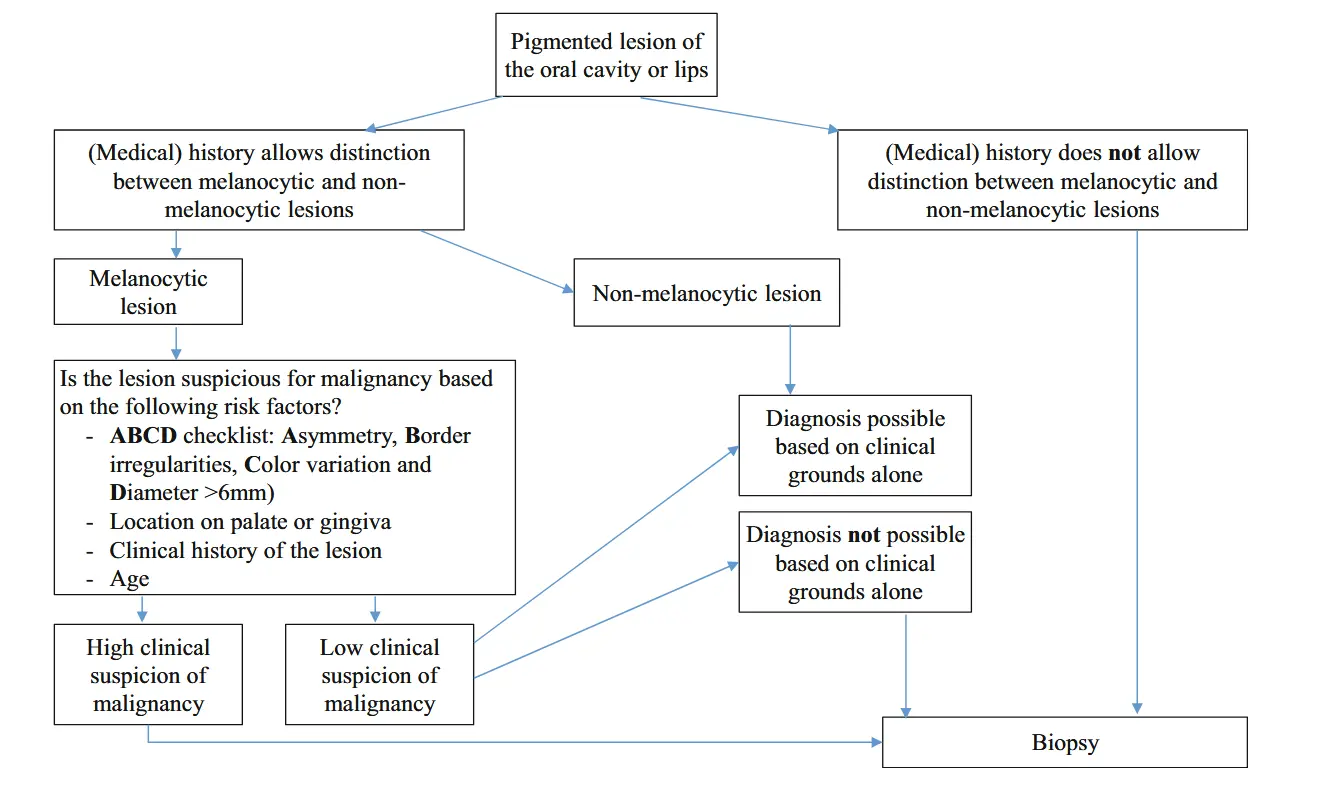
“Fig. 2 Flowchart for pigmented lesions. (Adapted from Meleti et al. 2008)” (“Contemporary Oral Medicine: A Comprehensive Approach to Clinical Practice”, 2019, p. 344) (pdf)
Indications to perform a biopsy
“Indications to perform a biopsy include:
- Lesions (especially manifesting as a red patch or having inflammatory features) persisting for more than 2 weeks and: (a) without apparent etiopathogenic factors, and/or (b) after removal of identifiable irritating factors, and/or (c) after empirical treatment has been attempted.
- All white hyperkeratotic lesions for which a cause is not obvious, especially speckled non-homogeneous white and red lesions.
- Any lesion suspicious of a neoplasm (presence of growth, ulceration, induration, with or without painful symptoms).
- Lesions interfering with function (e.g., fibroepithelial polyps).
- Tissue from draining sinus tracts when there is no probable cause.
- Lesions in the jaw bones that cannot be diagnosed radiographically, especially if they cause symptoms, such as pain or altered sensations.” (“Contemporary Oral Medicine: A Comprehensive Approach to Clinical Practice”, 2019, p. 345) (pdf)
Contraindications to perform a biopsy
“There are no absolute contraindications for performing a biopsy, yet one should keep in mind several issues that may help mitigate unwanted complications.” (“Contemporary Oral Medicine: A Comprehensive Approach to Clinical Practice”, 2019, p. 345) (pdf)
“. In case of acute pyogenic or viral infection, one should refrain from biopsy until the patient has recovered or after a course of medical treatment.” (“Contemporary Oral Medicine: A Comprehensive Approach to Clinical Practice”, 2019, p. 345) (pdf)
“anticoagulation therapy or suffers from a serious bleeding disorder” (“Contemporary Oral Medicine: A Comprehensive Approach to Clinical Practice”, 2019, p. 345) (pdf)
“The utmost attention should be exercised when performing a biopsy on a lesion involving the hard palate mucosa, so as not to puncture the greater palatine artery (a. palatina major).” (“Contemporary Oral Medicine: A Comprehensive Approach to Clinical Practice”, 2019, p. 345) (pdf)
When to perform a biopsy
“Thus, one should firstly collect as much etiological and clinical data as possible.” (“Contemporary Oral Medicine: A Comprehensive Approach to Clinical Practice”, 2019, p. 346) (pdf)
Who should Perform a biopsy?
“typically, general dental practitioners do not have sufficient knowledge of oral diseases nor sufficient clinical experience in undertaking biopsies to routinely perform them and interpret their results for the ultimate benefit of the patien” (“Contemporary Oral Medicine: A Comprehensive Approach to Clinical Practice”, 2019, p. 347) (pdf)
“In countries where oral medicine is a recognized specialty, it would be best that these specialists undertake biopsies for suspected oral lichen planus and other non-malignant mucosal disease.” (“Contemporary Oral Medicine: A Comprehensive Approach to Clinical Practice”, 2019, p. 347) (pdf)
What to Do Before the biopsy
“Contemporary Oral Medicine: A Comprehensive Approach to Clinical Practice”, 2019, p. 348
“1. Size: How large is the lesion? Does it cross anatomical boundaries? Has it changed size over time or since it was noted? 2. Shape: Has the lesion changed in shape? For example, has a blister become an ulcer? This may signify autoimmune vesicullobulous condition. 3. Progression: Is the lesion static, or has it changed over time? Has this occurred slowly or quickly, alternatively has it regressed? Have there been alternating periods of increase or decrease in size, or quiescent periods? If a lesion progresses rapidly, then it may be more sinister. This information is biased by the patient’s perception. 4. Duration: How long has the lesion been present? If the lesion has been present for years, it is more likely to be benign in nature.” (“Contemporary Oral Medicine: A Comprehensive Approach to Clinical Practice”, 2019, p. 348) (pdf)
“his provides determination of morphological and topographical features, which should be provided to the reporting pathologist. 1. Look: Determine site, shape, morphology, color, sharpness, and regularity of border, and whether the lesion is single or multiple. 2. Feel: Determine consistency (soft, firm, hard), description of surface (smooth, lobulated, irregular, verruciform), tenderness, associated symptoms (e.g., discharge) and pulsations (e.g., vascular lesions). 3. Move: Determine tethering (fixation to mucosa, skin or underlying structures). 4. Measure: Determine size (using a ruler or calipers for exact measurement). 5. Examine: Determine presence of head and neck lymph nodes (necessary part of every oral examination, always performed before biopsy). 6. Photography: Record the exact appearance and size for future reference. 7. Radiography: Perform if possible dental or bony involvement is suspected.” (“Contemporary Oral Medicine: A Comprehensive Approach to Clinical Practice”, 2019, p. 348) (pdf)