Oral Pathology
Introduction to Oral Pathology1
Oral pathology is the specialty of dentistry and discipline of pathology that addresses the nature, identification, and management of diseases affecting the oral and maxillofacial regions. It is a science that investigates the causes, processes, and effects of these diseases.
Clinical Manifestations and Diagnosis
The practice of oral pathology involves the clinical examination, radiographic interpretation, and microscopic analysis of tissues to reach a definitive diagnosis. Key areas of focus include:
- Developmental Disorders: Abnormalities occurring during the growth and development of the oral structures.
- Infectious Diseases: Pathologies caused by bacterial, viral, or fungal agents.
- Neoplastic Conditions: Benign and malignant tumors of the soft tissues and jawbones.
- Immune-Mediated Diseases: Conditions where the immune system targets oral mucosa.
Role of the Pathologist
Pathologists work closely with clinicians to provide essential diagnostic information that guides patient treatment plans. This includes the evaluation of biopsy specimens and the correlation of clinical findings with laboratory data to ensure comprehensive patient care.
Clinical Categories and Disease Classifications2
- Developmental Conditions
- Mucosal Lesions—Reactive
- Mucosal Lesions—Infections
- Mucosal Lesions—Immunologic Diseases
- Mucosal Lesions—Premalignant
- Mucosal Lesions—Malignant
- CT Tumors—Benign
- CT Tumors—Malignant
- Salivary Gland Diseases—Reactive
- Salivary Gland Diseases—Benign
- Salivary Gland Diseases—Malignant
- Lymphoid Neoplasms
- Odontogenic Cysts
- Odontogenic Tumors
- Bone Lesions—Fibro-Osseous
- Bone Lesions—Giant Cell
- Bone Lesions—Inflammatory
- Bone Lesions—Malignant
- Hereditary Conditions
Developmental Conditions
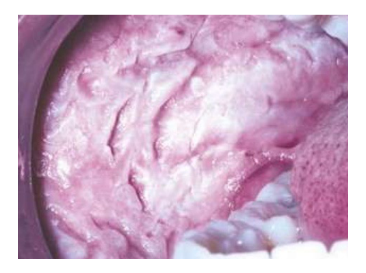
- Autosomal dominant
- Asymptomatic spongy white buccal mucosa
- Diagnostic Feature: Unlike pseudomembranous candidiasis, this white lesion cannot be wiped off with gauze.
White Sponge Nevus3
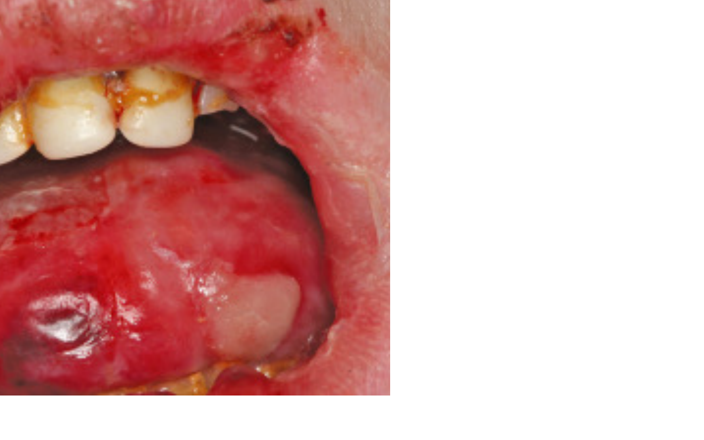
Epidermolysis Bullosa4
- Autosomal dominant or recessive
- Causes skin and mucosa to be fragile and blister easily
- Clinical Appearance: The presentation can resemble erythema multiforme but involves more extensive blistering across the skin and mucosa.
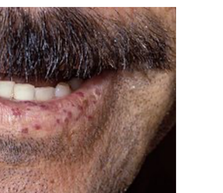
Hereditary Hemorrhagic Telangiectasia
Clinical Characteristics5
- Autosomal dominant
- Also known as Olser-Weber-Rendu Syndrome
- Telangiectasia: Red macule or papule representing a dilated or broken capillary
- Abnormal capillary formation on skin, mucosa, and viscera
- Diascopy/Blanching: Unlike extravascular lesions (petechiae, purpura, hematoma), these are intravascular; they will blanch when pressure is applied and refill when released.
- Associated with iron-deficiency anemia
- Epistaxis (nosebleeds) is a frequent presenting sign
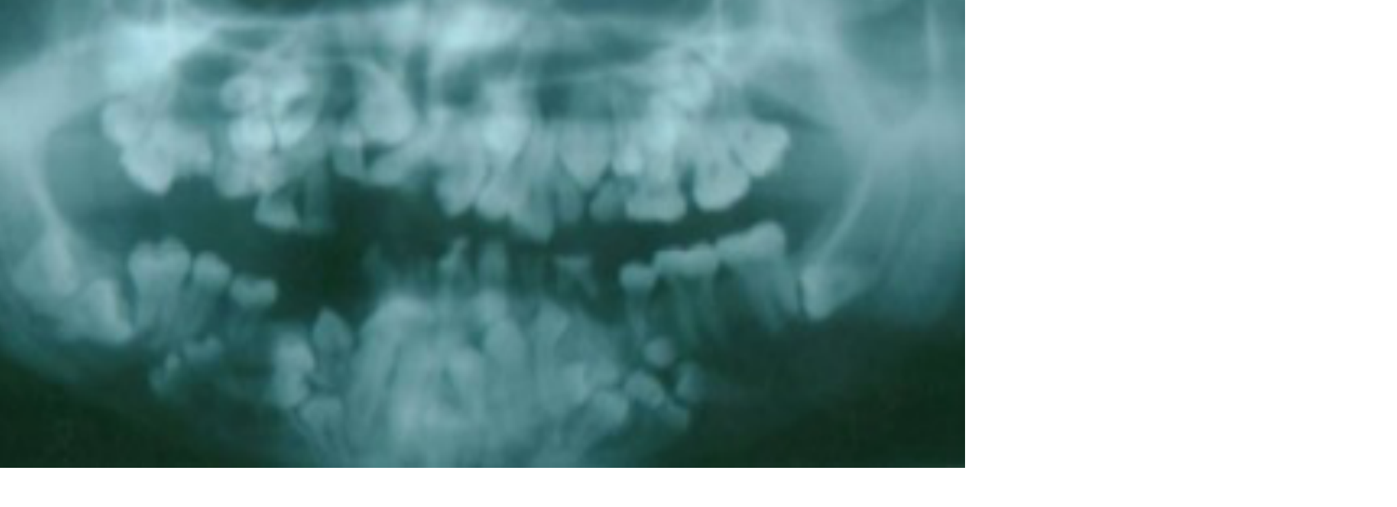
Cleidocranial Dysplasia6
- Autosomal dominant
- Missing clavicles
- **Supernumerary teeth
- Treatment: Patients often require a series of extractions and may need dentures.**
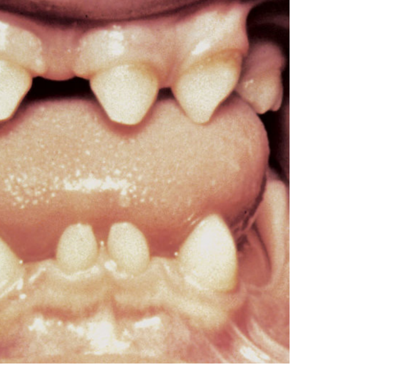
Ectodermal Dysplasia7
- X-linked recessive
- Missing teeth
- **Hypoplastic hair or nails
- Clinical Presentation: May also present with balding.**
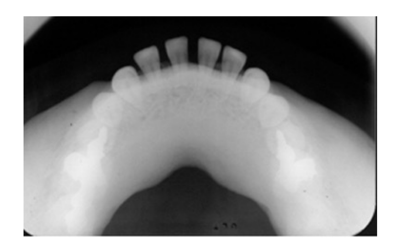
Osteopetrosis8
- Autosomal dominant or recessive
- Also called Albergss-Schonberg disease and marble bone disease
- Lack of bone remodeling and resorption leads to “stone bone”
- Radiographic Features: Unlike localized radiopaque lesions, the entire bone is affected by increased density.
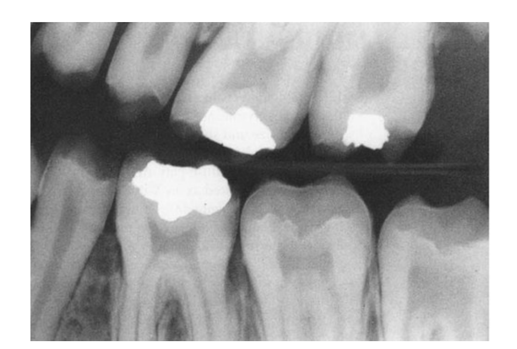
Amelogenesis Imperfecta
Clinical Presentation9
- Autosomal dominant, recessive, or X-linked
- Intrinsic alteration of enamel
- All teeth from both dentitions are affected
- Thin to no enamel, but dentin and pulp are normal
- Radiographic Features: Enamel appears very thin or completely absent; there is a lack of the usual radiodensity contrast between enamel and dentin.
Treatment
- Full-coverage crowns for cosmetics
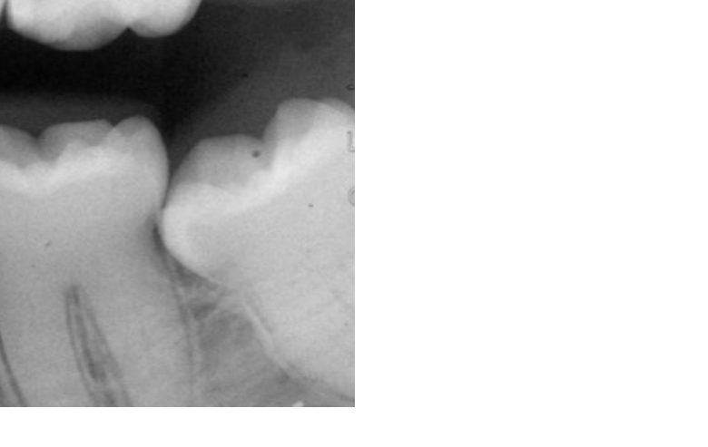
Dentinogenesis Imperfecta
Clinical and Radiographic Features10
- Autosomal dominant
- Intrinsic alteration of dentin
- All teeth from both dentitions are affected
- Short roots, bell-shaped crowns, and obliterated pulps
- Bulbous crowns visible in radiographs due to constricted DEJ
- Associated with **blue sclera
- Systemic Association: Blue sclera is also a sign linked to osteogenesis imperfecta.**
Treatment
- Full-coverage crowns for cosmetics
Dentin Dysplasia
Clinical Features11
- Autosomal dominant
- Intrinsic alteration of dentin
- All teeth from both dentitions are affected
- **Chevron pulps and short roots
- Classification: Type 1 is associated with short roots (one finger); Type 2 is associated with chevron pulps (two fingers in a "V" shape).**
- Teeth are not good candidates for restoration
- Clinical Considerations: Poor candidates for crowns due to a poor crown-to-root ratio and risk of pulp exposure during preparation.
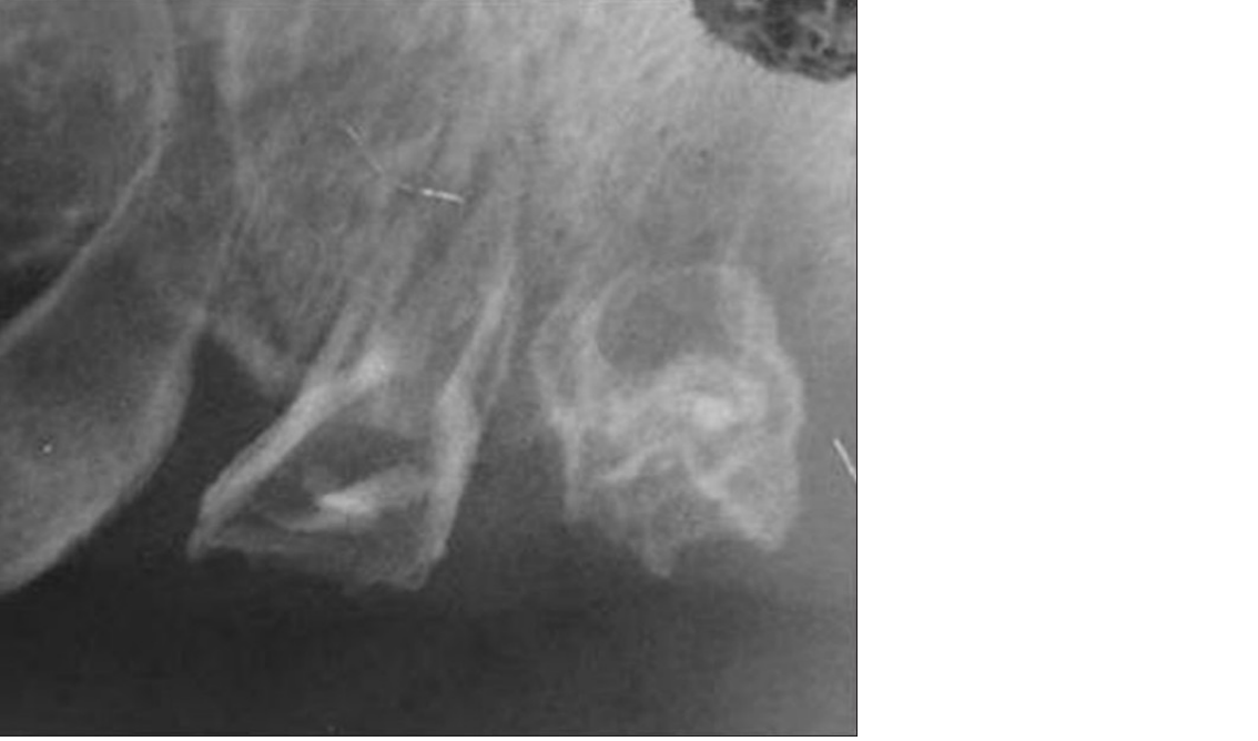
Regional Odontodysplasia
Clinical Features12
- Quadrant of teeth exhibit short roots, open apices, and enlarged pulp chambers
- Characterized as Ghost teeth
Treatment
- Extract affected teeth
Audio Appendix
Additional Audio Content
The following sections from the lecture audio did not correspond to any heading in the main document.
Anomalies of Tooth Shape and Number
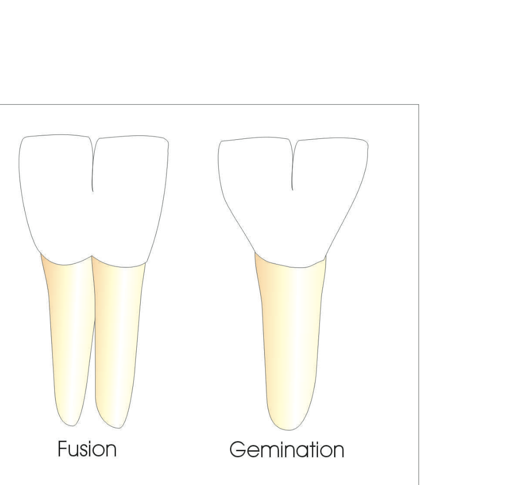
Fusion
- Definition: Two separate tooth buds merge into one large tooth.
- Clinical Presentation: The patient will have one less tooth than normal in the dental arch because two teeth have joined to form one crown and fused roots.
Gemination (Twinning)
- Definition: One tooth bud attempts to divide into two crowns.
- Clinical Presentation: A single root with a crown that appears split or multifaceted.
- Tooth Count: The tooth count is normal because the “double” crown is supported by a single root.
Footnotes
-
Original PDF page 1: Oral Pathology - 19 - Hereditary Conditions, p.1 ↩
-
Original PDF page 2: Oral Pathology - 19 - Hereditary Conditions, p.2 ↩
-
Original PDF page 3: Oral Pathology - 19 - Hereditary Conditions, p.3 ↩
-
Original PDF page 4: Oral Pathology - 19 - Hereditary Conditions, p.4 ↩
-
Original PDF page 5: Oral Pathology - 19 - Hereditary Conditions, p.5 ↩
-
Original PDF page 6: Oral Pathology - 19 - Hereditary Conditions, p.6 ↩
-
Original PDF page 7: Oral Pathology - 19 - Hereditary Conditions, p.7 ↩
-
Original PDF page 8: Oral Pathology - 19 - Hereditary Conditions, p.8 ↩
-
Original PDF page 9: Oral Pathology - 19 - Hereditary Conditions, p.9 ↩
-
Original PDF page 10: Oral Pathology - 19 - Hereditary Conditions, p.10 ↩
-
Original PDF page 11: Oral Pathology - 19 - Hereditary Conditions, p.11 ↩
-
Original PDF page 12: Oral Pathology - 19 - Hereditary Conditions, p.12 ↩