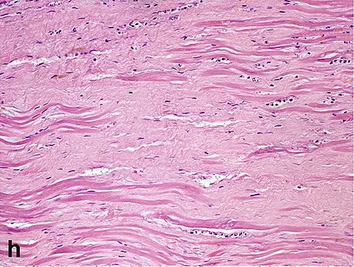
how to determine fibrosis histologically
Fibrosis is determined histologically by identifying the abnormal deposition of collagen and other extracellular matrix components within tissues. The assessment of fibrosis varies depending on tissue type (e.g., lung, liver, oral mucosa), but several core microscopic features are universally recognized.
Key Histological Features of Fibrosis
- Increased Collagen Deposition: Dense, pink (eosinophilic) bands of collagen are seen in the stroma on hematoxylin and eosin (H&E) stains. In special stains like Masson’s trichrome, collagen appears blue or green and is easily distinguished from normal tissue elements.12
- Replacement of Normal Architecture: Normal parenchyma is replaced, in whole or part, by fibrous connective tissue, leading to architectural distortion or loss of tissue organization.31
- Fibrous Septa/Bands: Formation of broad, sometimes delicate septa or bands of collagen that may segment parenchyma (e.g., bridging fibrosis or cirrhosis in the liver; band-like or nodular patterns in the lungs or oral tissues).2
- Reduced Cellular Elements: Over time, fibrotic tissue may contain fewer fibroblasts and inflammatory cells, especially as fibrosis matures and collagen becomes more densely packed and acellular.1
- Associated Findings: Evidence of prior injury—such as ductular reaction, inflammatory infiltrates, or myofibroblast proliferation—may be present depending on etiology and location.32
Special Stains and Quantification
- Masson’s Trichrome or Sirius Red stains are used to highlight collagen fibers and help distinguish fibrosis from other tissue elements.
- Quantitative scoring systems (e.g., METAVIR or Scheuer for liver, Ashcroft for lung) can provide a standardized assessment of the extent and pattern of fibrosis.2
- Advanced methods such as fractal morphometry or morphometric digital analysis can provide objective measurement of fibrotic texture and extent in research and specialized practice.3
Practical Example
In idiopathic pulmonary fibrosis, the histological hallmark is “usual interstitial pneumonia” (UIP), marked by patchwork fibrosis alternating with normal tissue and areas of architectural distortion. In the liver, cirrhosis or advanced chronic hepatitis shows dense, acellular, thin, or broad collagen septa with nodular regeneration.123
In summary, fibrosis is diagnosed by recognizing excessive, dense collagen that replaces or disrupts normal tissue structure, with confirmation by special stains and, if needed, quantitative or morphometric assessment.231 456
Footnotes
-
https://www.nature.com/articles/s41379-021-00889-5 ↩ ↩2 ↩3 ↩4 ↩5
-
https://pmc.ncbi.nlm.nih.gov/articles/PMC5760001/ ↩ ↩2 ↩3 ↩4 ↩5 ↩6
-
https://pmc.ncbi.nlm.nih.gov/articles/PMC10216340/ ↩ ↩2 ↩3 ↩4 ↩5
-
https://www.sciencedirect.com/science/article/pii/S0954611110001319 ↩
-
https://pro.boehringer-ingelheim.com/us/insights-in-ild/diseases/ipf-histopathology-features ↩