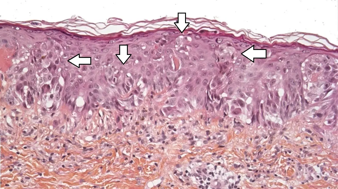
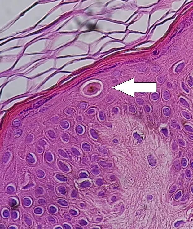
Histologically, “pagetoid appearance” refers to the upward or scattered spread of atypical cells within an epithelium, often singly or in small clusters, resembling the pattern seen in Paget’s disease of the breast. This term describes a distinctive growth pattern where abnormal cells are dispersed within the normally organized layers of cells, sometimes described as “buckshot scatter.” It is often associated with malignant or precancerous conditions like melanoma in situ or Paget’s disease.12
In the context of cementoblastoma, which is a benign odontogenic tumor composed of cementum-like tissue attached to tooth roots, pagetoid appearance is less commonly described but can be referenced to identify any unusual spread of tumor cells within odontogenic epithelium or surrounding tissues if present. Typically cementoblastomas do not show pagetoid spread because they are well-circumscribed tumors. However, if pagetoid cells are referenced in cementoblastoma histology, it would imply scattered or isolated tumor cells infiltrating or spreading within the epithelium or adjacent tissues, which might suggest an active or aggressive growth pattern, or could be a description of cellular arrangement within the lesion in rare atypical cases.31
Summary
- Pagetoid appearance means abnormal cells scattered or spreading upwards within an epithelial layer.
- It is commonly seen in cancers like melanoma in situ and Paget’s disease but is not typical of cementoblastoma.
- In cementoblastoma, if mentioned, it would indicate an unusual histological feature possibly implying scattered cell spread or atypia, which might warrant closer pathological evaluation.
If needing to interpret the term specifically for cementoblastoma histology, the pagetoid pattern would be an atypical descriptive feature highlighting scattered tumor cells in the epithelium or nearby tissues.21
If more detailed or image-based histological examples are needed for pagetoid growth, especially in dermatopathology or other cancers, references can be provided. 456