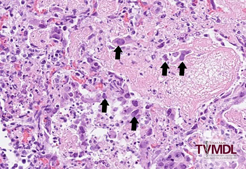
Viral inclusion bodies appear as distinct intranuclear or intracytoplasmic structures in infected cells, often diagnostic for specific viruses. Here are direct representative micrographs showing typical appearances of inclusion bodies from several major viral infections:12
Herpesvirus Inclusion Bodies
Cells infected with HSV or VZV exhibit intranuclear inclusions (Cowdry type A): eosinophilic deposits with a surrounding clear halo, multinucleation, and margination of nuclear chromatin.12

Micrograph of a tuberculous lymph node showing a caseating granuloma.
Cytomegalovirus (“Owl’s Eye” Inclusions)
CMV-infected cells display large, basophilic intranuclear inclusions with a halo—known as “owl’s eye” inclusions. Cytoplasmic inclusions may also be present.2
Molluscum Contagiosum Bodies
In molluscum contagiosum, cells show large, rounded, eosinophilic intracytoplasmic inclusions (Henderson-Paterson bodies) that displace the nucleus peripherally.2
Poxvirus (Guarnieri Bodies)
Poxvirus-infected epithelial cells contain sharply defined, eosinophilic, round intracytoplasmic inclusions (Guarnieri bodies).2
These representative micrographs show the hallmark features of viral inclusion bodies useful for pathology diagnosis. For specific, named viral inclusion photos, consult authoritative pathology textbooks or digital archives with labeled histological images.12