Furcation Diagnosis and Management in Multirooted Teeth1
Dr. Pradeep Koppolu
Dr. Anna Hughes / Dr. Tina Choo
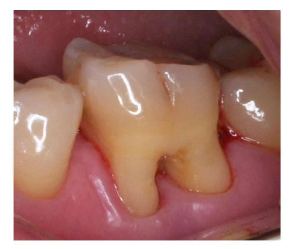
Significance of Furcation Involvement2
- Furcation involvement is a significant risk factor for tooth loss.
- Risk of tooth loss increases with increasing bone loss in the furcation area.
- Diagnosis of furcation involvement is challenging.
- Hence thorough examination and early diagnosis is essential.
- Multirooted teeth have a significant role in our dentition and have unique anatomical features.
- Due to their posterior position, access is challenging for professional and self performed cleaning.
Definition
Furcation involvement occurs when there is bone loss in the area between the roots of multirooted teeth. Normally, this area is covered by bone.
Clinical Implications
It is often inadequately addressed by general dentists, who may favor extraction and replacement with bridges or implants. Periodontists focus on treating and retaining these teeth.
- Etiology:
- Bone Loss: Primary cause due to periodontal disease.
- Trauma: Hard toothbrushing techniques can cause recession, leading to plaque accumulation below the cemento-enamel junction (CEJ).
- Anatomic Factors: Cervical enamel projections (CEP) or enamel pearls can retain plaque and calculus.
- Endodontic Factors: Accessory canals can transmit infection from the pulp to the furcation area.
Anatomy and Terminology
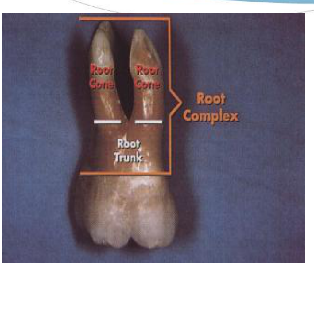
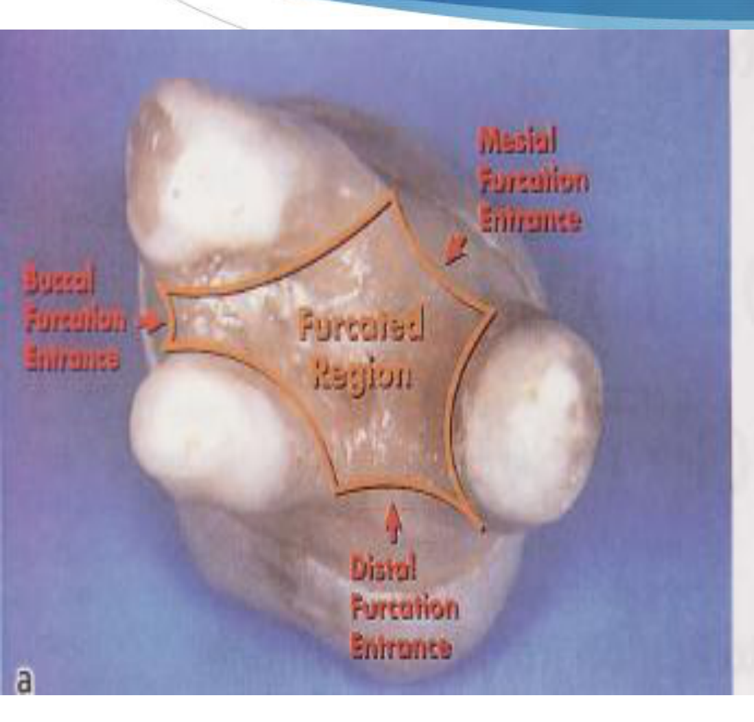
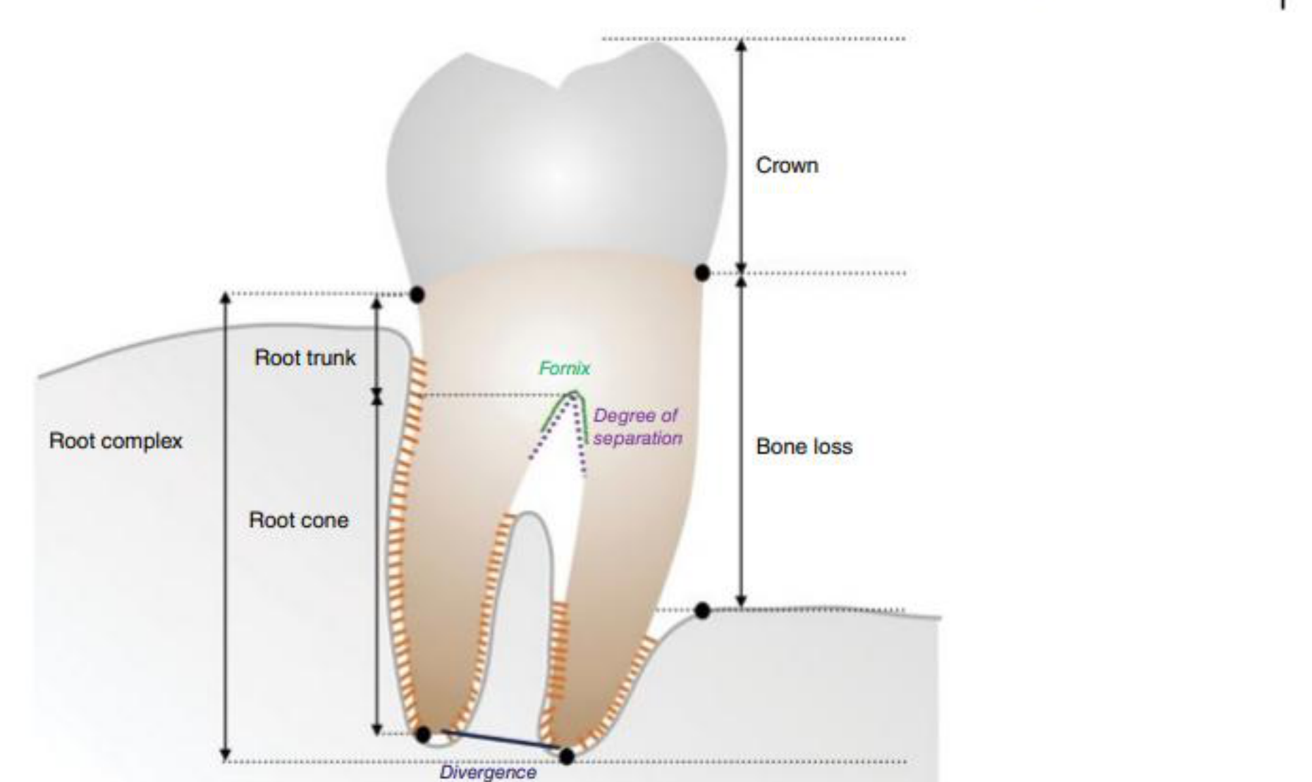
Root Complex and Terminology345678
Root Complex
- The entire distance from the CEJ to the end of the root.
Root Trunk
- The distance from the CEJ to the entrance of the furcation.
Root
Cone
- The distance from the furcation entrance to the end of the root.
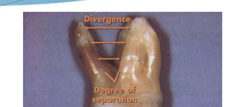
Divergence
Degree of separation
- The distance between roots. If roots are close together, instrumentation becomes difficult.
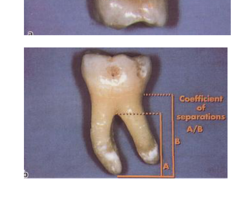
Coefficient of separations A/B
Figure 1.1 Drawing of mandibular molar with furcation involvement, showing the main anatomical features, including root trunk (part of the root from the cemento-enamel junction [CEJ] to the furcation entrance) and root cones, and pointing at root divergence and degree of separation between roots. The ‘bone loss’ is schematically indicated as the distance between the CEJ and the most apical part of the bone. Source: Courtesy of Dr Aliye Akcali.
Crown-to-Root Ratio
Ideal is 1:3; the minimum accepted ratio is 1:1.
 |  |  |
 |  |  |
 | 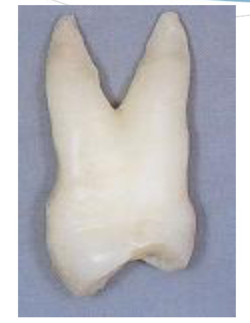 | 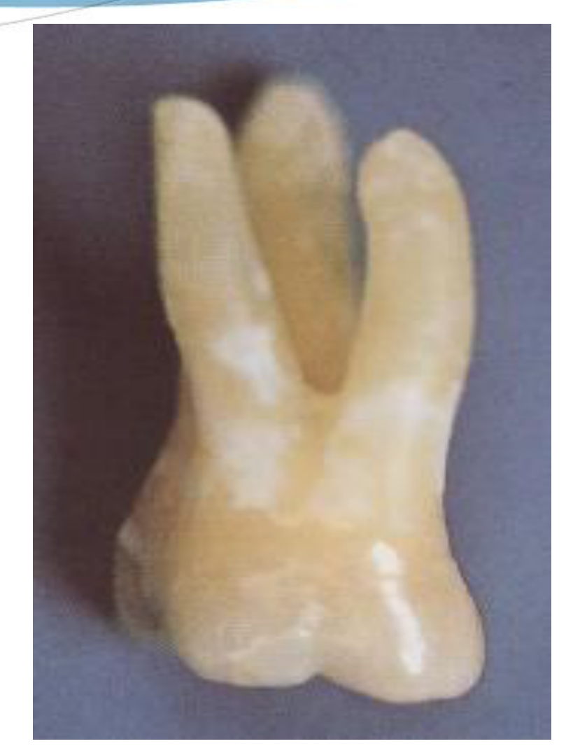 |
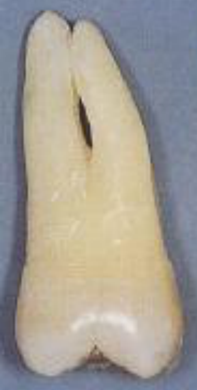 | 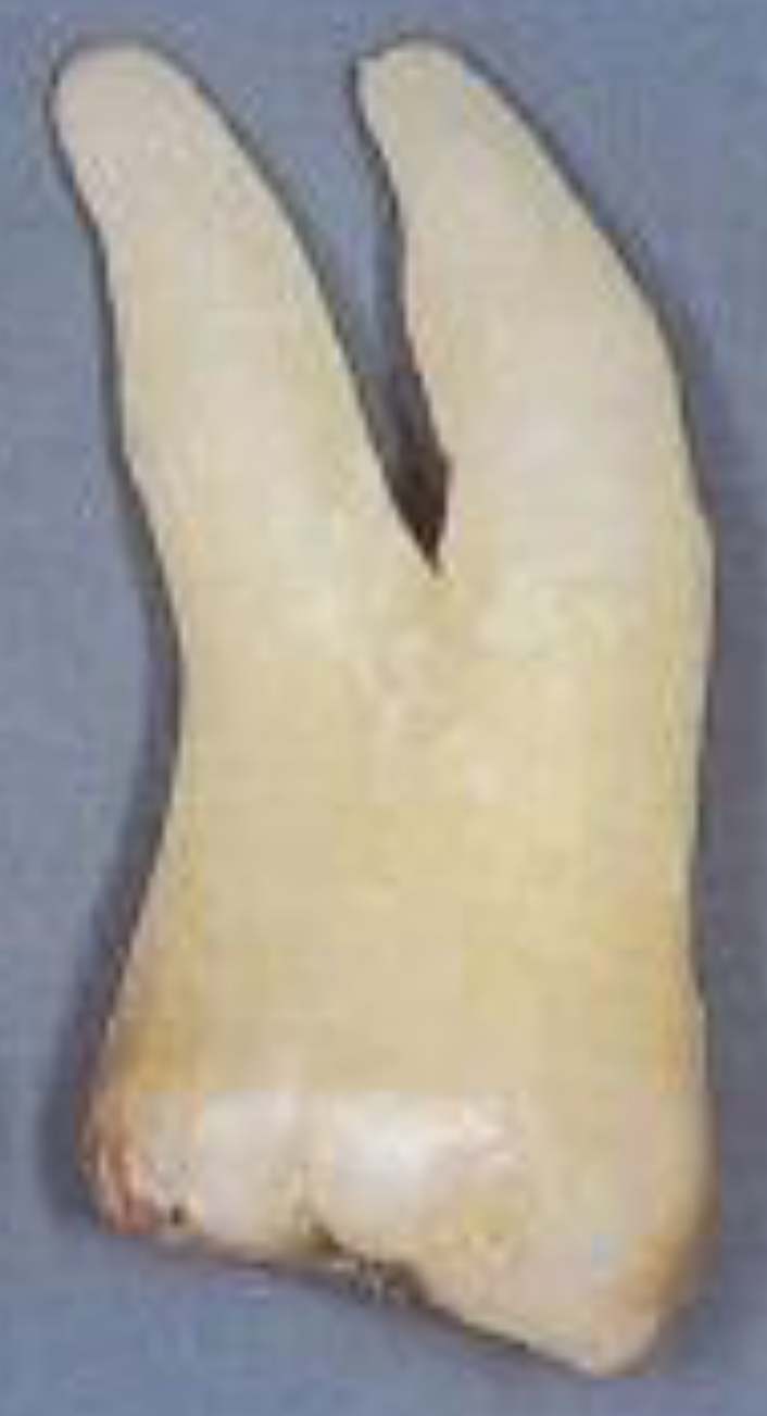 |  |
Anatomy of Maxillary First Premolars910
Maxillary first premolars
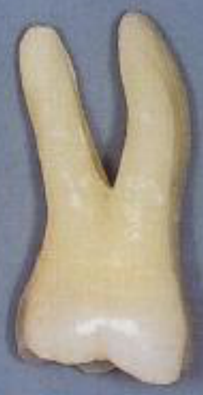
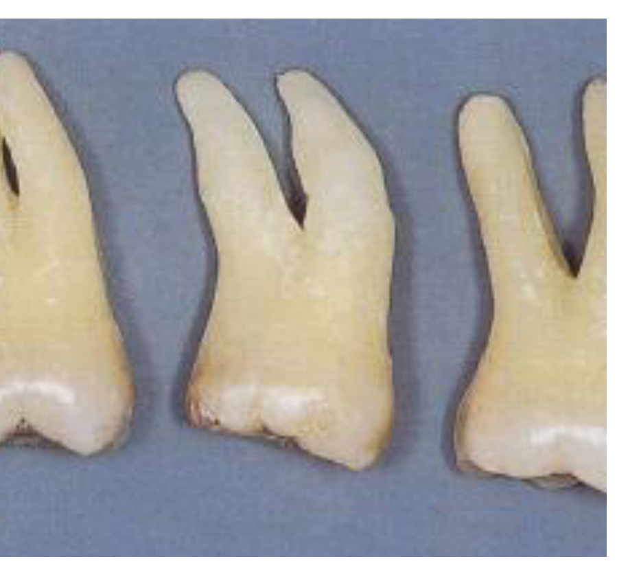
- 40% of maxillary premolars have 2 roots and furcation @ 8mm from CEJ
- Mesial Groove: A developmental groove on the mesial side caused by contact with the canine during eruption. This groove, combined with the furcation, makes treatment difficult.
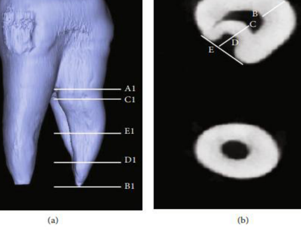
FIGURE 2: Measurement of the buccal root with furcation grooves in maxillary first premolars using micro-CT. (a) Vertical measurement. (b) Horizontal measurement. A1: root furcation position; B1: apical position; C1: the start point of furcation
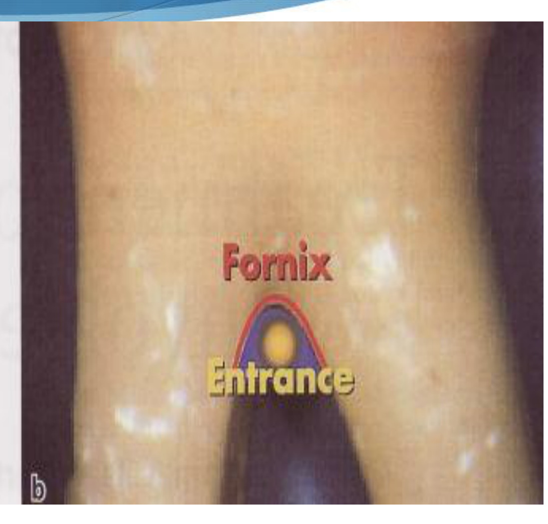
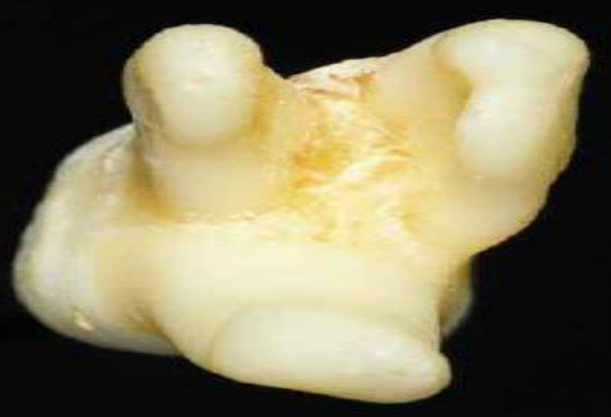
Fornix
-
The entrance or “roof” of the furcation. groove; D1: the end point of furcation groove; E1: the middle point of CD connection. A1B1: buccal root length; C1D1: furcation groove length; A1C1: length from root bifurcation to the beginning of furcation groove; B1D1: length from apical position to the end point of furcation groove. AB: buccal root canal wall thickness; CD: palatal root canal wall thickness; DE: furcation groove depth (depth of root invagination).
-
Root surface area and shape
-
Bifurcation ridges
-
Enamel projections/pearls – connective tissue does NOT adhere to enamel
-
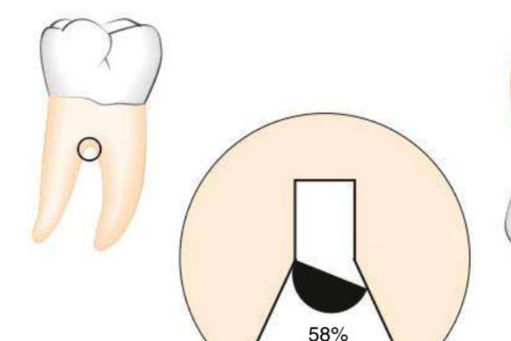
Horizontal component of furcations = 4.6–6.9 mm wide
-
Accessory canals
-
Iatrogenic – overhangs, caries, fractures
Furcation Entrance and Enamel Projections111213
Furcation entrance width
- Almost all less than 1 mm
- 50% less than 0.75 mm
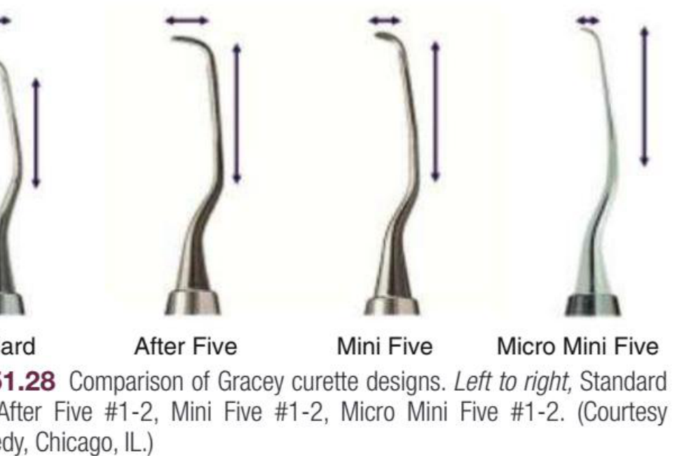
- Average curette width is 1 mm, thin US tip 0.5 mm
Furcation entrance distances from CEJ (Root trunk length)
Maxillary molars
- Buccal – 4.3 +/- 1 mm
- Distal – 4.8 +/- 0.8 mm
- MP – 3.6 +/- 0.8 mm
Mandibular molars
- Buccal – average 4.15 mm
- Lingual – average 3.4 mm
Hence at 6 mm from CEJ – all furcation entrances involved
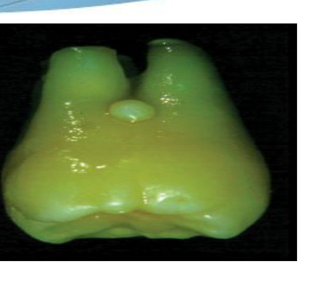
Enamel projecting to furcation
- Projections / enamel pearls
- Risk factor for furcation involvement
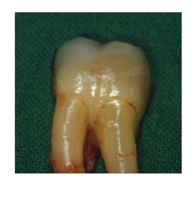
Enamel Pearl
An excess deposition of enamel during development that retains plaque.
 |  |
 |
Diagnosis and Classification
Clinical and Radiographic Examination1415161718
- History and examination—clinical and radiographic.
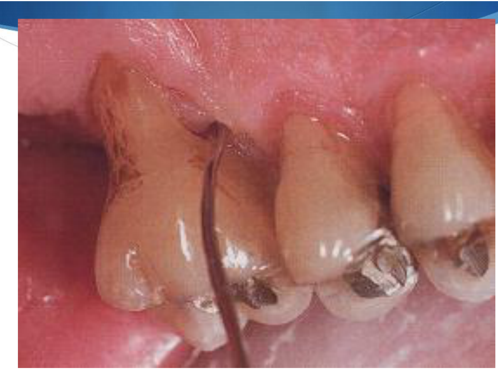
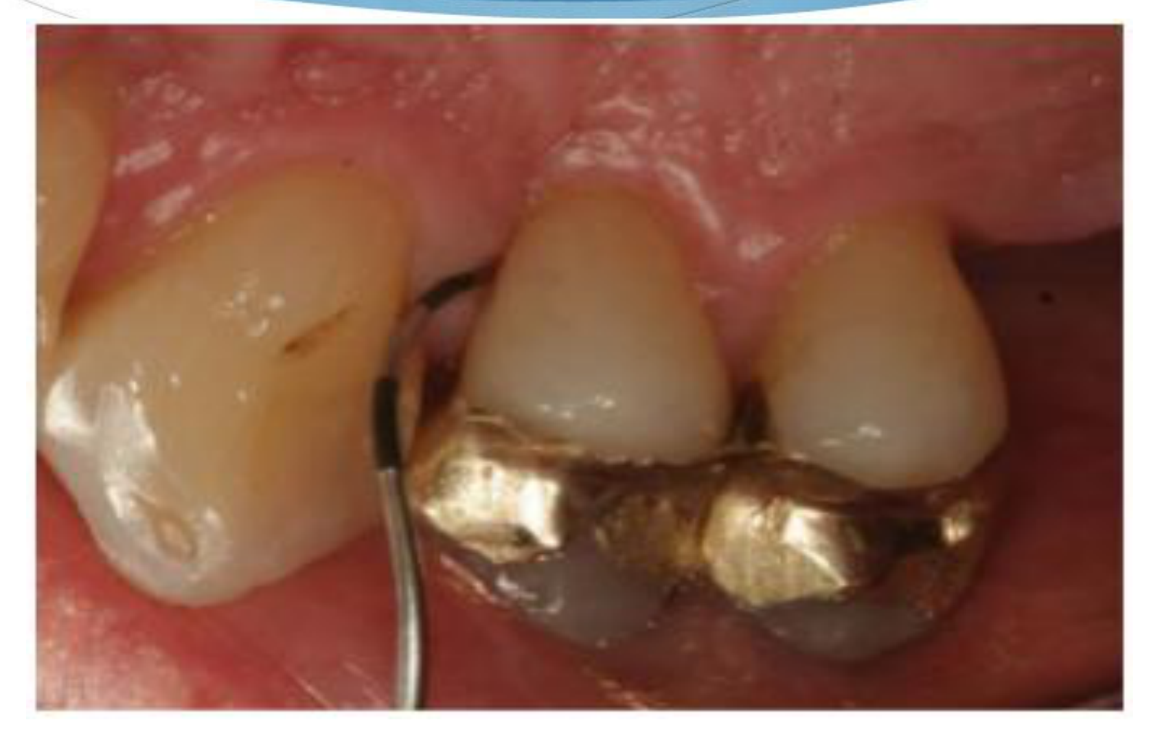
- Colour coded Nabers probe, Perio probe.
- The Nabers probe is the primary instrument for detecting furcations and features markings at 3, 6, 9, and 12 mm increments.
- Radiographs—parallel periapicals/vertical bitewings but limitations of overlapping roots etc, esp upper molars and premolars.
- Standard periapical X-rays and OPGs have about 65% accuracy, as overlapping roots in 2D images can hide furcation involvement.
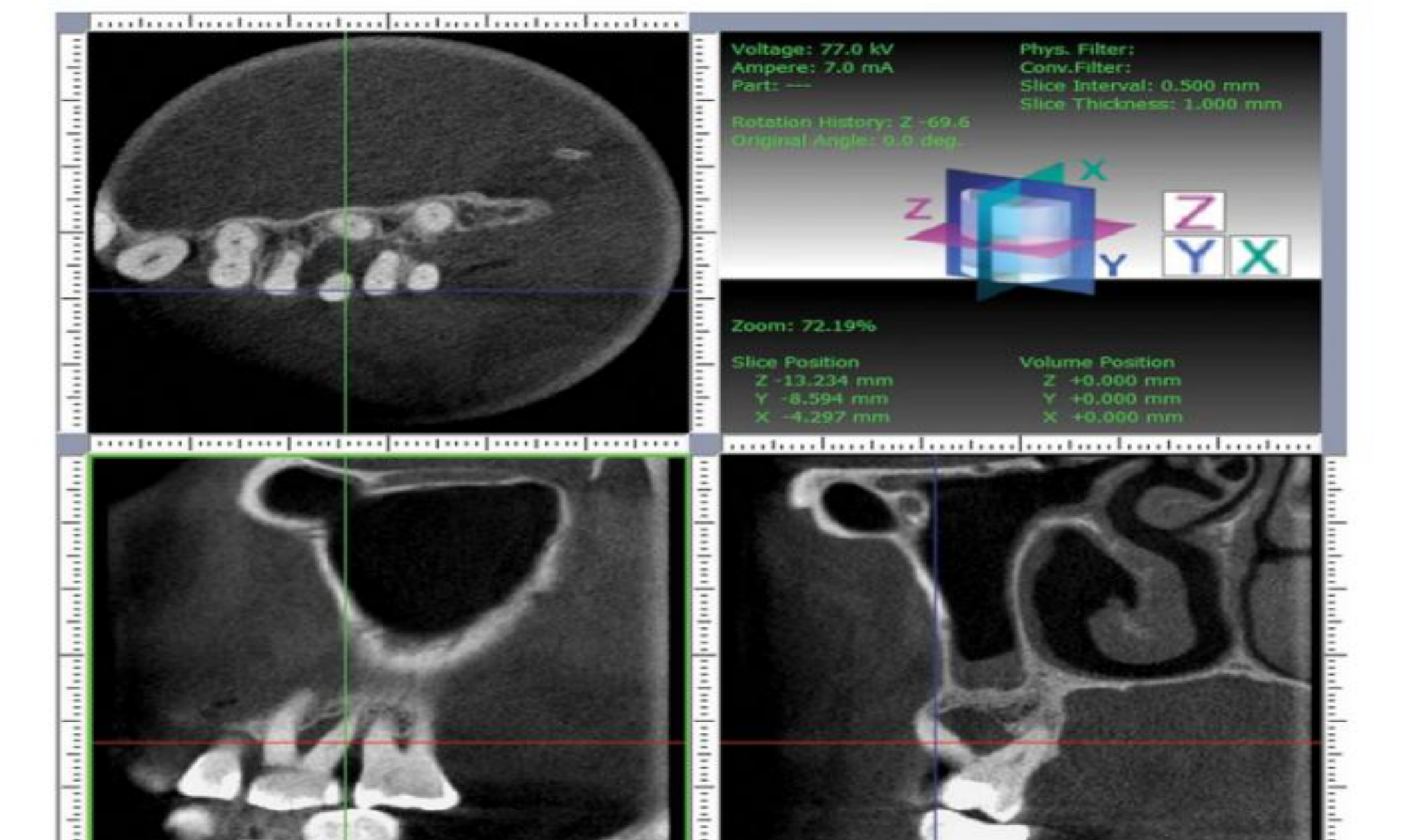
- Consider CBCT if tooth is strategic in treatment planning.
- CBCT is used for Grade II or III involvements to provide 3D views (sagittal and frontal sections) to identify the exact site of bone loss. Grade, Class, Degree
 |  |
 |  |
Classification Systems and Subclasses1920
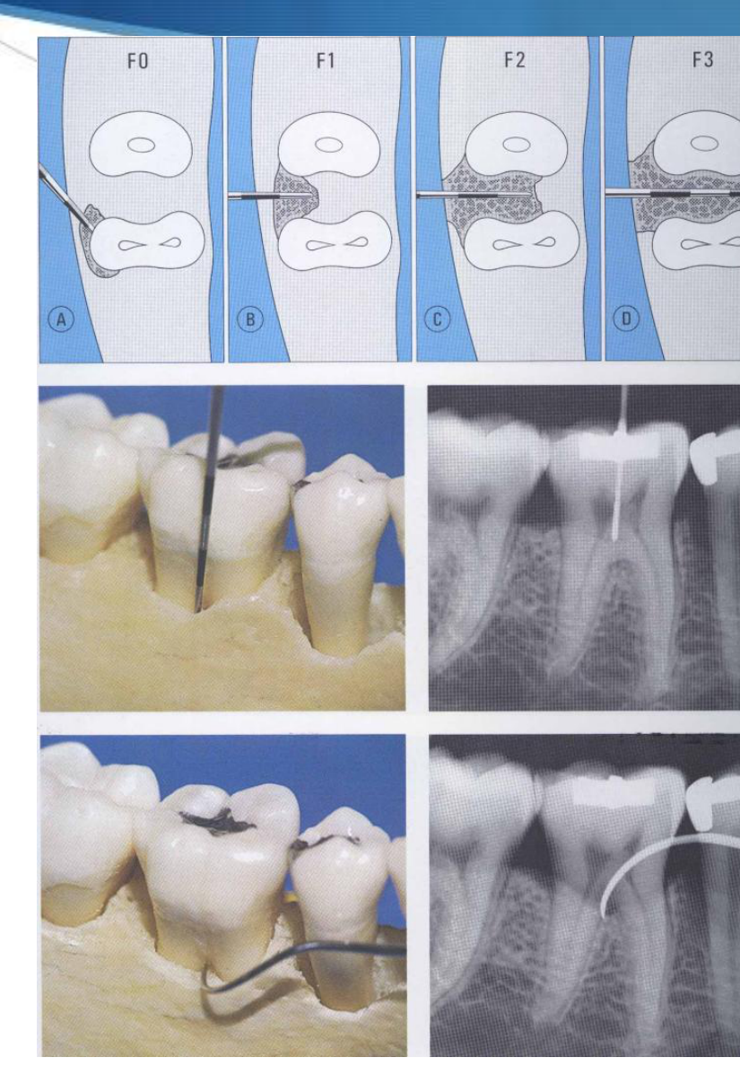
Diagnosis & Classification of furcation involvement
-
F0
-
F1
-
F2
-
F3
-
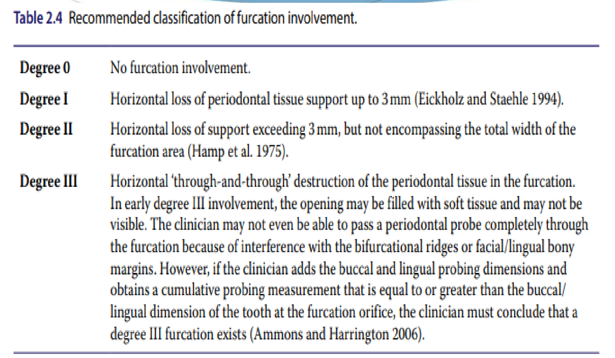
Degree 0: No furcation involvement.
-
Degree I: Horizontal loss of periodontal tissue support up to 3 mm (Eickholz and Staehle 1994).
-
Degree II: Horizontal loss of support exceeding 3 mm, but not encompassing the total width of the furcation area (Hamp et al. 1975).
- Degree 2 is often referred to as a “Cul-de-sac” involvement.
-
Degree III: Horizontal ‘through-and-through’ destruction of the periodontal tissue in the furcation. In early degree III involvement, the opening may be filled with soft tissue and may not be visible. The clinician may not even be able to pass a periodontal probe completely through the furcation because of interference with the bifurcational ridges or facial/lingual bony margins. However, if the clinician adds the buccal and lingual probing dimensions and obtains a cumulative probing measurement that is equal to or greater than the buccal/lingual dimension of the tooth at the furcation orifice, the clinician must conclude that a degree III furcation exists (Ammons and Harrington 2006).
Sources: Hamp et al. (1975); Eickholz and Staehle (1994); Ammons and Harrington (2006).
Glickman’s Classification (1964)
- Grade 1: Incipient involvement (a “catch” with the probe).
- Grade 2: Probe enters but does not pass through.
- Grade 3: Through-and-through, but furcation is covered by soft tissue.
- Grade 4: Through-and-through, and the furcation is clinically visible due to recession.
 |  |
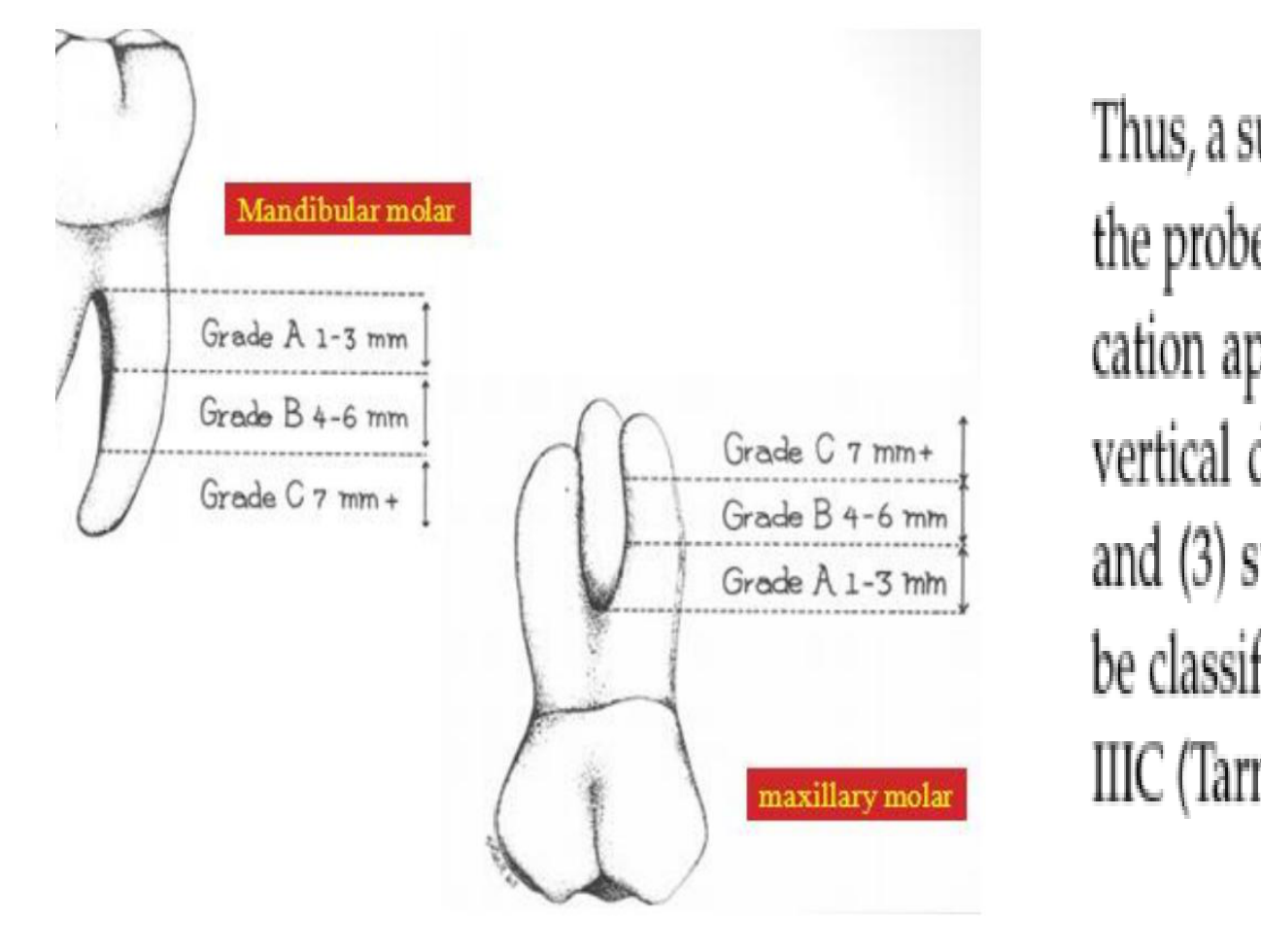
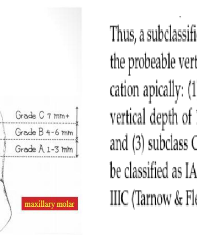
Vertical component-subclasses affecting prognosis21
Mandibular molar
- Grade A: 1–3 mm
- Grade B: 4–6 mm
- Grade C: 7 mm+
Maxillary molar
- Grade C: 7 mm+
- Grade B: 4–6 mm
- Grade A: 1–3 mm
Thus, a subclassification was proposed that measures the probeable vertical depth from the roof of the furcation apically: (1) subclass A indicates a probeable vertical depth of 1–3 mm, (2) subclass B of 4–6 mm, and (3) subclass C of ≥7 mm. Furcations would thus be classified as IA, IB, IC, IIA, IIB, IIC and IIIA, IIIB, IIIC (Tarnow & Fletcher 1984).
Clinical exam: Nabers Probe
Clinical Charting Symbols
In clinical software, furcations are typically marked with triangles:
- Open triangle: Class 1
- Lightly filled/shaded: Class 2
- Completely dark/filled: Class 3
 |  |
Diagnostic Accuracy and Considerations22232425
- Clinical assessment alone detects 3% of maxillary molars and 9% of mandibular molars.
- 2D radiographs and clinical assessment combined—65% of maxillary molars and 23% of mandibular molars.
- WHY?
Angulation of radiographs, superimposition of roots, high mandibular bone density…
 |  |  |
 |  |
Important diagnostic considerations26
- Horizontal/vertical bone loss in furcation area
- Length of root trunk
- Length/divergence/shape of roots
- Fusion of roots
- Residual bone support
- Endodontic/restorative/caries status
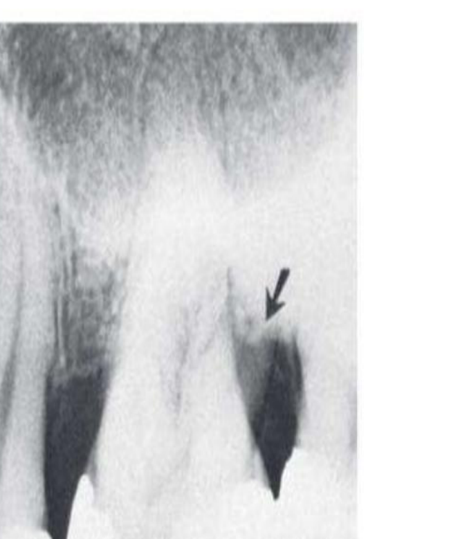
Radiographs: Furcation “Arrows”
Voltage: 77.0 kV Ampere: 7.0 mA Part: --- Phys. Filter: Conv.Filter: Slice Interval: 0.500 mm Slice Thickness: 1.000 mm Rotation History: Z -69.6 Original Angle: 0.0 deg.
Zoom: 72.19% Slice Position Z -13.234 mm Y -8.594 mm X -4.297 mm Volume Position Z +0.000 mm Y +0.000 mm X +0.000 mm
Figure 2.8 Diagnosis and treatment planning using cone-beam computed tomography (CBCT). CBCT images with horizontal, sagittal, and transversal sections of first and second left maxillary molars. According to the bone loss around the disto-buccal root and the remaining periodontal attachment around the mesio-buccal and palatal root, it was decided to extract the distobuccal root. Source: Walter et al. (2010).
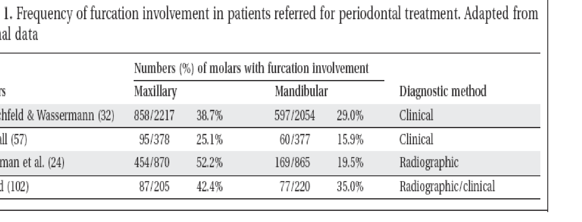
| Authors | Numbers (%) of molars with furcation involvement | Diagnostic method |
|---|---|---|
| Maxillary | Mandibular | |
| Hirschfeld & Wassermann (32) | 858/2217 (38.7%) | 597/2054 (29.0%) |
| McFall (57) | 95/378 (25.1%) | 60/377 (15.9%) |
| Goldman et al. (24) | 454/870 (52.2%) | 169/865 (19.5%) |
| Wood (102) | 87/205 (42.4%) | 77/220 (35.0%) |
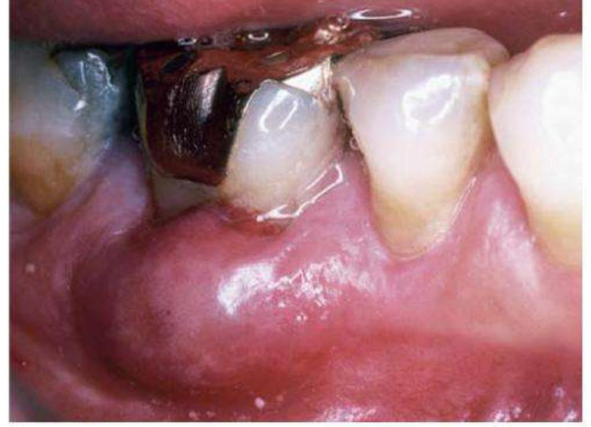
Fig. 38.24 Periodontal abscess associated with a furcation-involved mandibular molar.
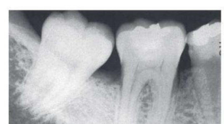
Fig. 39.24 Early furcation involvement suggested by fuzziness in the furcation of the mandibular first molar, particularly when associated with bone loss on the roots.
Fig. 39.25 Furcation involvement of mandibular first and second molars indicated by thickening of the periodontal space in the furcation area. The furcation of the third molar is also involved, but the thickening of the periodontal space is partially obscured by the external oblique line.
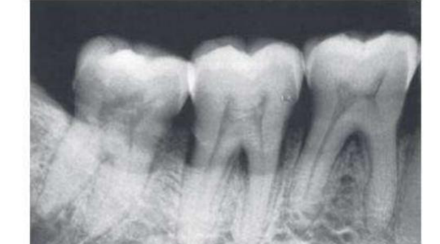
Fig. 39.26 Furcation involvement of the first molar partially obscured by the radiopaque lingual root. The horizontal line across the distobuccal root demarcates the apical portion (arrow), which is covered by bone, from the remainder of the root, where the bone has been destroyed.
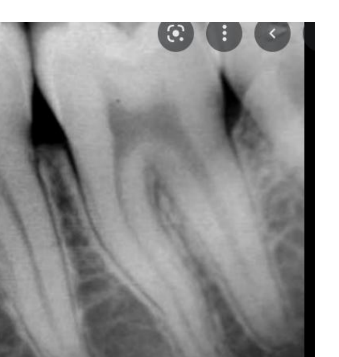
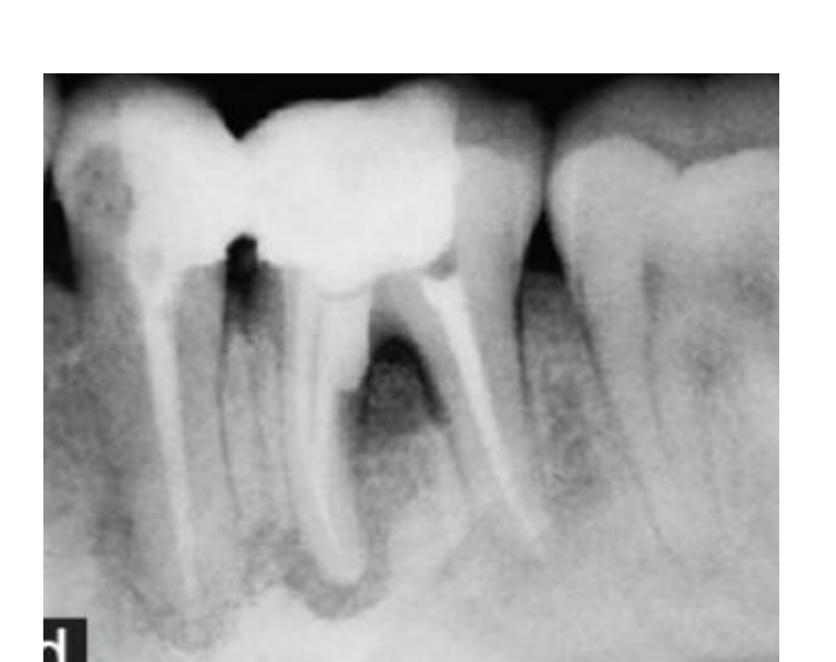
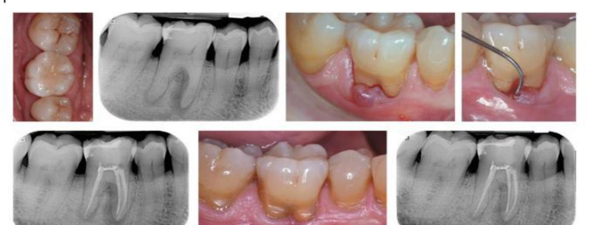
Differential Diagnosis and Endodontic Considerations27282930
-
Endodontic infection
-
Trauma from occlusion
-
Iatrogenic damage
-
Periodontal Abscess: Characterized by a pocket, vital tooth, and infection confined to the attached gingiva.
-
Pulpal/Periapical Abscess: Associated with a non-vital tooth.
-
Perio-Endo Lesions:
- Primary Endo, Secondary Perio: Starts with caries/pulp infection.
- Primary Perio, Secondary Endo: Infection travels from a deep pocket through the apical foramen to the pulp.
- Combined: Origin is unclear.
-
Treatment Sequence: In Australia, the standard is to treat the Endo first, then the Perio.
Endodontic infection – may take up to 5 years of radiographic healing
Endodontic considerations for FI teeth
- Pulp changes can be induced by periodontal disease – can be reparative or degenerative changes. Pulp necrosis can occur if apical NV bundle involved.
- Endodontic infections (e.g., through accessory canals) can mimic FI Class III – pulp testing important.
- If both periodontal disease and necrotic pulp present – medicate canals first.
Endodontic considerations for FI teeth
- A vital pulp appears to hinder bacterial migration from the pocket to the pulp chamber – immune cells and fluid flow in tubules.
- If tooth is vital and root resection is performed – RCT must be done within 2 weeks of resection.
- Preventative RCT not recommended for surgical and regenerative therapy unless pulp necrosis/infection is present.
Elective Root Canal
While some countries perform elective RCT if bone loss reaches the apex of a vital tooth, Australian practice generally avoids RCT unless the tooth becomes non-vital.
 |  |
 |
Management of Furcation Involved Teeth
Treatment Objectives and Terminology3132
Essentially 3 objectives:
- Eliminate microbial plaque from the exposed surfaces of the root complex
- Establish an anatomy of the affected surfaces that facilitates proper self performed plaque control (e.g., tunnelling)
- Eliminate the furcation (resection or regeneration)
Odontoplasty – reshaping of the tooth to aid cleansability
Osteoplasty – reshaping of the bone to aid cleansability
- Osteectomy – complete removal of bone
Flap procedures – OFD (open flap debridement) +/- reshaping, resection or repositioning the soft tissues
Regeneration – aims to restore the original architecture (and close the furcation) with new bone, cementum and PDL
Non-Surgical Management and Instrumentation333435
 |  |  |
 |  |
Non-surgical treatment36
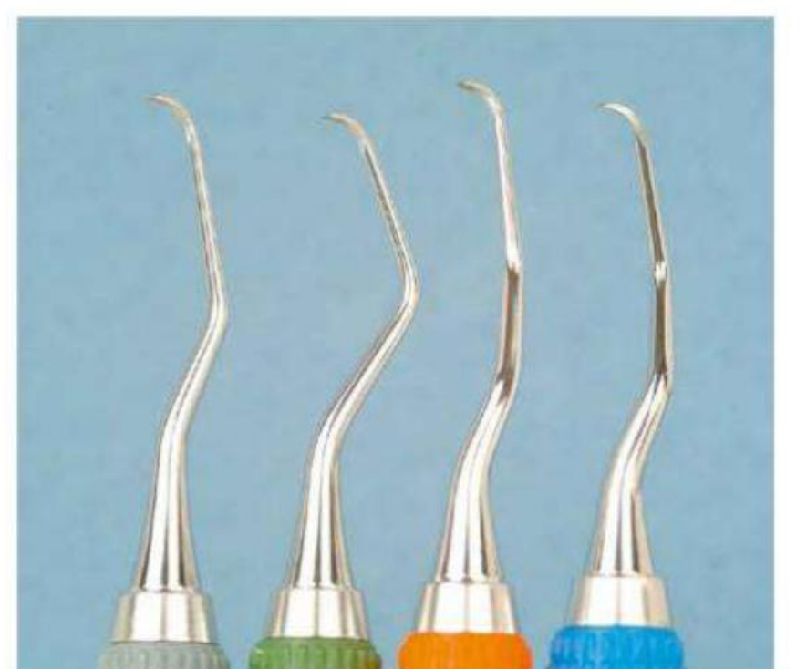
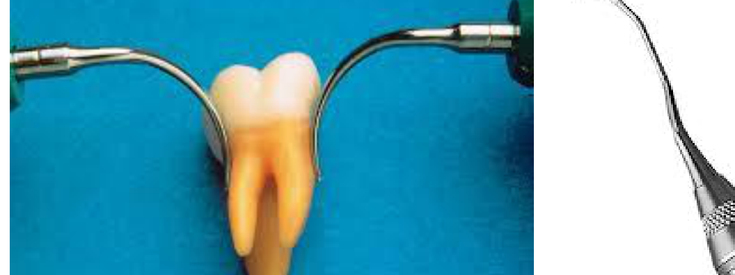
- Standard Curette width: 0.8–1.06 mm
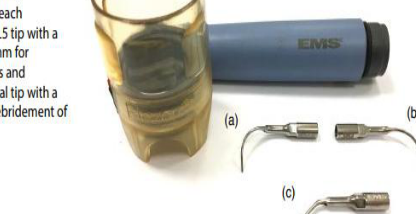
- Standard Ultrasonic tip: 0.88 mm measured 1 mm from the tip and 1.06 mm measured 2 mm from the tip
- Furcation opening < 1 mm
- Difficult to access furcation
- Initial approach for all cases is non-surgical treatment
- Standard Ultrasonic slightly better than standard curettes in narrow furcation areas
- Grade I furcation relatively stable with SRP
- Grade II and III furcation relapse or deteriorated over 2 years
Success rates of calculus removal from furcations37
- Experienced vs inexperienced
- Less experienced / closed approach: only 8% of surfaces were calculus free
- Closed vs open access
- Experienced periodontists, open approach: 68% calculus free furcation surfaces achieved
- Experienced, closed approach: 44% calculus free
PL1 = 0.5, PS = 0.6, PL5 & 6 = 0.8
Many different US tips38
-
Straight and thin (e.g., PS1)
-
Curved and thin (e.g., PL1/PL2)
-
Ball ended – for furcation roof and grooves (PL5/PL4)
-
Diamond coated – very fast and efficient but removes cementum and dentine too
-
Some systems (e.g., Kavo) have inbuilt light (LED) to improve visibility
-
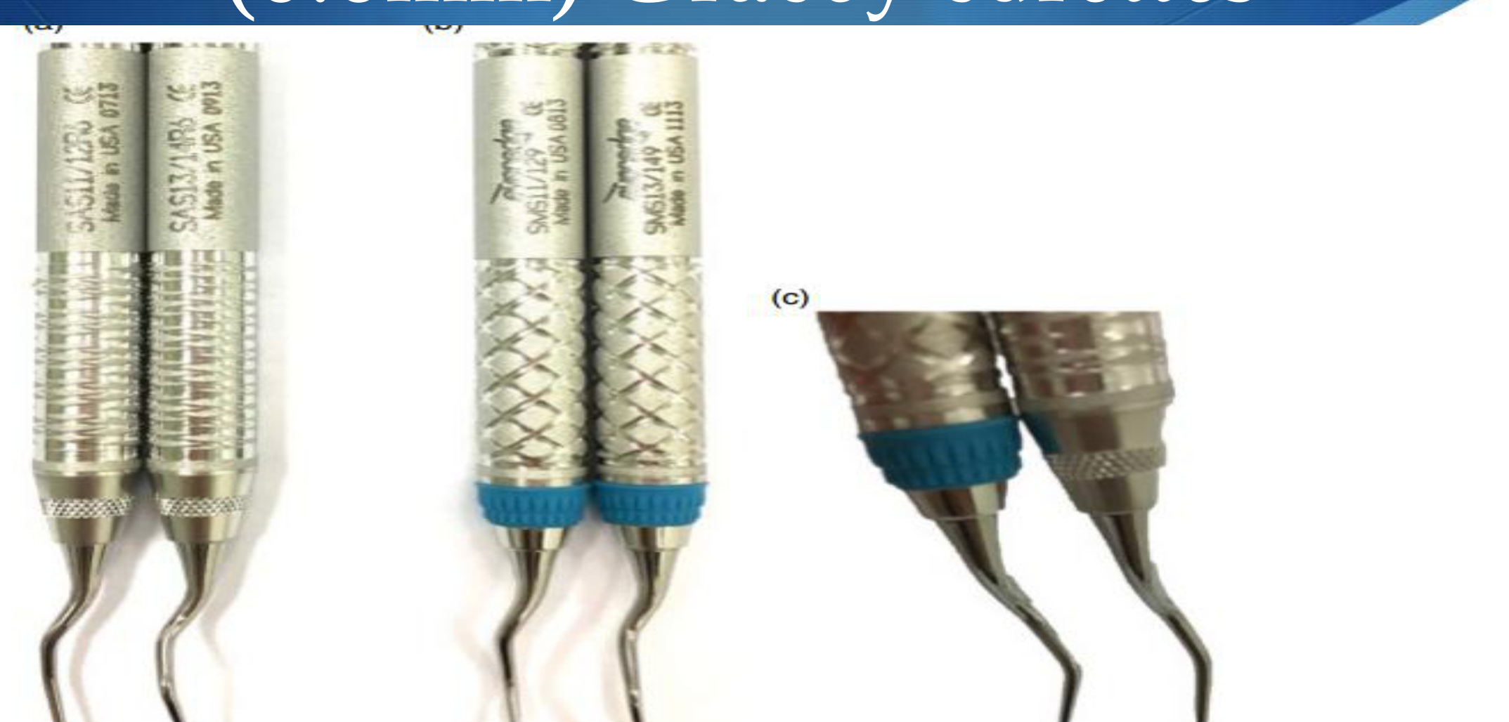
Quittene Furcation Curette – a specialized instrument specifically for furcation debridement
-
Mini-Five/Micro-Mini Five – features thinner and longer blades for better access
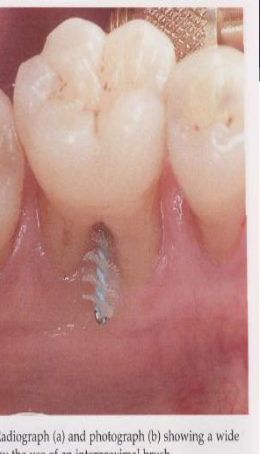
Self-Performed Biofilm Control39
Current evidence suggests40
- Oscillating rotating electric toothbrush
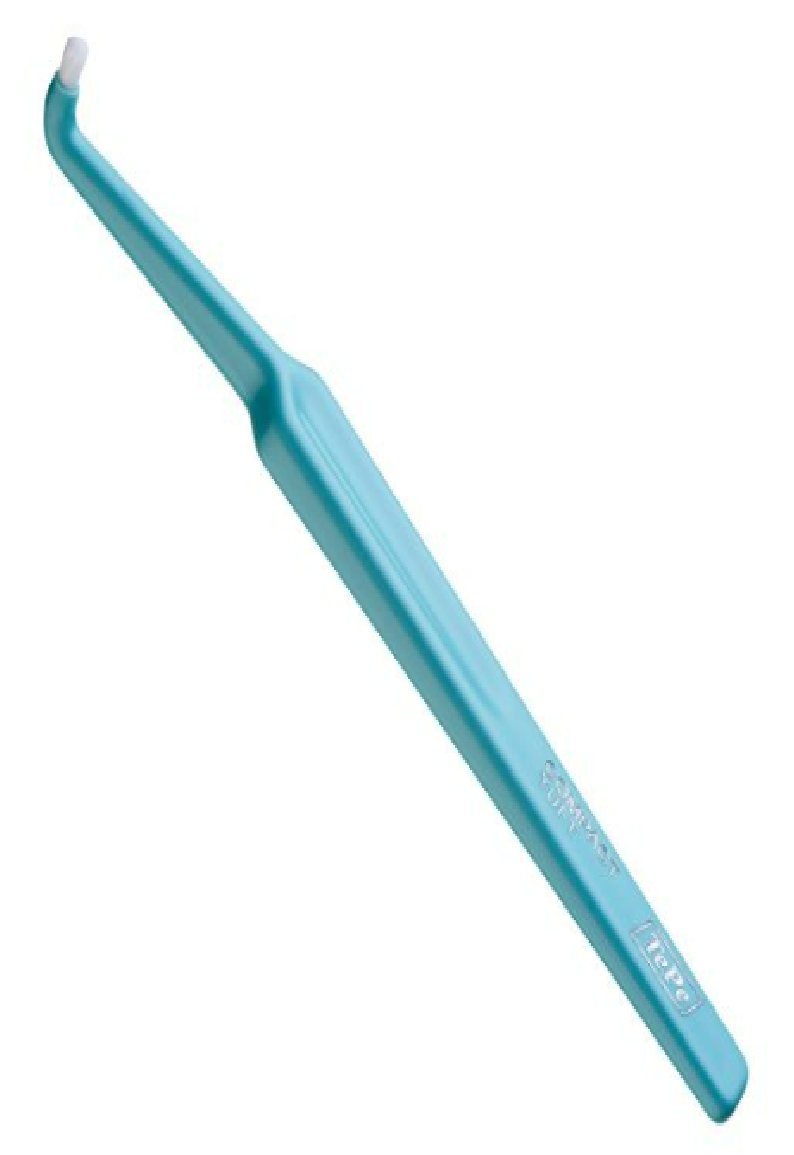
- End or compact tufted brush
- Cylindrical interdental brushes
- WaterPik?
Waterpik Caution
Use Waterpiks on a case-by-case basis as high pressure may damage soft tissue.
TePe Compact Tuft Toothbrush – $4.00
This special toothbrush has a small, dome-shaped tuft and short, extra soft filaments. The stable handle and dense, firm tuft make this brush ideal for precision cleaning of difficult to reach areas.
The toothbrush is suitable for cleaning around fixed orthodontic appliances, attachments for overdentures or along the gum line. It is also ideal for brushing children’s erupting molars.
Colours may vary.
Surgical Management Strategies
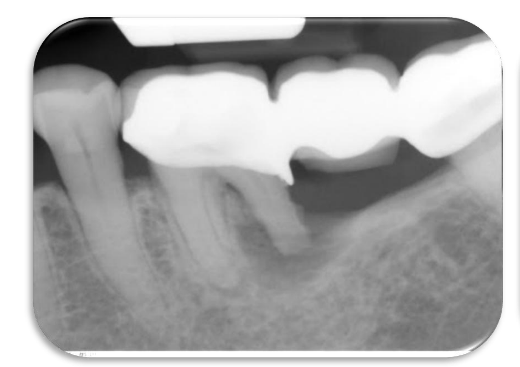
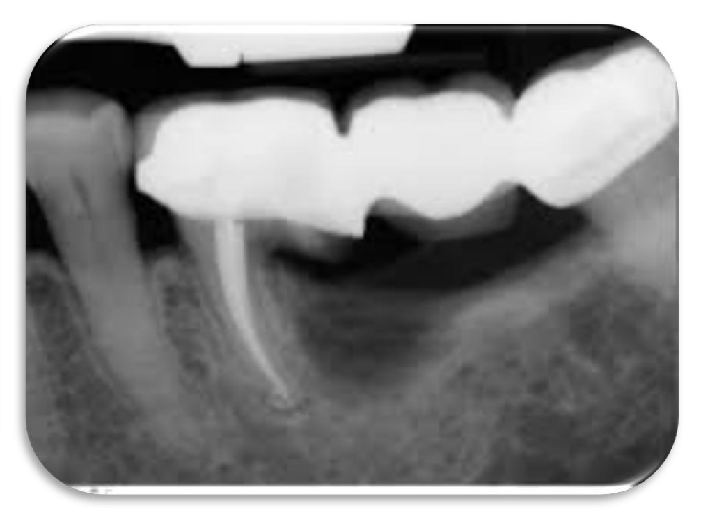
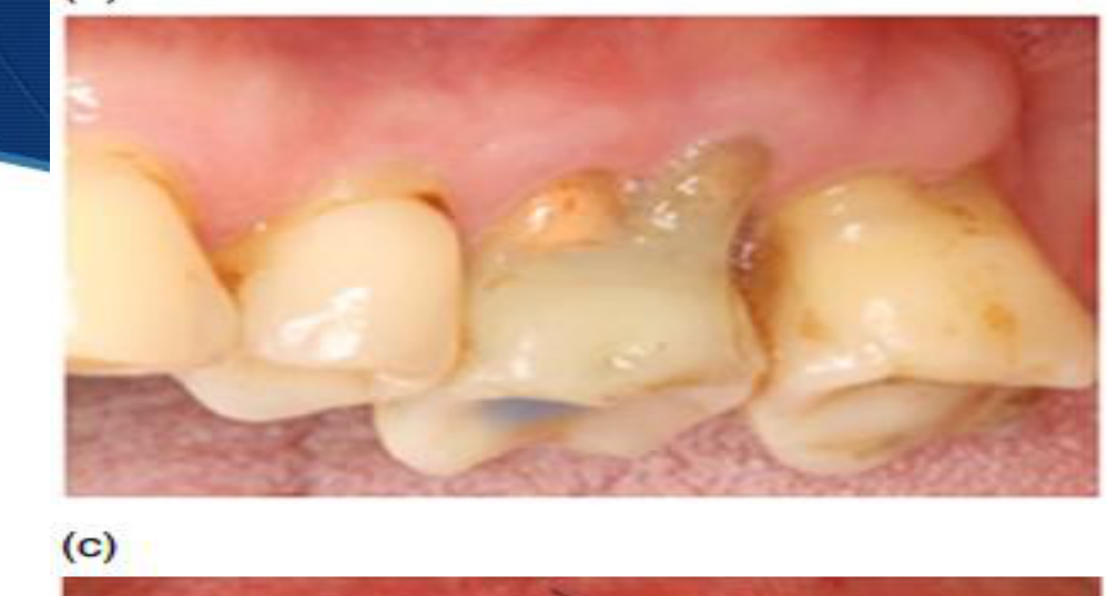
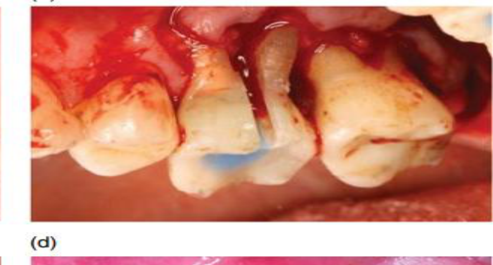
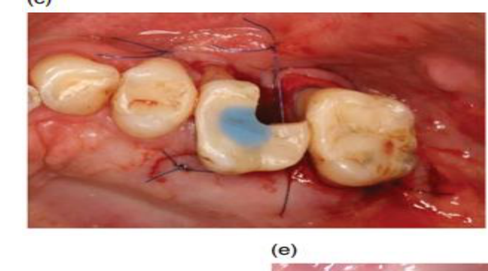
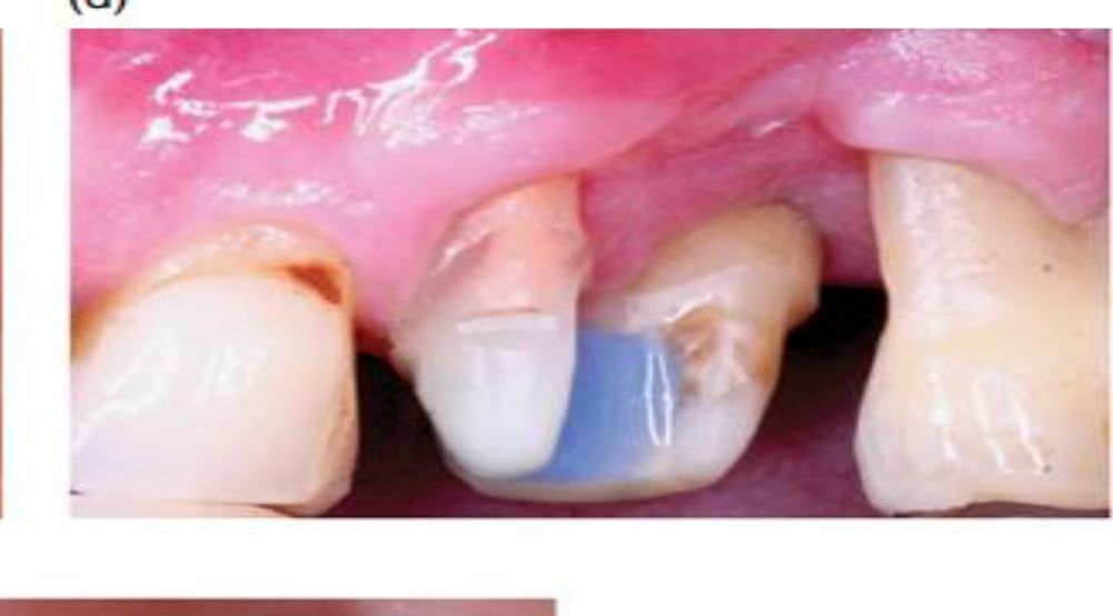
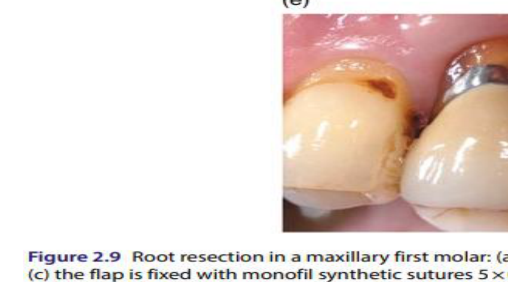
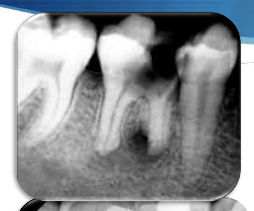
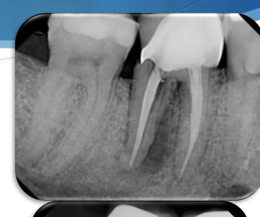
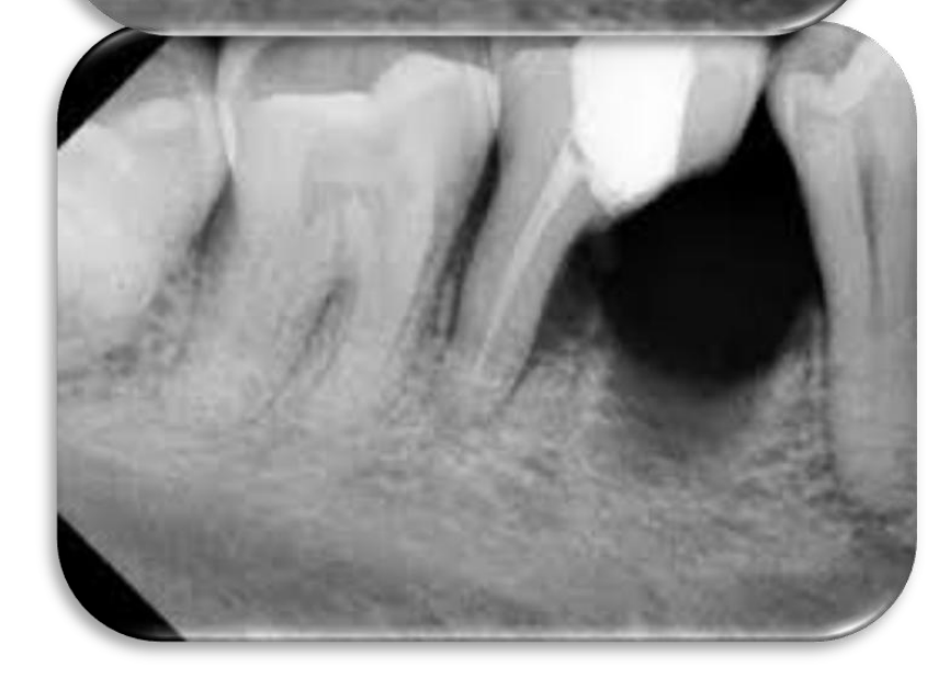
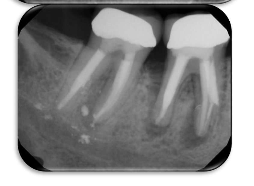
Root Resection4142434445
- The surgical removal of all or a portion of the root.
- Can eliminate an involved furcation
- Popularity of root resection has declined since 2010 in favor of other treatments.
The ‘ideal’ candidate:
- Class II or III furcation involved molar
- Severe bone loss around just 1 root ideally or occasionally 2 roots
- If there is a localised root fracture, perforation or resorption
- Remaining roots should have adequate bony support - favourable crown-root ratio
- Sound endodontic & restorative prognosis
- Tooth has minimal mobility (Gr 1 mobile or nil)
- Good oral hygiene
Contraindications:
- Teeth with a poor crown-root ratio on remaining roots
- Inadequate bone support on roots to be retained
- Long root trunks
- Fused roots
- Teeth where endodontic & restorative treatment not feasible on remaining roots
- Poor surgical access & inability to perform oral hygiene procedures
F
 |  |  |
 |  |  |
 |  |  |
 |  |  |
 |
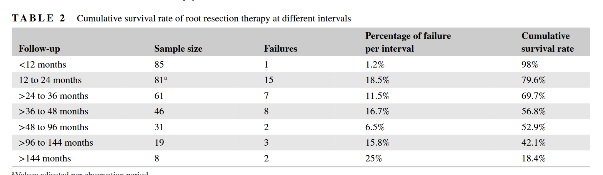
Success of Root Resection46
outcomes of root resection therapy up to 16.8 years: A retrospective study in an academic setting
Madi Alassadi | Musa Qazi | Andrea Ravidà Rafael Siqueira | Carlos Garaicoa-Pazmiño | Hom-Lay Wang
| Follow-up | Sample size | Failures | Percentage of failure per interval | Cumulative survival rate |
|---|---|---|---|---|
| <12 months | 85 | 1 | 1.2% | 98% |
| 12 to 24 months | 811 | 15 | 18.5% | 79.6% |
| >24 to 36 months | 61 | 7 | 11.5% | 69.7% |
| >36 to 48 months | 46 | 8 | 16.7% | 56.8% |
| >48 to 96 months | 31 | 2 | 6.5% | 52.9% |
| >96 to 144 months | 19 | 3 | 15.8% | 42.1% |
| >144 months | 8 | 2 | 25% | 18.4% |
Received: 21 January 2019 | Revised: 6 May 2019 | Accepted: 13 May 2019 DOI: 10.1002/JPER.19-0033
Methods: Patient-related demographic data, medical history information, and relevant data pertaining to the root-resected teeth performed from January 1990 to September 2017 were reviewed through electronic and paper chart.
Conclusions: Root resection therapy remains a treatment solution for molars with furcation defects. In an academic setting, >50% of teeth remained functional after 9 years of root resection therapy.
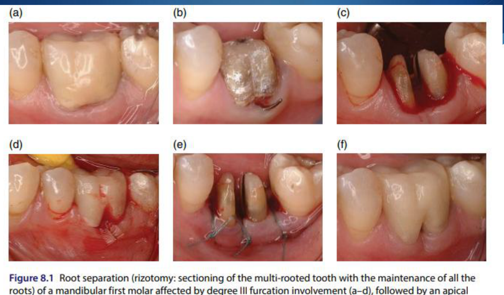
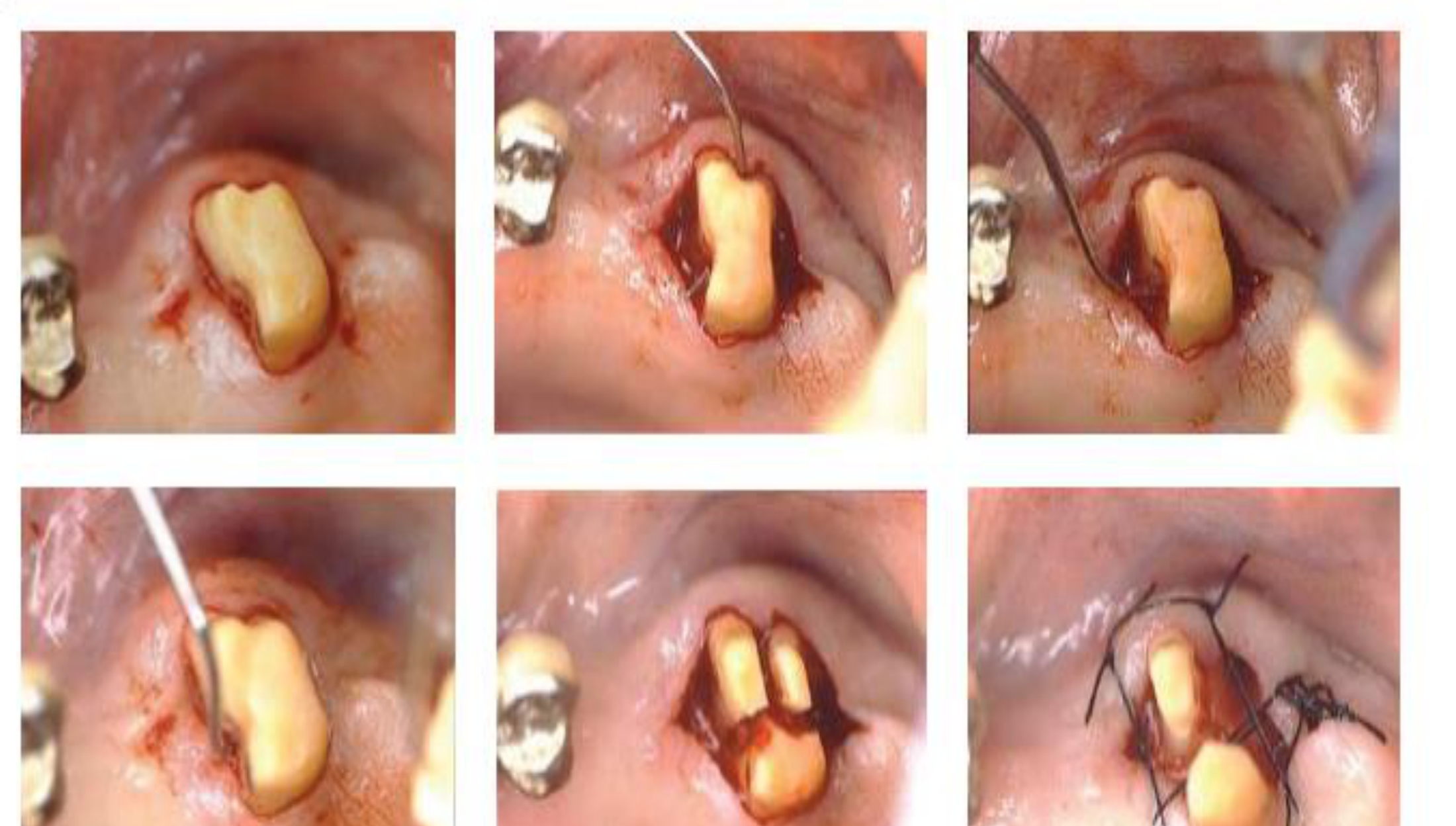
Root Separation and Premolarization474849
- All roots retained
- Indications: caries/perforations of root trunk, deep Class II or III, usually mandibular molars where it is termed premolarization
- Rarely, can be used on maxillary molars +/- root resection as well
 |  |
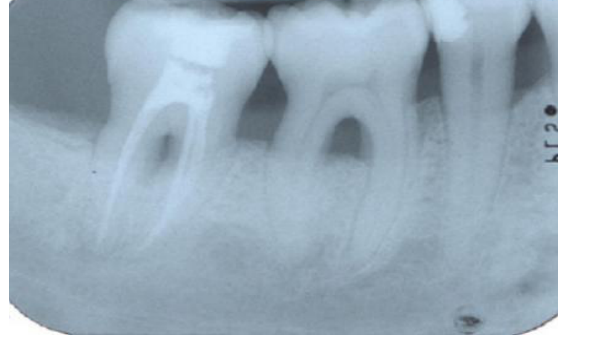
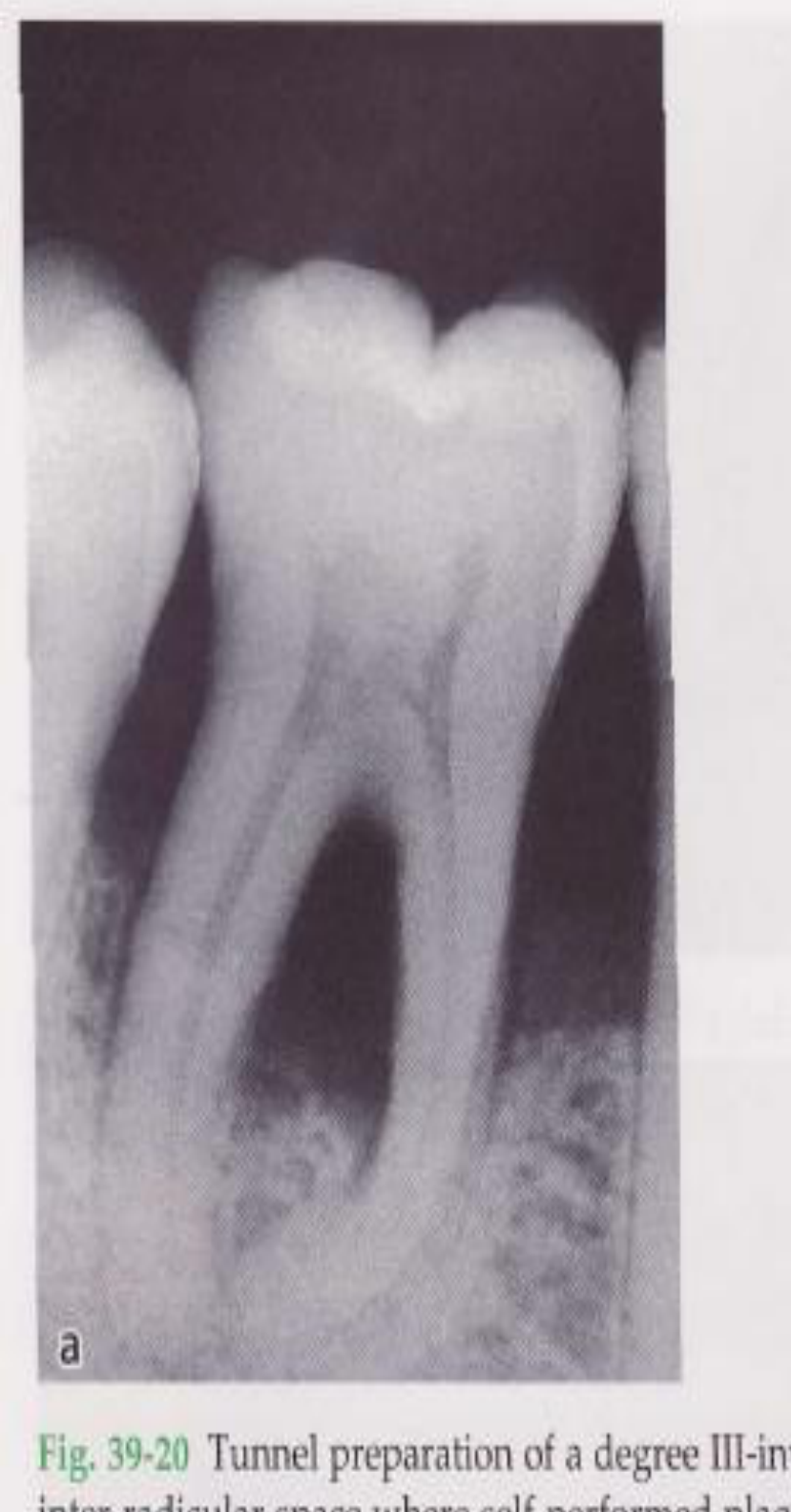
Tunneling Procedures50515253
| TUNNELING PROCEDURES | |
Beware – caries risk & tooth sensitivity
Hamp et al - 7 molars with degree III furcation defects undergone tunnel preparation. After 5 years, caries in 4 teeth (57%).
Little et al over 5 years - 18 class II or III molar furcations that underwent tunneling procedures. Crestal bone levels and attachment levels in the furcations were maintained for this time period. During follow-up, 3 of the 18 teeth experienced root caries (17%).
Hellden et al - 149 teeth received tunnel preparation, over avg 3 years. 24% of the teeth - caries. Of the 17 teeth that had to be extracted or further treated by root resection, root caries in 70%.
 |  |
 |
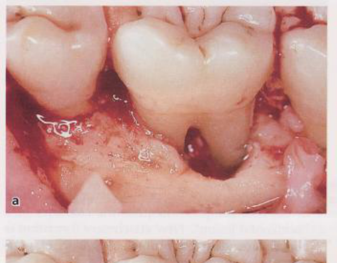
Regenerative Treatment54
Management: Surgery55
Surgical treatment:
- Open flap debridement
- Placing bone grafts to fill the furcation and prevent plaque retention.
Regenerative treatment:
- GTR (Guided Tissue Regeneration) +/- bone grafts
- And/or EMD (Enamel Matrix Derivative)
- Not for class III furcations
- Most suited for lower molars class II buccal (and lingual) & upper buccal furcation class II but complete closure NOT PREDICTABLE
Important Factors for Success of Regeneration56
- Controlled systemic factors
- Good OH
- Thicker phenotype / wide band of keratinized tissue / no recession
- Good interproximal bone height
- Good access
- Complete coverage of defect and membrane - more important than defect size
Treatment Planning and Maintenance
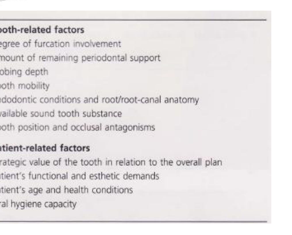
Selection of Treatment Modality57585960
Which treatment is best?
- Depend on tooth-related factors: degree of furcation involvement, root length & amount of bony support, access, phenotype
- Factors include residual bone support, root length, and whether roots are fused. Fused roots limit treatment options.
- Patient-related factors: smoking, level of diabetic control, other systemic factors, caries risk
Which treatment is best?
Degree of furcation involvement:
- Class I – NST, odontoplasty
- Class II – NST, odontoplasty, OFD, osteoplasty, regeneration e.g. GTR /EMD, Tunnel Preparation, root resection
- Class III – SRD, OFD, Tunnel Preparation, Root resection, extraction
Non-surgical treatment depends on patient compliance to attend regular maintenance visits.
Maintenance is important.
Less furcation-involved molars were lost in well maintained group (difference >10%) across all studies.
Importance of Long-Term Maintenance61
The importance of Maintenance
- Perio treatment and maintenance reduced tooth loss rate of FI molars in all studies vs no treatment (not many studies on no treatment!)
- Tooth loss rate is approx. double for FI molars (Class II) vs non-FI molars over a 15 year period.
- Degree of FI involvement influences tooth loss – x3 risk of tooth loss for Class III.
Classification of Furcation Involvement and Treatment options
Class I
- NST, odontoplasty
Class II
- NST, odontoplasty
- OFD, regeneration, Tunnel Preparation
Class III
- SRD, OFD, Tunnel Preparation, Root resection, extraction
Treatment options depend on patient compliance to attend regular maintenance visits
Important issues to be discussed – cost, time, consent
- Extraction
- Implant
- Long term maintenance and unpredictability of treating periimplantitis
- OFD
- Emdogain
- GTR + Bone
- Root resection / tunnelling
- +/- RCT
- +/- Crown
- Long term maintenance
Summary and Conclusions6263
Summary of Evidence
- Teeth with FI can be retained in the long term but at higher cost than teeth without FI
- The higher degrees of FI, bone loss and mobility, higher cost of maintaining the tooth
- However removing and replacing teeth with implant supported crowns does not seem to cost less in the long term
Conclusions
- Furcations can be very difficult to manage
- Class I – non-surgical approach with ultrasonics / mini curettes
- Class II and III – more effective debridement with a surgical approach
- Regenerative approach more successful with mandibular Class II buccal & lingual furcations; maxillary Class II buccal molars
- GTR is more effective than OFD in managing furcation defects, but results can be variable due to operator skill, patient & tooth selection
- Tunnel procedures & Root resection both effective & successful treatment modalities long-term for Gr II + III furcation involvements
- Regular maintenance is crucial to success of treatment (only 30% of degree III FI lost after 15 years of regular maintenance – Nibali et al, 2019)
OSCE Preparation
- Students must identify the Neighbors probe (3mm increments) and demonstrate its use.
- Knowledge of specific instruments like the Quittene curette and the ability to choose the correct Gracey curette for specific surfaces (e.g., disto-palatal of 2 6) is required.
References and Literature Review6465666768
Diagnosis and Treatment of Furcation-Involved Teeth
Edited by Luigi Nibali
Senior Clinical Lecturer Centre for Immunobiology and Regenerative Medicine Centre for Oral Clinical Research, Institute of Dentistry Barts and the London School of Medicine and Dentistry Queen Mary University of London (QMUL), London, UK Honorary Associate Professor, University of Hong Kong
References for those interested
Review > J Periodontol. 2015 Feb;86(2 Suppl):S108-30. doi: 10.1902/jop.2015.130677.
Periodontal regeneration – furcation defects: a systematic review from the AAP Regeneration Workshop
Gustavo Avila-Ortiz, Juan G De Buitrago, Michael S Reddy
Conclusions: On the basis of the reviewed evidence, the following conclusions can be drawn.
- Periodontal regeneration has been demonstrated histologically and clinically for the treatment of maxillary facial or interproximal and mandibular facial or lingual Class II furcation defects.
- Although periodontal regeneration has been demonstrated histologically for the treatment of mandibular Class III defects, the evidence is limited to one case report.
- Evidence supporting regenerative therapy in maxillary Class III furcation defects in maxillary molars is limited to clinical case reports.
- In Class I furcation defects, regenerative therapy may be beneficial in certain clinical scenarios, although most Class I furcation defects may be successfully treated with non-regenerative therapy.
- Future research efforts should be primarily directed toward the conduct of clinical trials to test novel regenerative approaches that place emphasis primarily on patient-reported outcomes and also on histologic demonstration of periodontal regeneration. Investigators should also focus on understanding the influence that local, systemic, and technical factors may have on the outcomes of regenerative therapy in furcation defects.
References for those interested
Review > J Clin Periodontol. 2009 Feb;36(2):164-76. doi: 10.1111/j.1600-051X.2008.01358.x.
The effect of periodontal therapy on the survival rate and incidence of complications of multirooted teeth with furcation involvement after an observation period of at least 5 years: a systematic review
Guy Huynh-Ba, Patrick Kuonen, Dominik Hofer, Jürg Schmid, Niklaus P Lang, Giovanni E Salvi
Conclusions: Good long-term survival rates (up to 100%) of multirooted teeth with furcation involvement were obtained following various therapeutic approaches. Initial furcation involvement (Degree I) could be successfully managed by non-surgical mechanical debridement. Vertical root fractures and endodontic failures were the most frequent complications observed following resective procedures.
— Department of Periodontology, University of Zurich
Received: 20 June 2021 | Revised: 16 August 2021 | Accepted: 8 September 2021 DOI: 10.1111/jcpe.13551 ORIGINAL ARTICLE | Journal of Clinical Periodontology | WILEY
Long-term prognosis of teeth with class III furcation involvement
Peter Eickholz, Maren Runschke, Bettina Dannewitz, Katrin Nickles, Hari Petsos, Dorothea Kronsteiner, Bernadette Pretzl
Objective: Evaluation of survival of teeth with class III furcation involvement (FI) ≥5 years after active periodontal treatment (APT) and identification of prognostic factors.
Conclusions: Subgingival instrumentation with adjunctive systemic antibiotics favours retention of class III furcation-involved teeth. Baseline RBL and PPD at T1 deteriorate long-term prognosis.
References
Lindhe (7th Ed):
Chapter 33
Footnotes
-
Original PDF page 1: L11 Furcation diagnosis, p.1 ↩
-
Original PDF page 2: L11 Furcation diagnosis, p.2 ↩
-
Original PDF page 3: L11 Furcation diagnosis, p.3 ↩
-
Original PDF page 4: L11 Furcation diagnosis, p.4 ↩
-
Original PDF page 5: L11 Furcation diagnosis, p.5 ↩
-
Original PDF page 6: L11 Furcation diagnosis, p.6 ↩
-
Original PDF page 7: L11 Furcation diagnosis, p.7 ↩
-
Original PDF page 8: L11 Furcation diagnosis, p.8 ↩
-
Original PDF page 9: L11 Furcation diagnosis, p.9 ↩
-
Original PDF page 10: L11 Furcation diagnosis, p.10 ↩
-
Original PDF page 11: L11 Furcation diagnosis, p.11 ↩
-
Original PDF page 12: L11 Furcation diagnosis, p.12 ↩
-
Original PDF page 13: L11 Furcation diagnosis, p.13 ↩
-
Original PDF page 14: L11 Furcation diagnosis, p.14 ↩
-
Original PDF page 18: L11 Furcation diagnosis, p.18 ↩
-
Original PDF page 19: L11 Furcation diagnosis, p.19 ↩
-
Original PDF page 22: L11 Furcation diagnosis, p.22 ↩
-
Original PDF page 23: L11 Furcation diagnosis, p.23 ↩
-
Original PDF page 15: L11 Furcation diagnosis, p.15 ↩
-
Original PDF page 16: L11 Furcation diagnosis, p.16 ↩
-
Original PDF page 17: L11 Furcation diagnosis, p.17 ↩
-
Original PDF page 20: L11 Furcation diagnosis, p.20 ↩
-
Original PDF page 24: L11 Furcation diagnosis, p.24 ↩
-
Original PDF page 25: L11 Furcation diagnosis, p.25 ↩
-
Original PDF page 26: L11 Furcation diagnosis, p.26 ↩
-
Original PDF page 21: L11 Furcation diagnosis, p.21 ↩
-
Original PDF page 27: L11 Furcation diagnosis, p.27 ↩
-
Original PDF page 28: L11 Furcation diagnosis, p.28 ↩
-
Original PDF page 29: L11 Furcation diagnosis, p.29 ↩
-
Original PDF page 30: L11 Furcation diagnosis, p.30 ↩
-
Original PDF page 31: L11 Furcation diagnosis, p.31 ↩
-
Original PDF page 32: L11 Furcation diagnosis, p.32 ↩
-
Original PDF page 35: L11 Furcation diagnosis, p.35 ↩
-
Original PDF page 37: L11 Furcation diagnosis, p.37 ↩
-
Original PDF page 38: L11 Furcation diagnosis, p.38 ↩
-
Original PDF page 33: L11 Furcation diagnosis, p.33 ↩
-
Original PDF page 34: L11 Furcation diagnosis, p.34 ↩
-
Original PDF page 36: L11 Furcation diagnosis, p.36 ↩
-
Original PDF page 40: L11 Furcation diagnosis, p.40 ↩
-
Original PDF page 39: L11 Furcation diagnosis, p.39 ↩
-
Original PDF page 41: L11 Furcation diagnosis, p.41 ↩
-
Original PDF page 42: L11 Furcation diagnosis, p.42 ↩
-
Original PDF page 43: L11 Furcation diagnosis, p.43 ↩
-
Original PDF page 44: L11 Furcation diagnosis, p.44 ↩
-
Original PDF page 45: L11 Furcation diagnosis, p.45 ↩
-
Original PDF page 46: L11 Furcation diagnosis, p.46 ↩
-
Original PDF page 47: L11 Furcation diagnosis, p.47 ↩
-
Original PDF page 48: L11 Furcation diagnosis, p.48 ↩
-
Original PDF page 49: L11 Furcation diagnosis, p.49 ↩
-
Original PDF page 50: L11 Furcation diagnosis, p.50 ↩
-
Original PDF page 51: L11 Furcation diagnosis, p.51 ↩
-
Original PDF page 52: L11 Furcation diagnosis, p.52 ↩
-
Original PDF page 53: L11 Furcation diagnosis, p.53 ↩
-
Original PDF page 56: L11 Furcation diagnosis, p.56 ↩
-
Original PDF page 54: L11 Furcation diagnosis, p.54 ↩
-
Original PDF page 55: L11 Furcation diagnosis, p.55 ↩
-
Original PDF page 57: L11 Furcation diagnosis, p.57 ↩
-
Original PDF page 58: L11 Furcation diagnosis, p.58 ↩
-
Original PDF page 60: L11 Furcation diagnosis, p.60 ↩
-
Original PDF page 61: L11 Furcation diagnosis, p.61 ↩
-
Original PDF page 59: L11 Furcation diagnosis, p.59 ↩
-
Original PDF page 62: L11 Furcation diagnosis, p.62 ↩
-
Original PDF page 63: L11 Furcation diagnosis, p.63 ↩
-
Original PDF page 64: L11 Furcation diagnosis, p.64 ↩
-
Original PDF page 65: L11 Furcation diagnosis, p.65 ↩
-
Original PDF page 66: L11 Furcation diagnosis, p.66 ↩
-
Original PDF page 67: L11 Furcation diagnosis, p.67 ↩
-
Original PDF page 68: L11 Furcation diagnosis, p.68 ↩