Acute Gingival and Periodontal Conditions123
The University of Western Australia
Dr Emma Perry | BDSc, AssocDeg DH, GradCert HPEd Lecturer (Clinical Dentistry) | UWA Dental School
Content courtesy of Dr Anna Hughes and Dr Tina Choo
Acknowledgement of country
On Noongar land. Our university is located alongside the Derbal Yerrigan (Swan River) on Whadjuk Noongar Boodja (land). The University of Western Australia acknowledges that we are situated on Noongar land and that Noongar people remain the spiritual and cultural custodians of their land and continue to practise their values, languages, beliefs and knowledge.
Artist: Dr Richard Barry Walley OAM
Everyone is welcome
 |  |  |
 |  |
Learning Outcomes and Lecture Outline45
Learning outcomes
- Diagnose and classify the most common acute gingival and periodontal conditions by recognising risk factors and clinical presentation
- Explain the aetiology of acute gingival and periodontal conditions
- Describe the rationale and methods for management of the most common acute gingival and periodontal conditions
Clinical Characteristics
Unlike chronic periodontal disease, acute conditions are characterized by rapid onset, pain, and tissue destruction.
Lecture outline
Necrotising periodontal diseases
- Necrotising gingivitis (NG)
- Necrotising periodontitis (NP)
- Viral lesions
- Acute Herpetic Gingivostomatitis
- Abscesses of the periodontium
- Gingival abscess
- Pericoronitis
- Periodontal abscess
- Other less common conditions
- Desquamative gingivitis and Neoplasia
- Endo-perio lesions covered in dedicated lecture from DENT4212 endodontics module
Characteristics of Acute Conditions67
- Rapid onset
- Patients seek urgent care because they are in significant pain and may have difficulty eating or drinking.
- Pain or discomfort
- Tissue destruction +/- infection
- Often involves the death of tissue (necrosis).
- Patients usually seek urgent care (pain)
- Underlying Causes: These conditions usually occur because a patient is compromised; they do not typically arise from simple short-term neglect (e.g., missing one day of brushing).
- Recurrence: If the underlying cause is not addressed, the condition is likely to recur.
It is important to identify and eliminate predisposing factors to prevent recurrence.
Periodontal Treatment Planning89
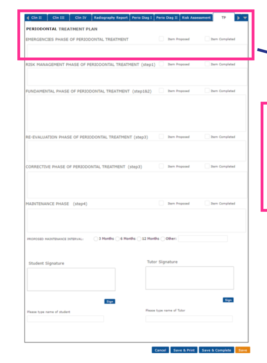
PERIODONTAL TREATMENT PLAN
EMERGENCIES PHASE OF PERIODONTAL TREATMENT Item Proposed Item Completed
RISK MANAGEMENT PHASE OF PERIODONTAL TREATMENT (step1) Item Proposed Item Completed
FUNDAMENTAL PHASE OF PERIODONTAL TREATMENT (step1&2) Item Proposed Item Completed
RE-EVALUATION PHASE OF PERIODONTAL TREATMENT (step3) Item Proposed Item Completed
CORRECTIVE PHASE OF PERIODONTAL TREATMENT (step3) Item Proposed Item Completed
MAINTENANCE PHASE (step4) Item Proposed Item Completed
PROPOSED MAINTENANCE INTERVAL
- 3 Months
- 6 Months
- 12 Months
- Other
Student Signature Tutor Signature
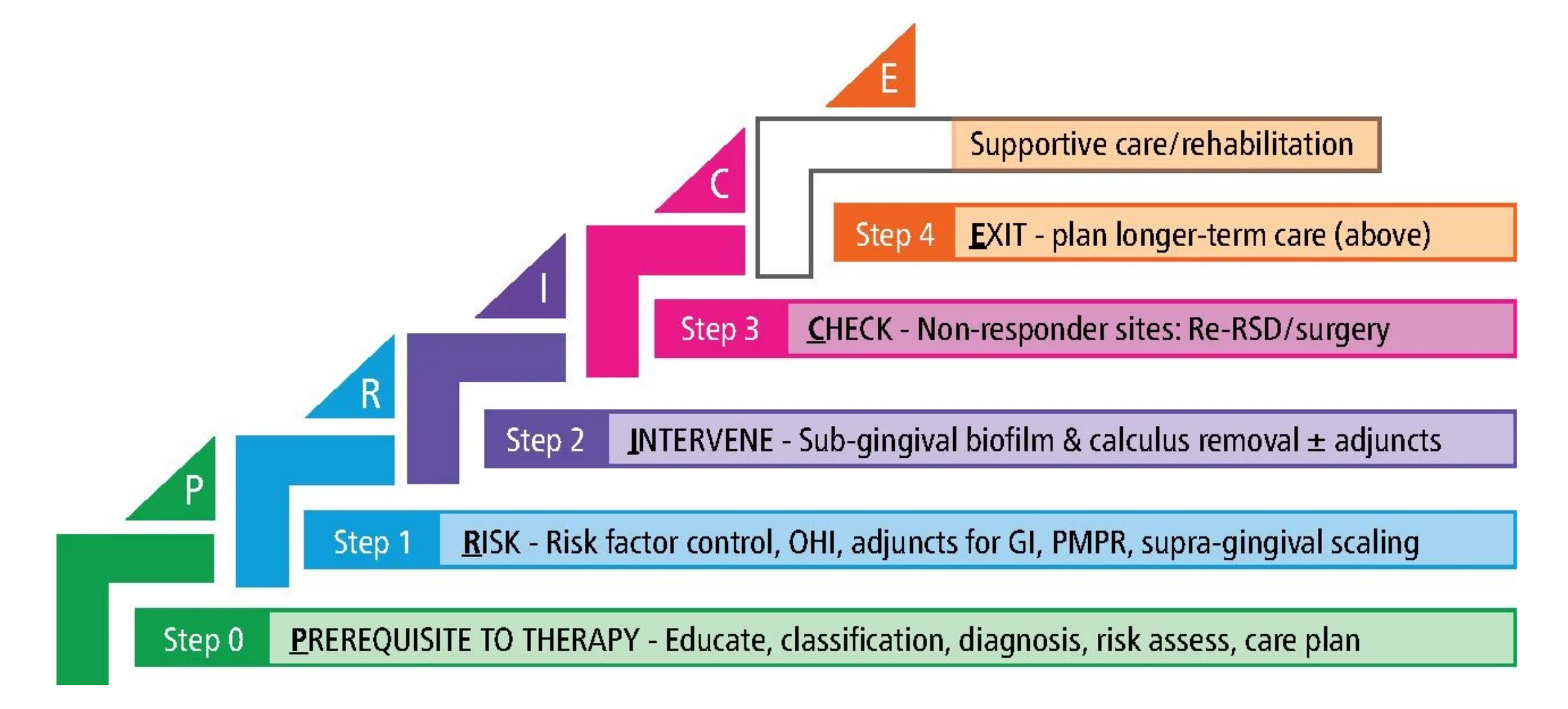
Periodontal Treatment Step
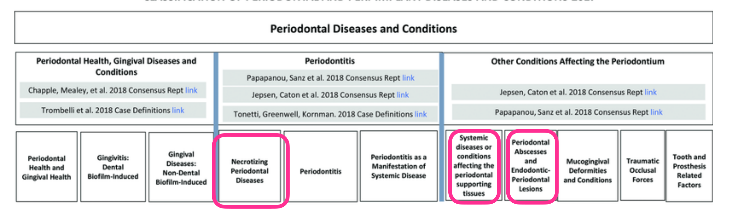
Step 0 PREREQUISITE TO THERAPY - Educate, classification, diagnosis, risk assess, care plan
Emergency Phase
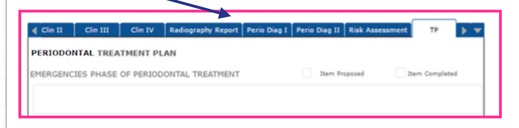
Acute conditions fall into the “emergency” or “palliative” phase of the treatment plan (Step 0). Steps 1 and 2 of periodontal therapy can often be performed simultaneously with Step 0. Most conditions require biofilm removal and risk factor control.
Step 1 RISK - Risk factor control, OHI, adjuncts for GI, PMPR, supra-gingival scaling
Step 2 INTERVENE - Sub-gingival biofilm & calculus removal ± adjuncts
Step 3 CHECK - Non-responder sites: Re-RSD/surgery
Step 4 EXIT - plan longer-term care (above)
C Supportive care/rehabilitation
E
 |  |
 |
Necrotising Periodontal Diseases1011
- NG and NP appear to be the same disease but at different stages.
Disease Progression
Necrotising diseases involve tissue death and are considered a progression of the same disease process. Unlike “garden variety” chronic gingivitis, necrotising gingivitis will usually progress to periodontitis if left untreated.
- Differentiated by:
- Severity
- Extent of tissue destruction
Glick et al 1994, Robinson et al 1998
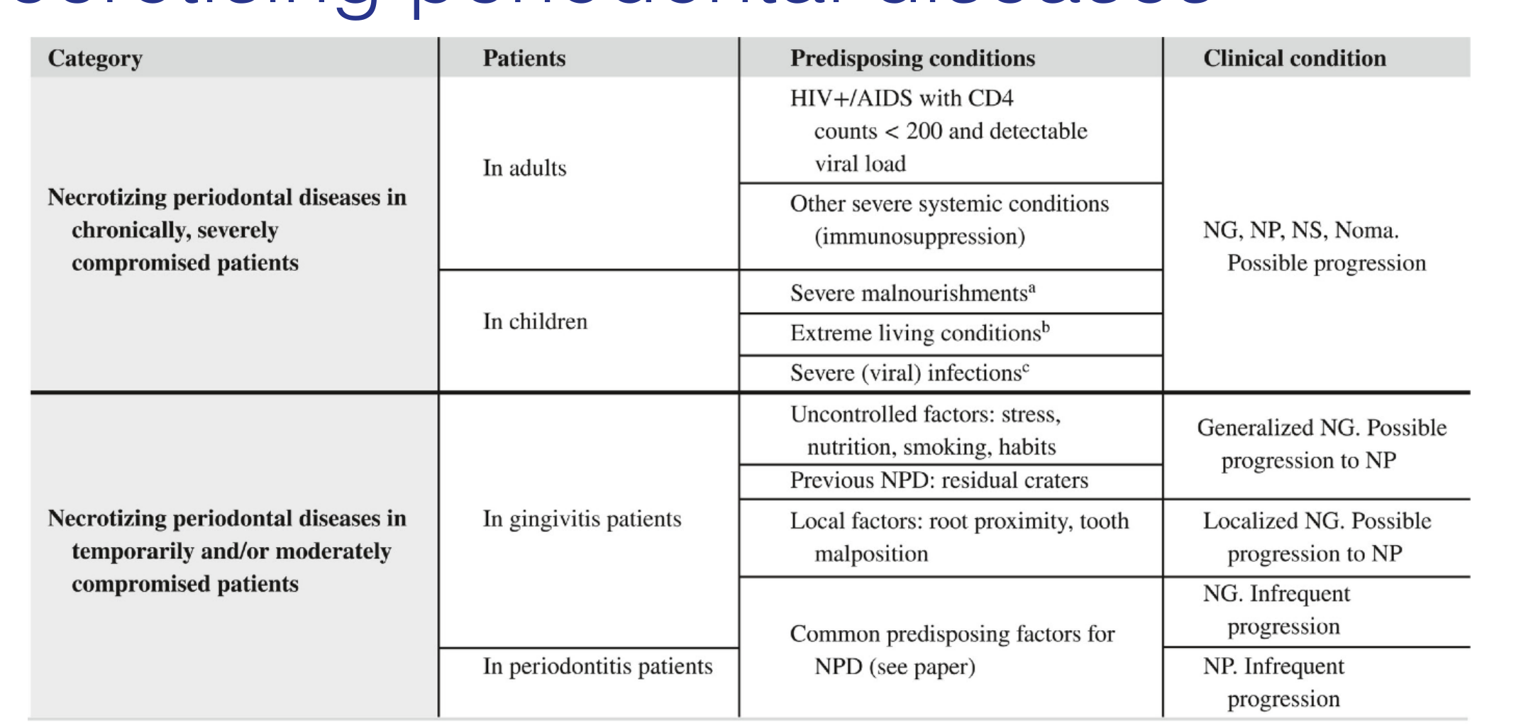
NG, necrotizing gingivitis; NP, necrotizing periodontitis; NS, necrotizing stomatitis
^a^ Mean plasma and serum concentrations of retinol, total ascorbic acid, zinc, and albumin markedly reduced, or very marked depletion of plasma retinol, zinc, and ascorbate; and saliva levels of albumin and cortisol, as well as plasma cortisol concentrations, significantly increased
^b^ Living in substandard accommodations, exposure to debilitating childhood diseases, living near livestock, poor oral hygiene, limited access to potable water and poor sanitary disposal of human and animal fecal waste
^c^ Measles, herpes viruses (cytomegalovirus, Epstein-Barr virus-1, herpes simplex virus) chicken pox, malaria, febrile illness
 |  |
Aetiology and Risk Factors
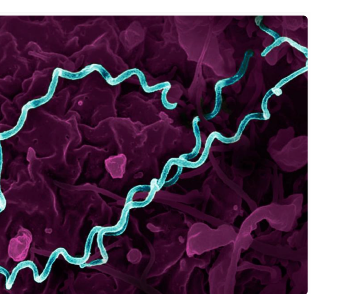
Aetiology12
- Opportunistic / dysbiotic microbiome in a susceptible host
- Fusiform bacteria, Prevotella intermedia, & spirochaetes
- Bacterial invasion of soft tissues occurs
- Intense release of cytokines and tissue destruction
Falkler et al 1987, Stevens et al 1984
Risk Factors13
- Poor oral hygiene
- Smoking
- Insufficient sleep & Psychological STRESS – inability to cope with stress
- Exhaustion and being “run down” (e.g., end of semester)
- HIV +ve patients & Immunosuppressed patients
- Severe malnutrition
- Developed countries - Young people (15-34yrs)
- Developing countries - children
Epidemiology14
- Unclear, but low in developed countries
- Higher in developing countries - 5-25%
- Higher in children - 54-68% are children <10 yrs
- HIV +ve patients 2% (30% if not on HAART)
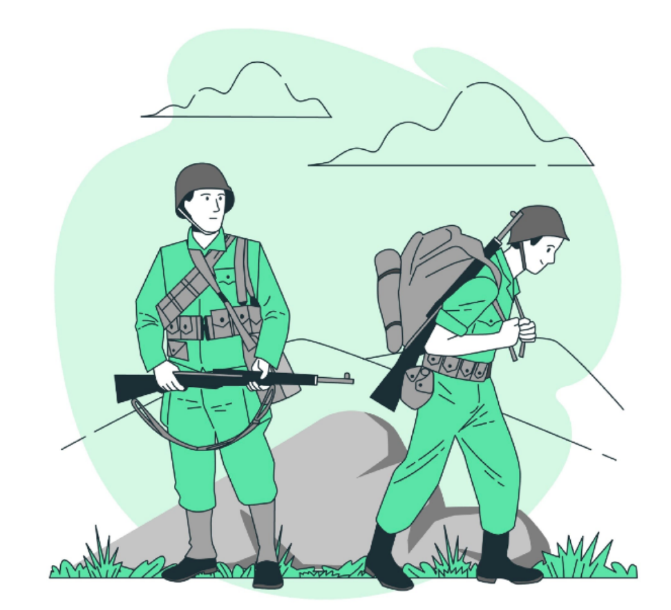
- Higher in military population
- High during WWI & WWII
- Historically known as “Trench Mouth” due to high prevalence in military populations during WWI and WWII (linked to stress, smoking, and poor hygiene)
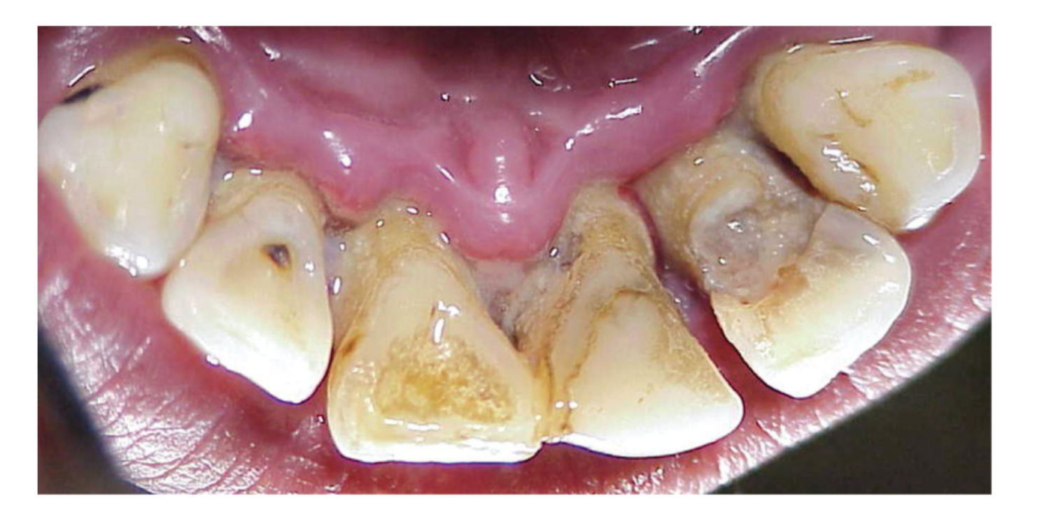
Necrotising Gingivitis Clinical Features1516
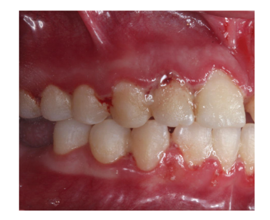
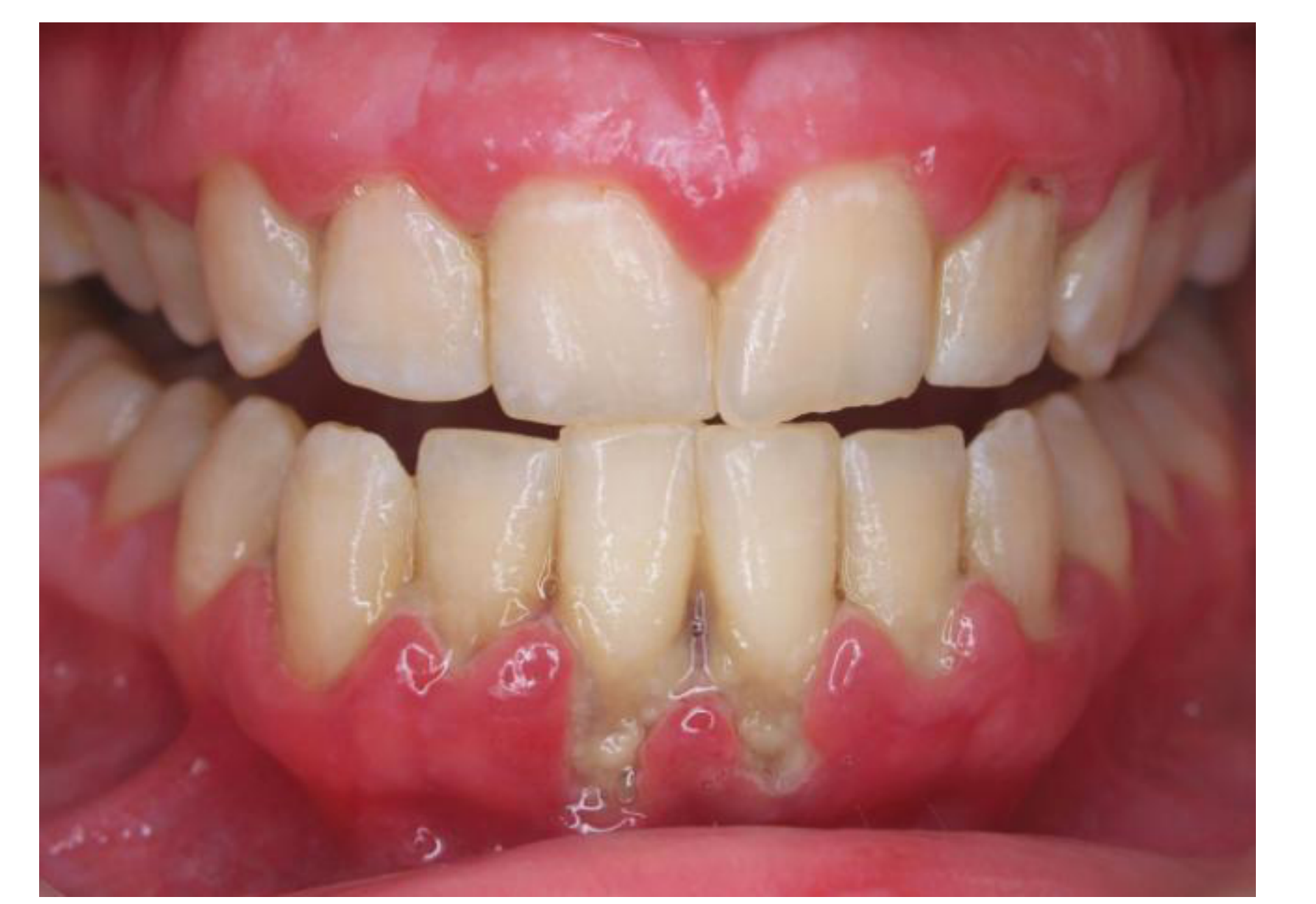
- Sudden Onset - Rapid development and painful, more common to affect anterior teeth
- Starts with ulceration of interdental papilla, leading to papillary necrosis
- “Punched out” papillae appearance
- Papillae may appear brown or “mushy”
- Fibrinous pseudomembrane
- +/- Fever, lymphadenopathy, malaise
- No bone loss
- Bleeding
- Malodour
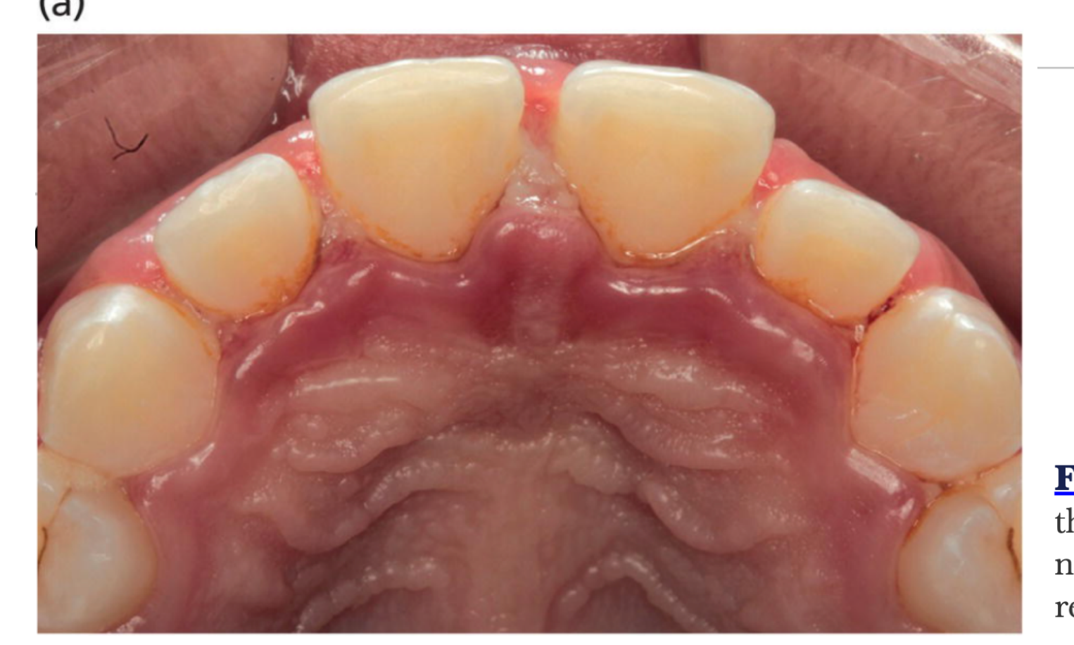
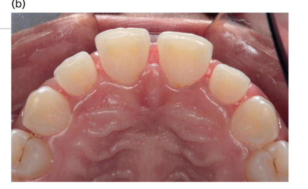
Fig. 19-9 Healing of necrotizing gingivitis lesions in the upper anterior sextant. (a) Active lesions with necrosis the interdental papillae. (b) Complete resolution after 60 days.
(Source: Courtesy of Dr. Nidia Castro dos Santos and Dr. Mauro Santamaria.)
 |  |
 |
Essential clinical features17
- Lesions are painful
- Lesions are gingival ulcers, punched-out crater-like affecting the interdental papilla and may involve marginal gingiva
- Gingival ulcers bleed spontaneously or readily
- The acute condition may occur from an existing chronic gingival condition or de novo
Non-essential clinical features
- “Pseudomembrane” of sloughed necrotic debris & bacteria covering the ulcerated area
- Bad breath
- Pungent, “metallic” smell
- Fever, malaise
- Lymphadenopathy – (submandibular & cervical)
Necrotising Periodontitis Clinical Features181920
- Lesions extend into alveolar bone
- Exposed bone, gingival recession, tooth mobility
- Malodour, lymphadenopathy, fever and malaise
- Severe pain and spontaneous bleeding
- Exfoliation of teeth within months of onset of NP
- Rapid destruction of the alveolar bone
- Visible bone fragments (sequestra)
- Teeth may become loose and “drop out” within months if untreated
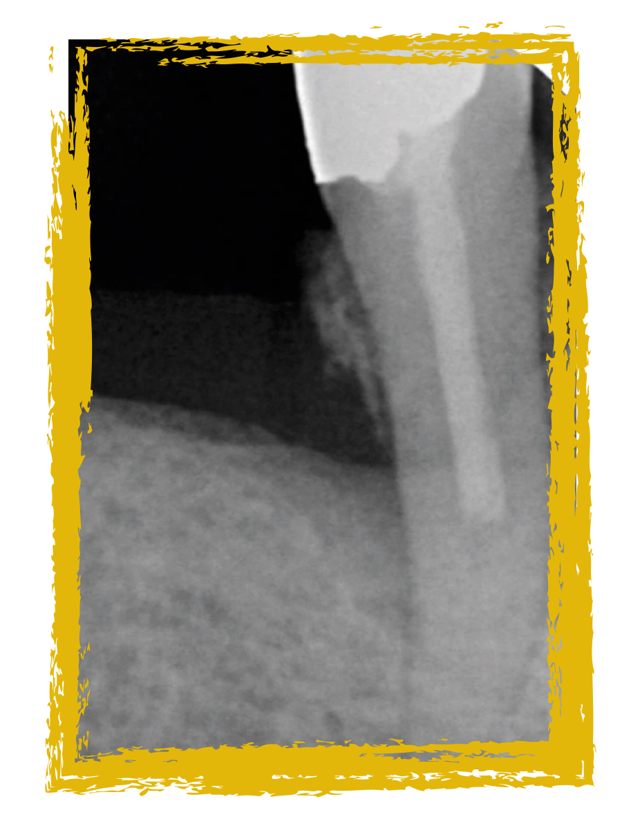
Fig. 19-8 Necrotizing periodontitis: presence of necrosis/ulcer of the interdental papillae.
(Source: Courtesy of Dr. Mauro Santamaria.)
 |  |  |
 |  |  |
Management of Necrotising Periodontal Diseases21222324
-
Needs urgent treatment
-
Disease can progress rapidly (necrotic bone & soft tissue)
-
If left untreated: NG → NP
-
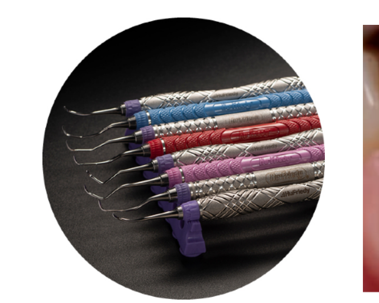
Gentle debridement with ultrasonic (with LA) to remove slough, plaque & necrotic tissues, saline irrigation
-
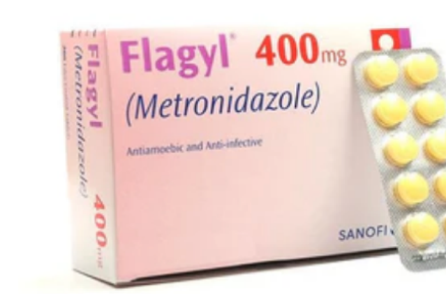
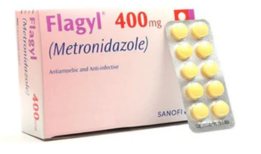
Rx metronidazole 400mg 12-hourly for 3-5 days, pain management, OHI (gentle)
-
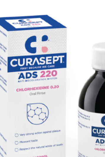
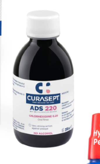
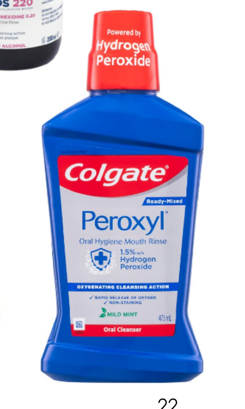
Mouthwash for 2-3 weeks, 2 x daily for 1 min
- CHX 0.2%
- OR
- H2O2 1.5%
-
Stop smoking!
-
Review 2-3 days later - further Rx may be needed
Robinson et al 1998
Access by University of Western Australia (CAUL) Therapeutic Guidelines > Oral and Dental > Necrotising periodontal disease
Necrotising periodontal disease
metronidazole 400 mg orally, 12-hourly for 3 to 5 days.
Metronidazole Contraindications
- Allergy
- First trimester of pregnancy
- Alcohol consumption (causes severe vomiting)
- Potential interaction with Warfarin
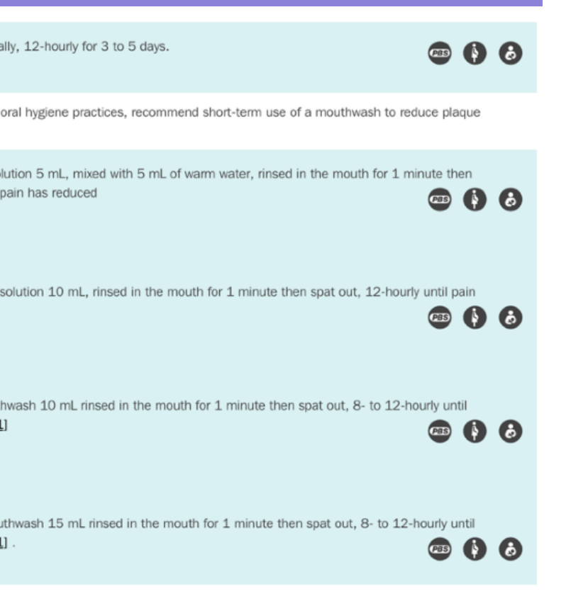
If pain and inflammation restrict oral hygiene practices, recommend short-term use of a mouthwash to reduce plaque formation; use:
- hydrogen peroxide 3% solution 5 mL, mixed with 5 mL of warm water, rinsed in the mouth for 1 minute then spat out, 12-hourly until pain has reduced OR
- hydrogen peroxide 1.5% solution 10 mL, rinsed in the mouth for 1 minute then spat out, 12-hourly until pain has reduced OR
- chlorhexidine 0.2% mouthwash 10 mL rinsed in the mouth for 1 minute then spat out, 8- to 12-hourly until pain has reduced [Note 1] OR
- chlorhexidine 0.12% mouthwash 15 mL rinsed in the mouth for 1 minute then spat out, 8- to 12-hourly until pain has reduced [Note 1]
Response to treatment is diagnostic
At 1-2 week review:
-
Subgingival debridement if necessary (earlier may hinder regenerative potential of tissues) and treat chronic disease if present
-
Reinforce oral hygiene
-
Review again - maintenance or needs surgery?
-
Failure to resolve may be due to incomplete debridement/ stress/ malnutrition/ smoking/ immunological conditions
-
Regimen shown to have 94% resolution within 1 week & 100% within 1 month
-
If not resolved, you need to rule other non-infective conditions that resemble NG, e.g. desquamative gingivitis
Robinson et al 1998
 |  |  |
 |  |  |
 |  | 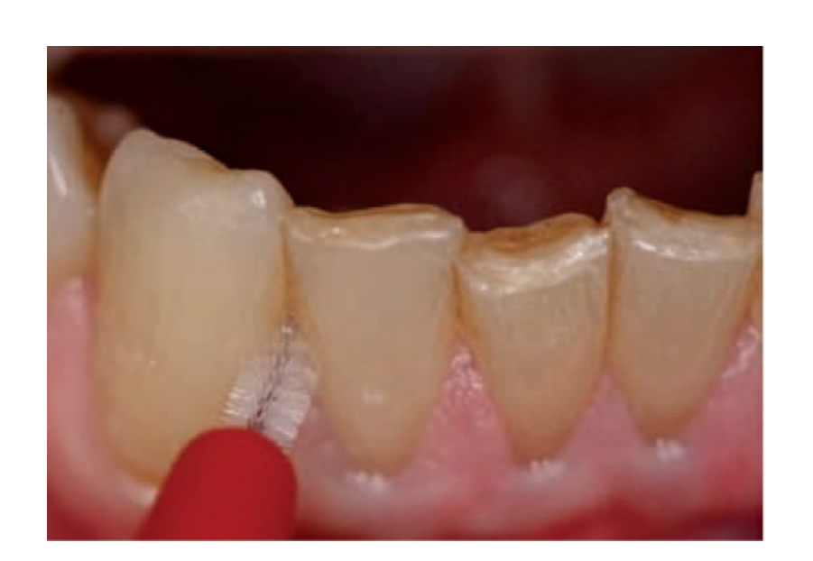 |
Longer term management2526272829
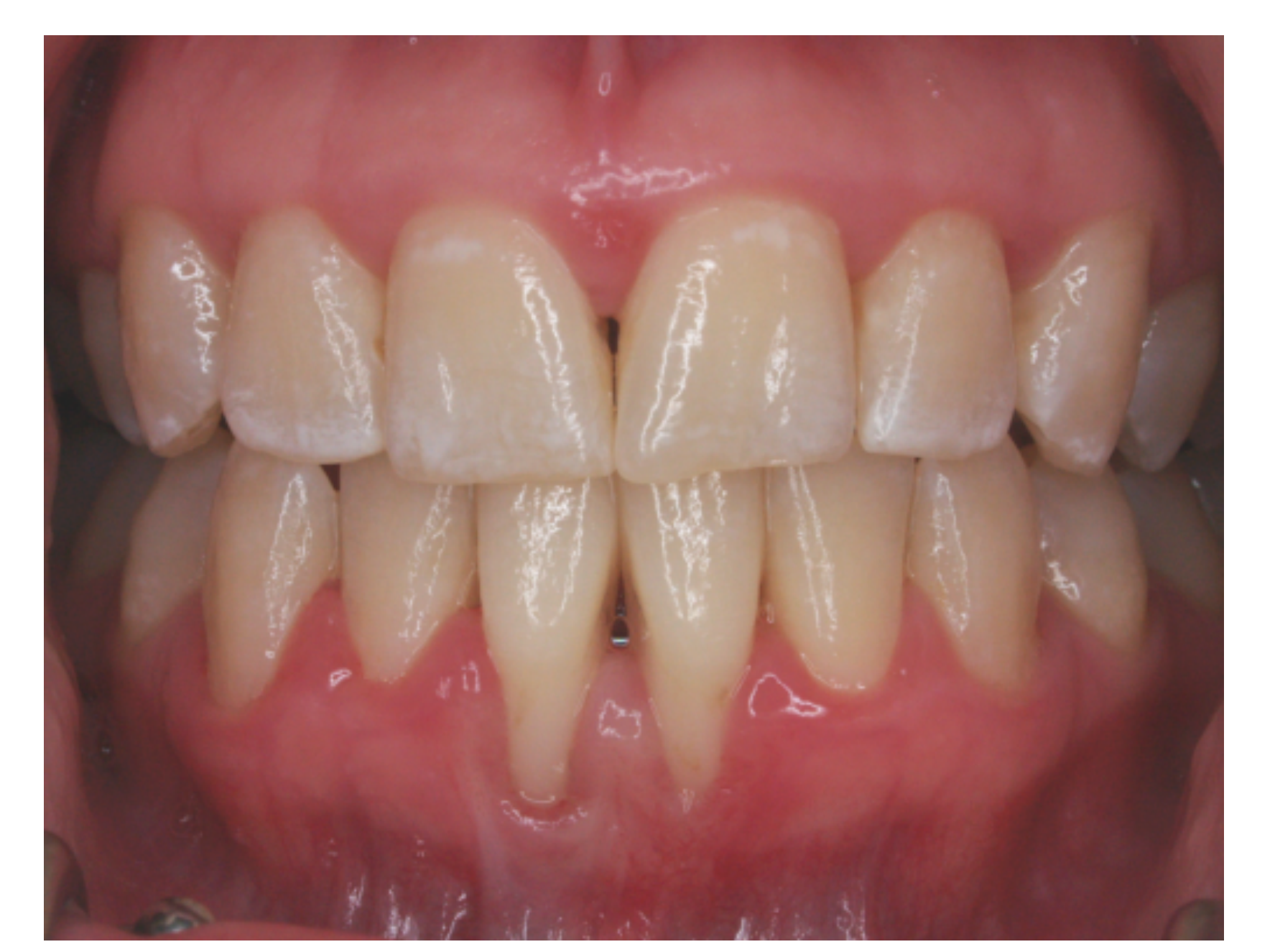
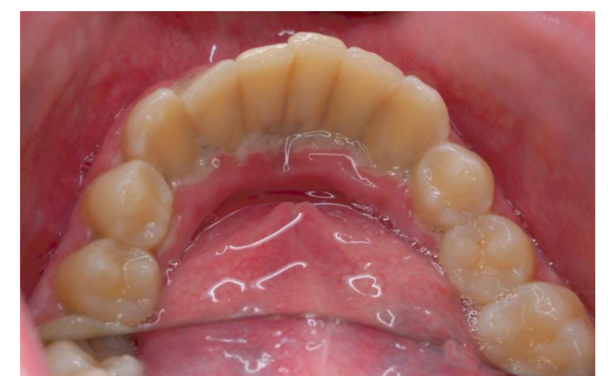
- Review at 1 month
- If resolved, place on 3/12 maintenance, (may need periodontal surgery if deformities present)
- If no resolution, refer to periodontist or oral medicine specialist, (blood test, microbiological sample & biopsy -> histopathology investigations)
- Up to 30% of patients will experience recurrence within 2 years
- Maintenance of meticulous OH and scaling/debridement
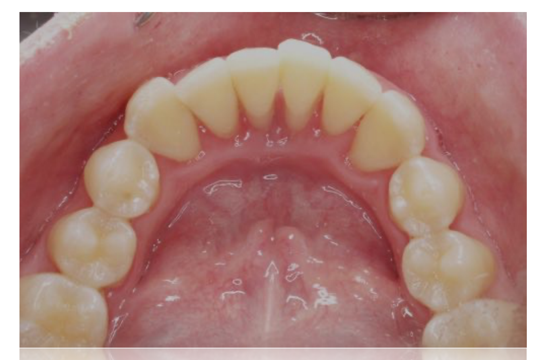
Image courtesy of Dr Anna Hughes
IPD-P03848 Eng-Patent (27)
 |  |  |
 |  |
Viral Lesions
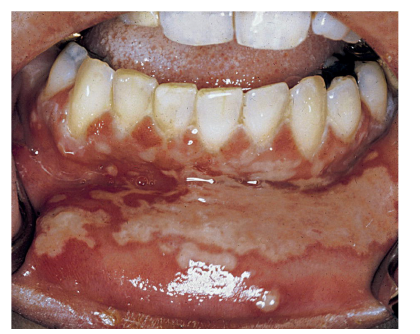
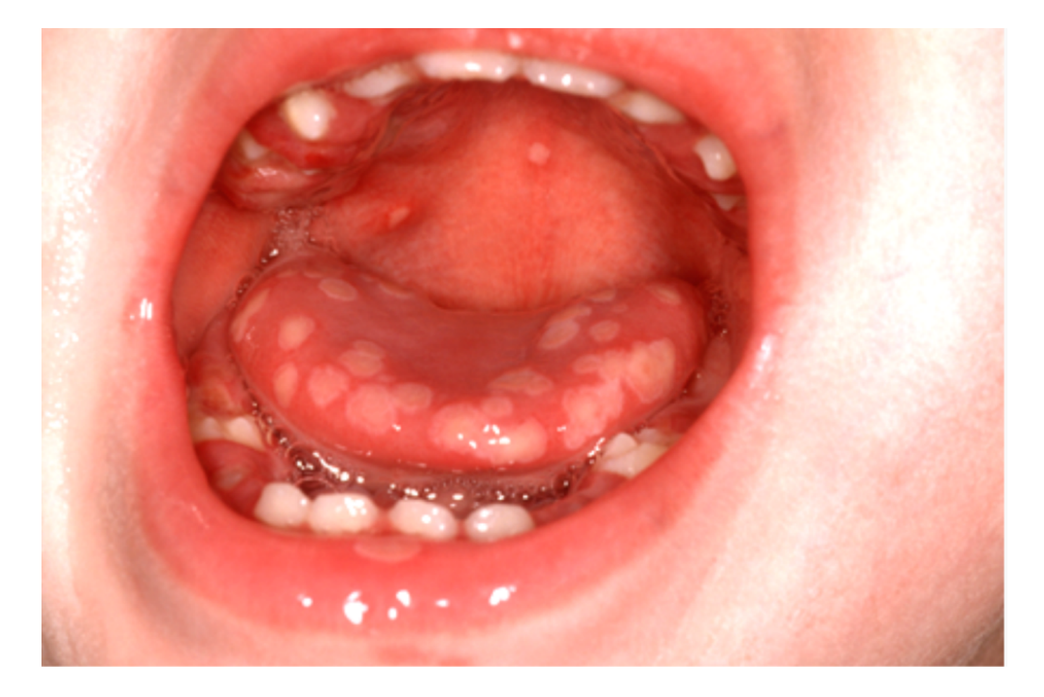
Acute Herpetic Gingivostomatitis303132
-
Herpes Simplex virus
-
70-80% of population exposed
-
Less in developed countries
-
Young adults & children
-
Affects whole mouth & gingiva
-
1-2mm blisters that rupture to form small, shallow, painful & irregular ulcers, 7-10 days
-
Ulcers covered by a yellowish-gray center & surrounded by a red halo
-
Ulcers may take up to 2 weeks to heal
-
Heal with no scarring
-
Rare to get gingival recession
-
Sore to touch, food & drinks
-
Worse with salty, acidic, hard foods
-
Fever & malaise
-
Ddx recurrent apthous ulcers?
- These are not seen on keratinised tissues
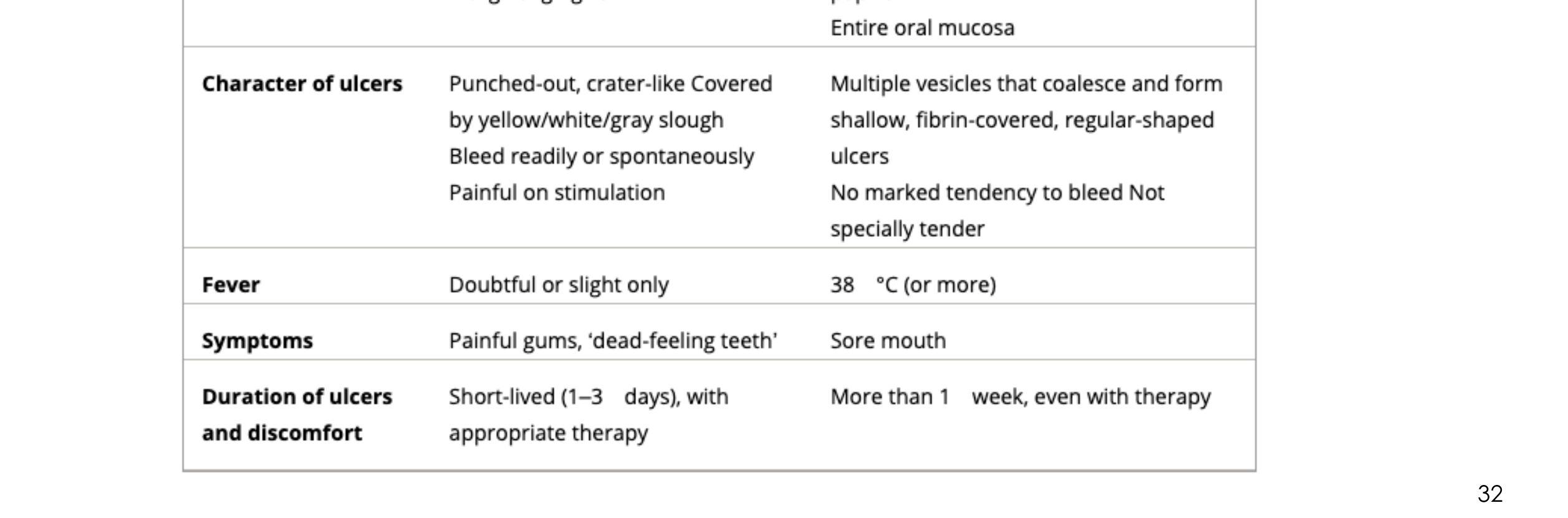
Table 4. Differences in clinical features of necrotizing ulcerative gingivitis (NUG) and acute herpetic gingivostomatitis (AHG)
Site of ulcers NUG: Interdental papilla, Marginal gingival AHG: Gingiva, no predilection for interdental papilla; Entire oral mucosa
Character of ulcers NUG: Punched-out, crater-like; Covered by yellow/white/gray slough; Bleed readily or spontaneously; Painful on stimulation AHG: Multiple vesicles that coalesce and form shallow, fibrin-covered, regular-shaped ulcers; No marked tendency to bleed; Not specially tender
Fever NUG: Doubtful or slight only AHG: 38 °C (or more)
Symptoms NUG: Painful gums, ‘dead-feeling teeth’ AHG: Sore mouth
Duration of ulcers and discomfort NUG: Short-lived (1–3 days), with appropriate therapy AHG: More than 1 week, even with therapy
 |  |
 |
Management33
- Pain relief: analgesics & topical anaesthetics rinses
- Advice to have adequate fluid intake & food
- Use of antiviral medication questioned: ACYCLOVIR, only if started immediately
- Systemic Acyclovir can be prescribed if started within the first 5 days.
- Inform - AHG is CONTAGIOUS!
- Dental treatment should be deferred to prevent self-inoculation, such as herpetic whitlow on fingers or ocular herpes in the eyes.
- Review in 2wks to confirm correct diagnosis
- Recurrence can occur - dental tx, stress, fever, trauma
Reactivation of Viruses34
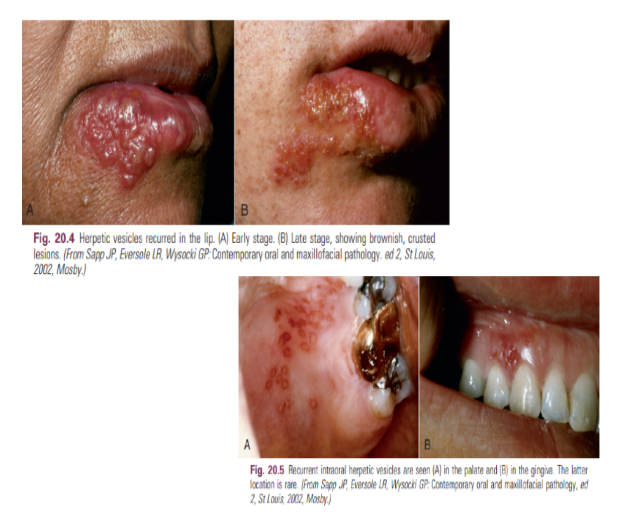
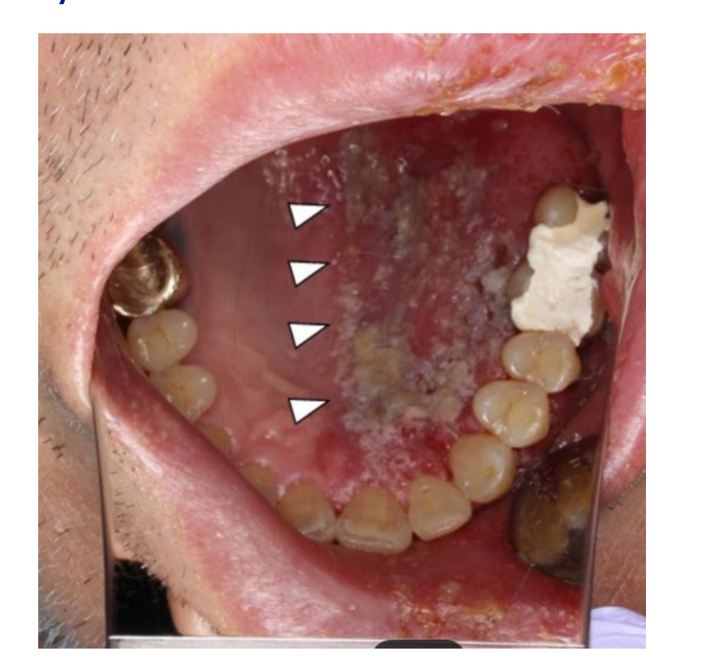
Herpes simplex virus Cold sores Varicella Zoster Shingles (from chicken pox)
Varicella Zoster Presentation
Reactivation of the chickenpox virus presents along dermatomes (nerve pathways) and is extremely painful. In the mouth, it may stop exactly at the midline of the palate.
Fig. 20.4 Herpetic vesicles recurred in the lip. (A) Early stage. (B) Late stage, showing brownish, crusted lesions. (From Sapp JP, Eversole LR, Wysocki GP. Contemporary oral and maxillofacial pathology. ed2, St Louis, 2002, Mosby.)
Fig. 20.5 Recurrent intraoral herpetetic vesicles are seen (A) in the palate and (B) in the gingiva. The latter location is rare. (From Sapp JP, Eversole LR, Wysocki GP. Contemporary oral and maxillofacial pathology, ed 2, St Louis, 2002, Mosby.)
 |  |
Abscesses of the Periodontium35
Definition
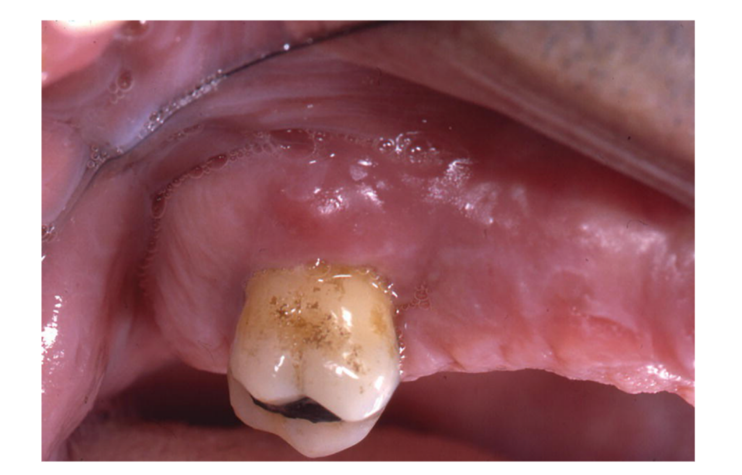
An abscess is a localized collection of pus (purulent exudate) involving a massive inflammatory response.
Classification of Abscesses3637
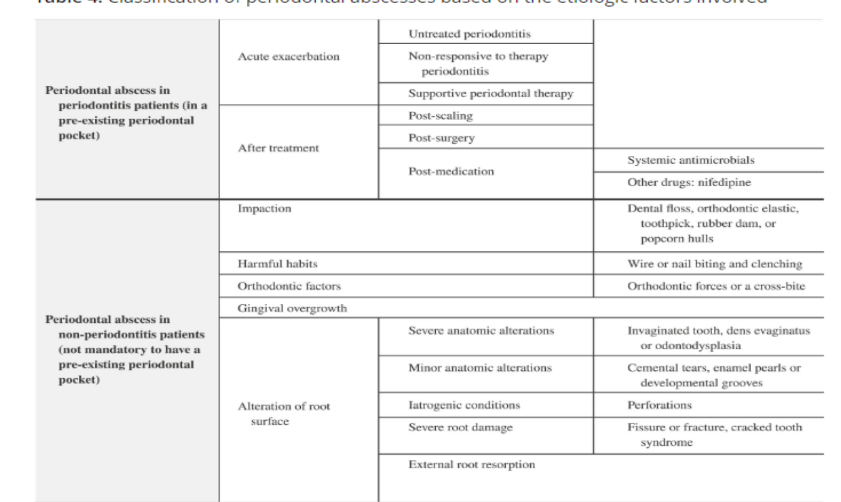
Classified based on aetiology
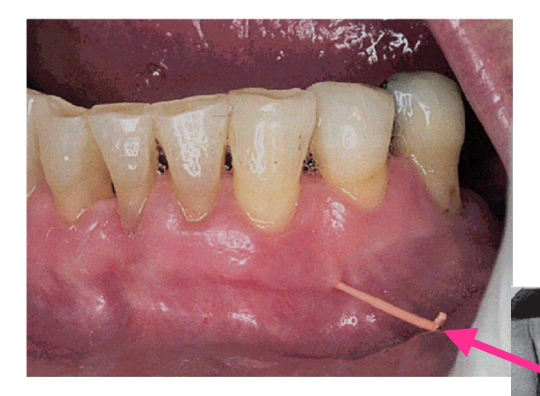
Periodontal abscess in periodontitis patients (in a pre-existing periodontal pocket)
- Acute exacerbation
- Untreated periodontitis
- Non-responsive to therapy periodontitis
- Supportive periodontal therapy
- After treatment
- Post-scaling
- Post-surgery
- Post-medication
- Systemic antimicrobials
- Other drugs: nifedipine
Periodontal abscess in non-periodontitis patients (not mandatory to have a pre-existing periodontal pocket)
-
Impaction
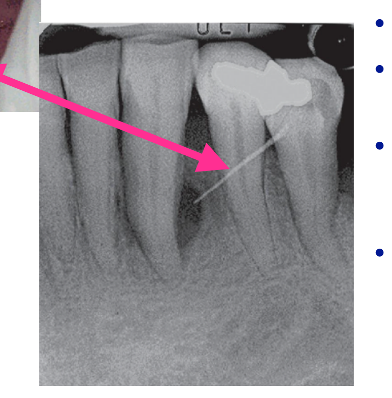
- Dental floss, orthodontic elastic, toothpick, rubber dam, or popcorn hulls
-
Harmful habits
- Wire or nail biting and clenching
-
Orthodontic factors
- Orthodontic forces or a cross-bite
-
Gingival overgrowth
-
Alteration of root surface
- Severe anatomic alterations
- Invaginated tooth, dens evaginatus or odontodysplasia
- Minor anatomic alterations
- Cemental tears, enamel pearls or developmental grooves
- Iatrogenic conditions
- Perforations
- Severe root damage
- Fissure or fracture, cracked tooth syndrome
- External root resorption
- Severe anatomic alterations
-
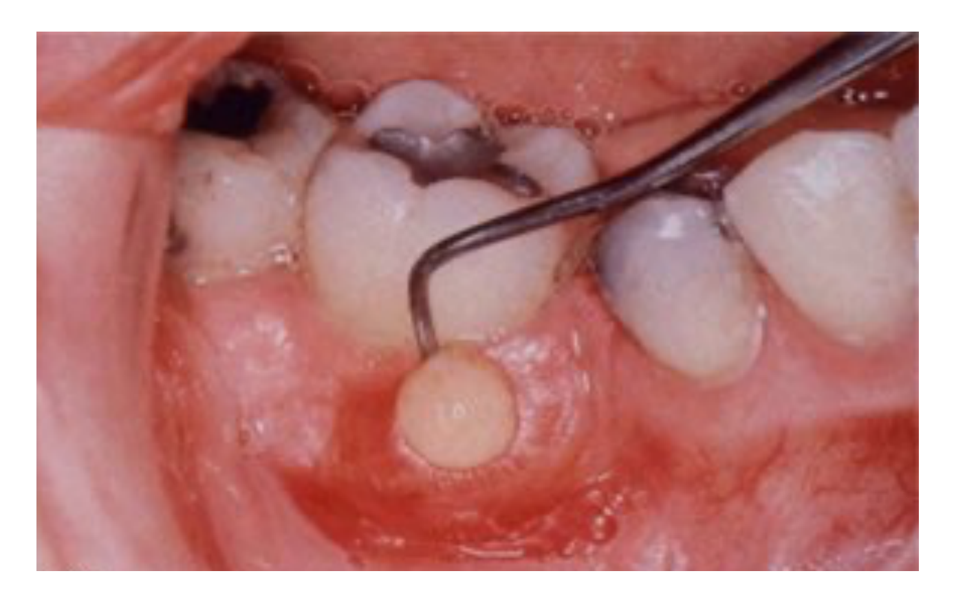
Pericoronal Abscess (Pericoronitis)
-
Gingival Abscess
-
Periodontal Abscess
- Combined perio-endo abscess
- Other acute abscesses
 |  |
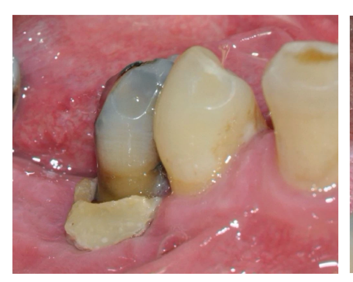
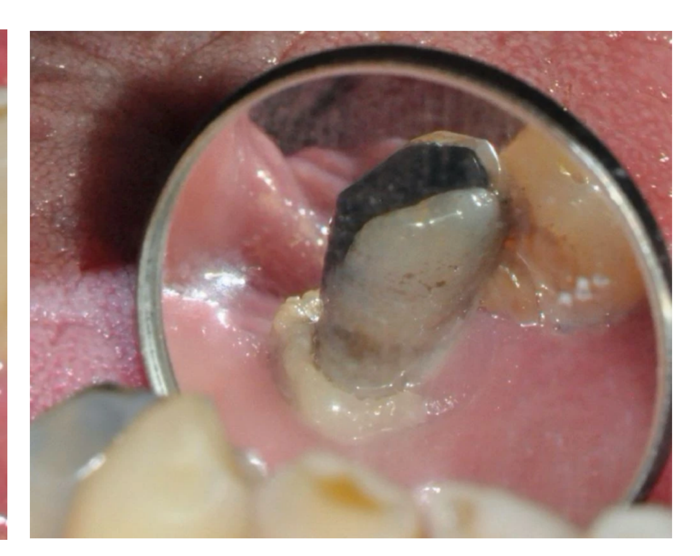
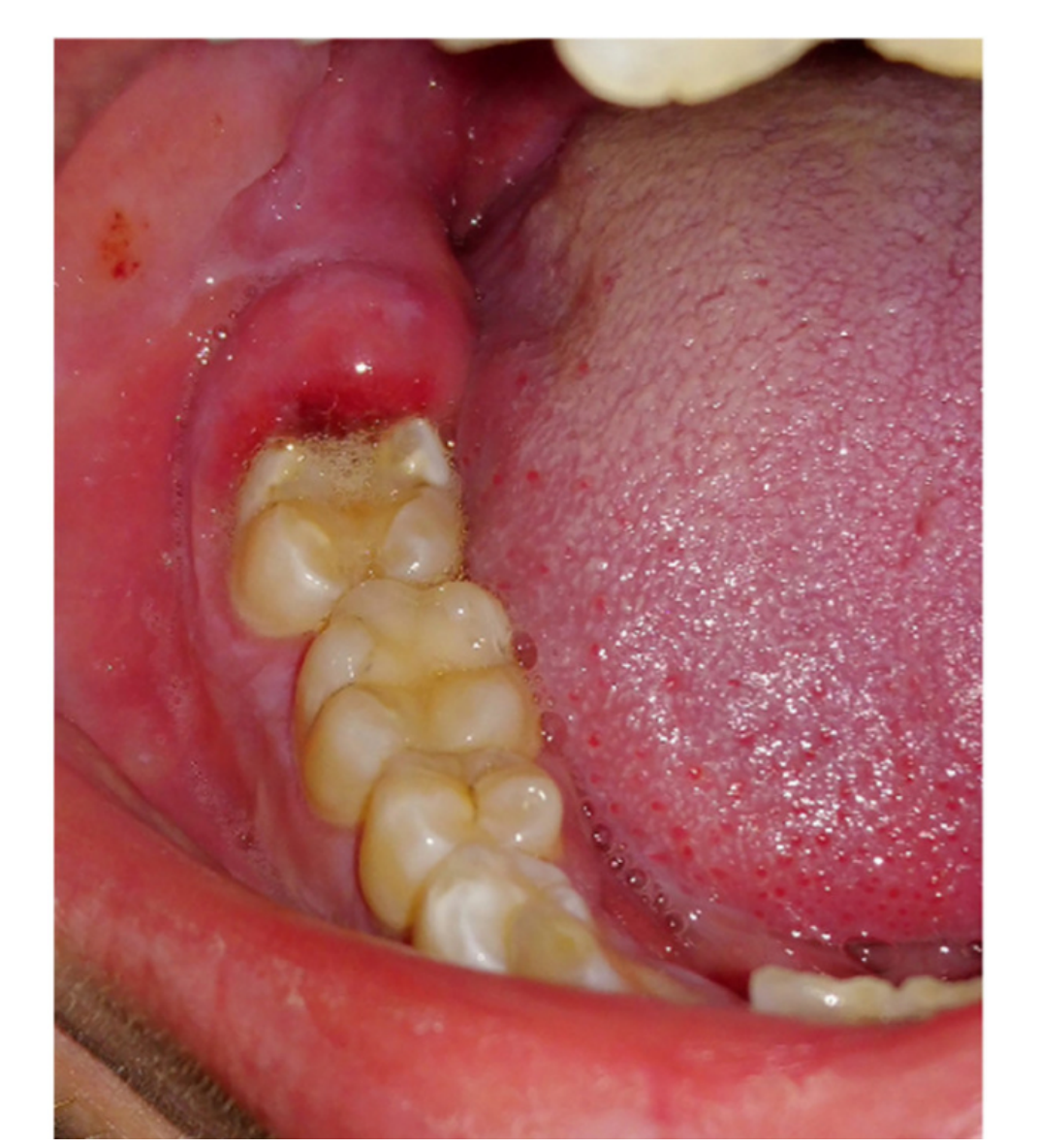
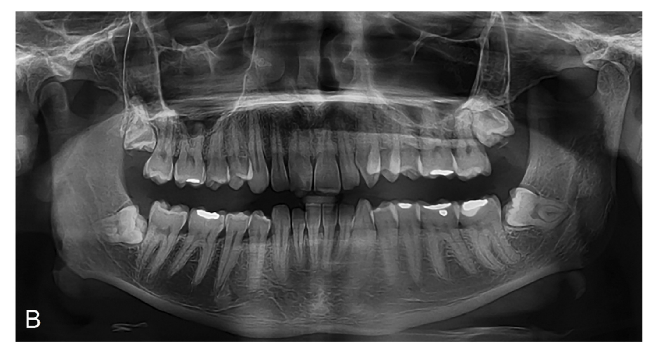
Pericoronitis38394041
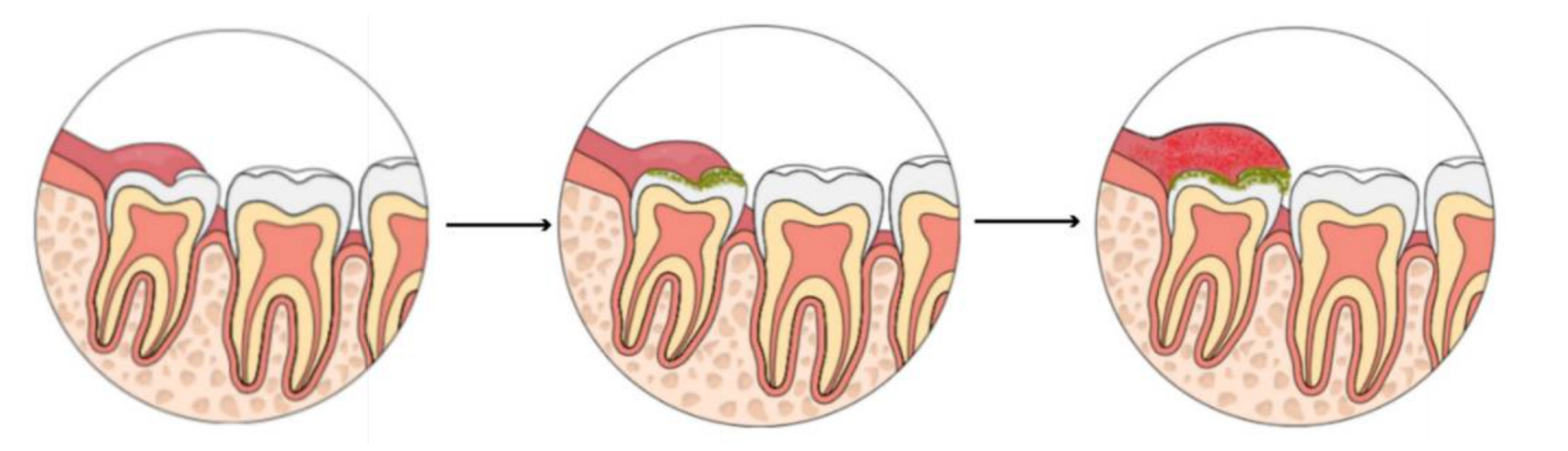
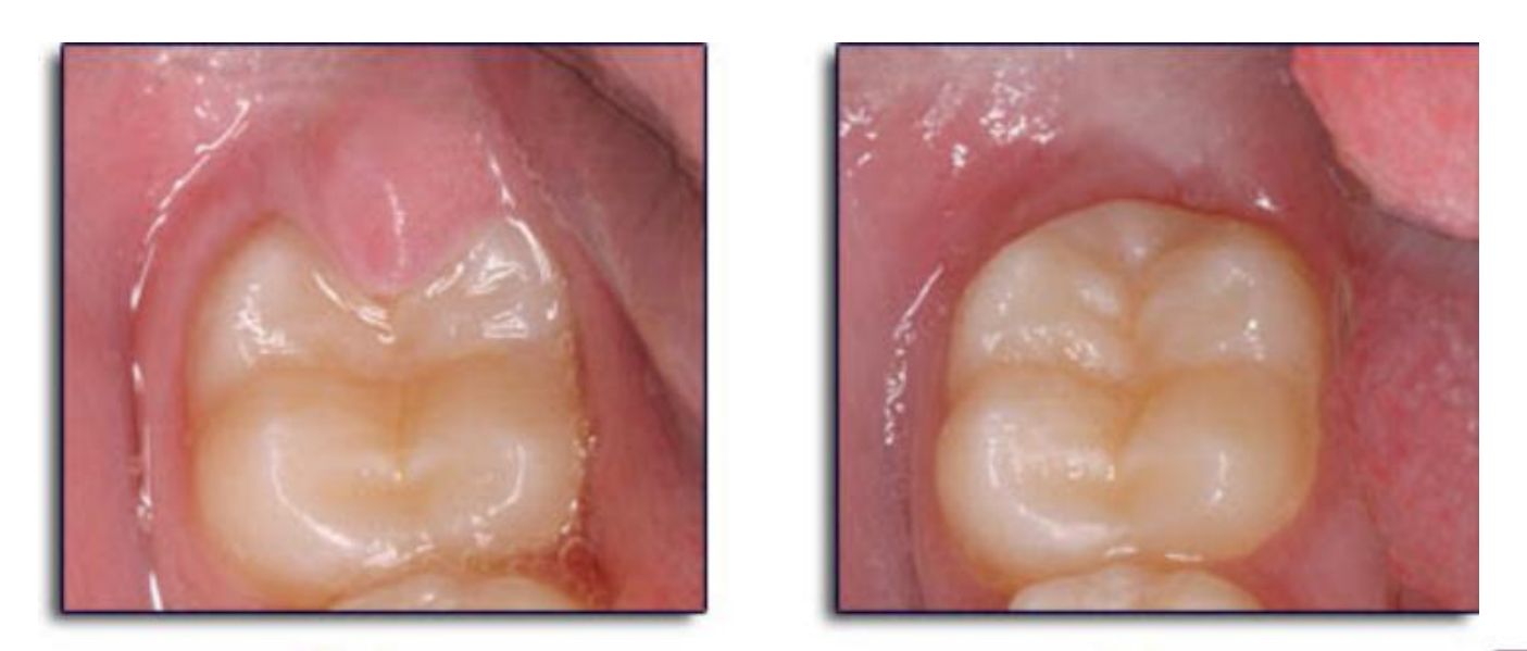
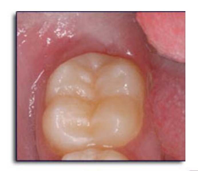
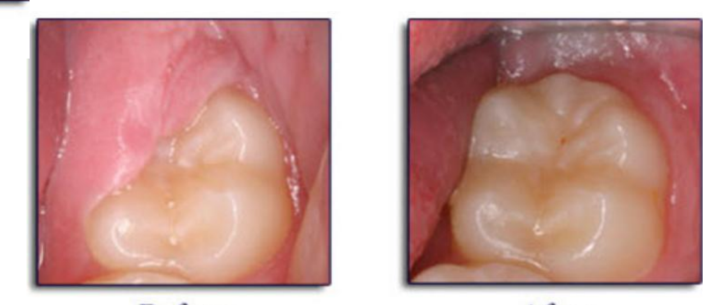
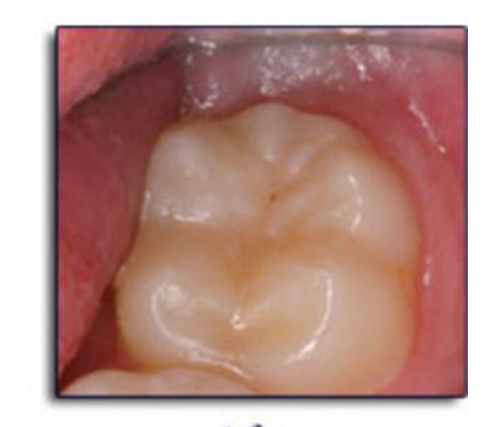
- Easy to diagnose as can see partially erupted tooth with inflamed gum flap
- Gingiva in 3rd molar area appear red & swollen, infection may be due to biofilm, trauma, food debris
- Location: Inflammation/infection of the operculum (gum flap) over a partially erupted tooth
- Danger: Can lead to difficulty swallowing or breathing, which requires immediate hospital referral
- Infection may spread posteriorly into the oropharyngeal area and medially to the base of the tongue and involve the regional lymph nodes, may have difficulty swallowing
- Patients may have a history of pericoronitis
- Gram-negative anaerobic pathogens
 |  |  |
 |  |  |
 |  |  |
 |
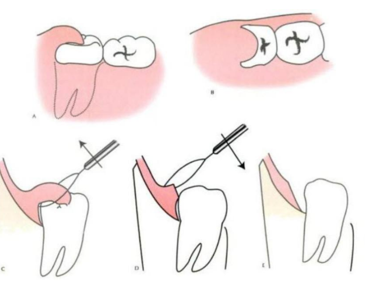
Management42
- Acute phase and initial Treatment:
- debride & irrigate under the pericoronal flap
- Rx. Antibiotics if systemic involvement (fever, lymphadenopathy), warm salt water mouthwash
- Manage underlying condition:
- operculectomy
- distal debridement
- extract third molar(s)
Gingival Abscess4344
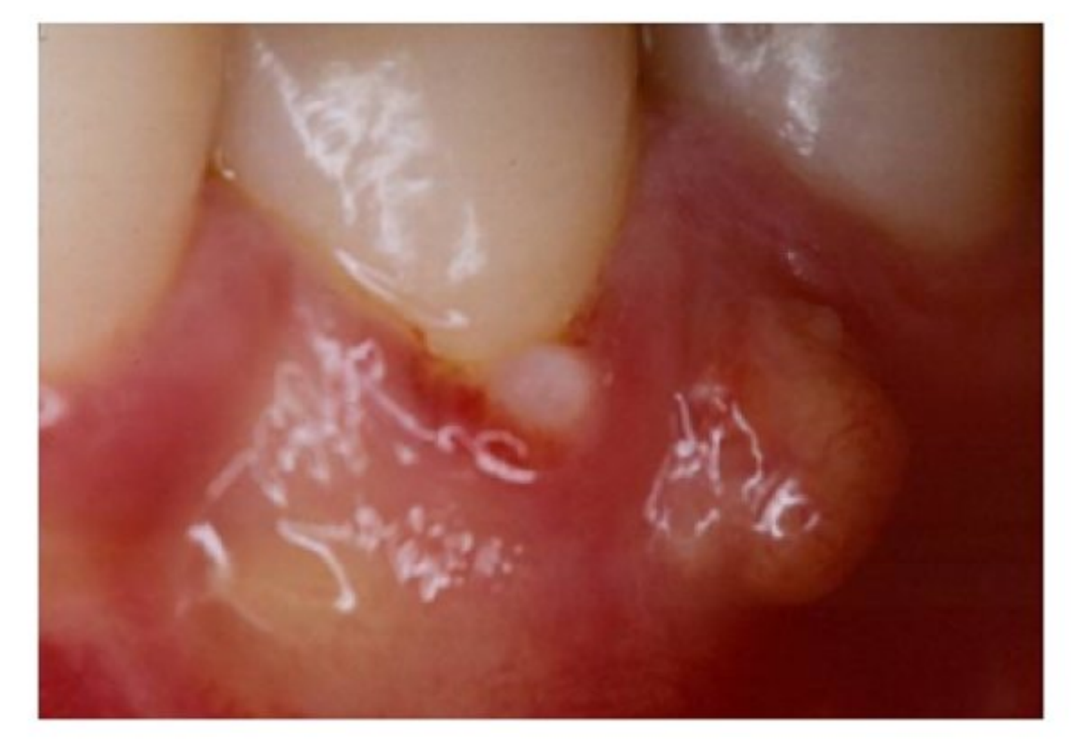
- Localised purulent infection - smooth, red, painful swelling
- Confined to marginal gingival tissue, often at previously non-diseased sites
- Usually an acute inflammatory response (1-2 days) to plaque, trauma or foreign substances forced into the gingiva (Piksters, dental floss, food)
- Etiology: Often caused by a local injury or foreign body, such as a popcorn husk, stuck in the gingiva
- If left untreated, lesion may ruptre spontaneously
- Diagnosis - find & retrieve the offending foreign material if applicable
- Treatment: debride & irrigate pocket
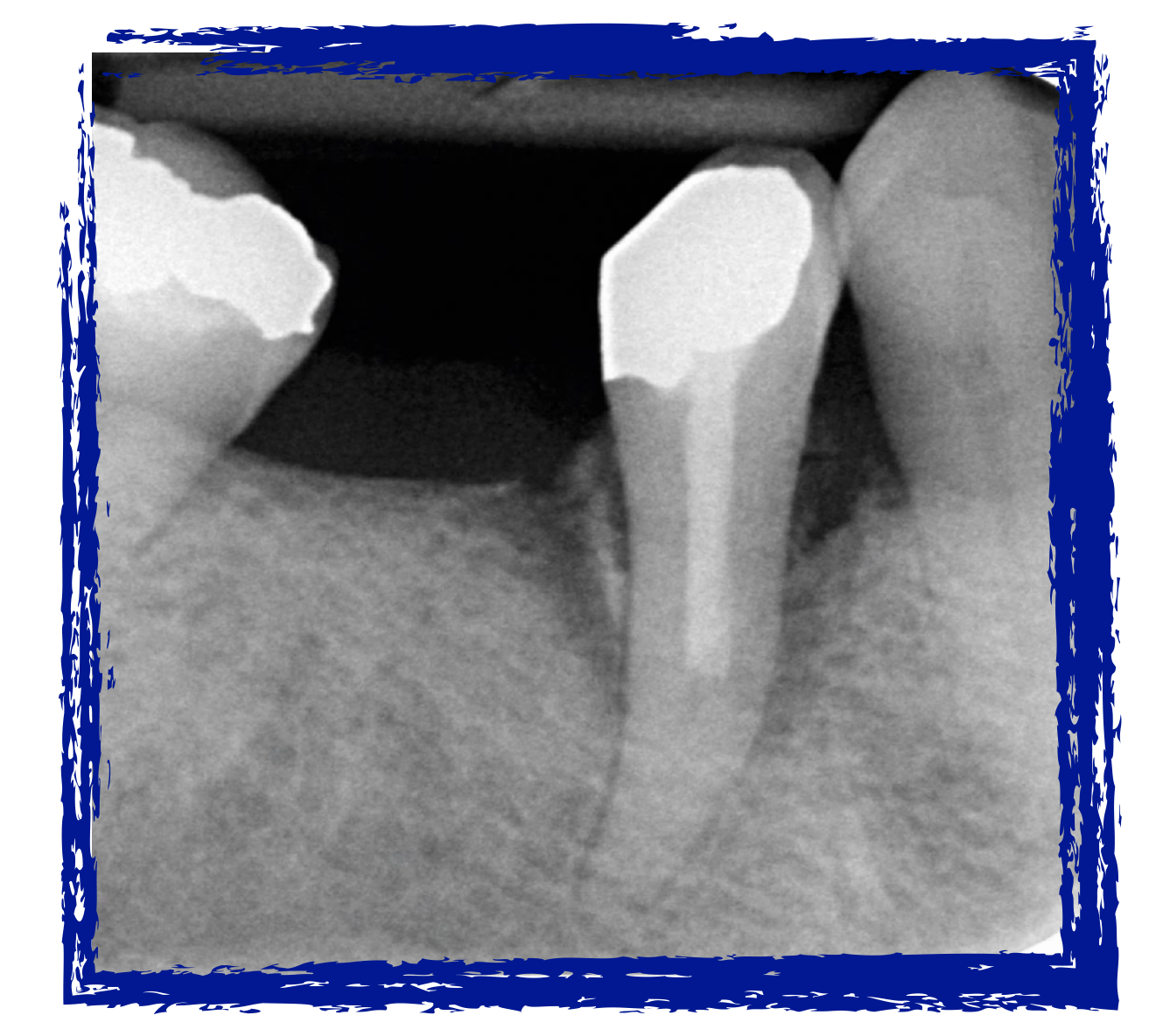
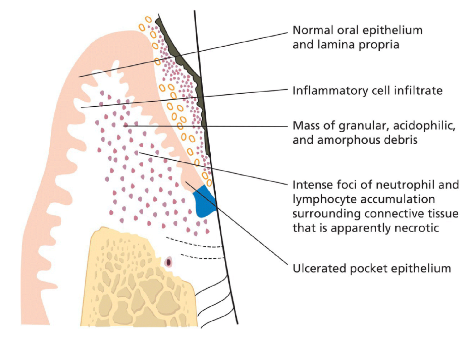
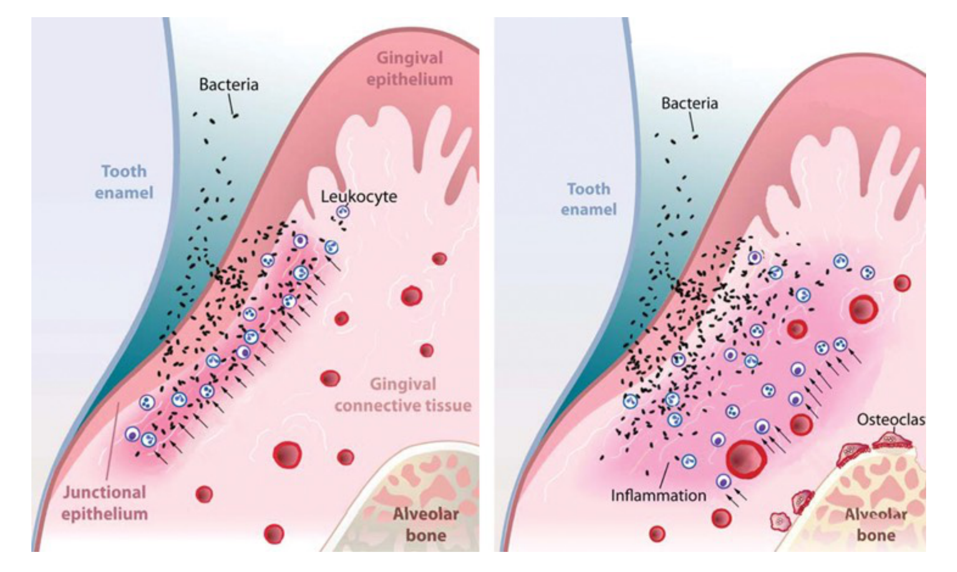
Periodontal Abscess45464748
-
3rd most common emergency in general practice (UK data)
-
8-14% of all dental emergencies
-
More common in patients with untreated gum disease (60%) vs patients undergoing active periodontal treatment (13.5%)
-
Patients in maintenance – 38% (5-29yrs follow up)
-
Usually around molars & lower incisors
-
Rapid destruction of periodontal tissues, with a negative effect on the prognosis of the affected tooth. Bacterial invasion of soft tissues ulceration of the PE occurs.
-
May lead to tooth loss
-
During SPT, main reason for tooth extraction
-
Teeth with repeated abscess formation – prognosis considered “hopeless”
-
Severe systemic consequences can occur
Herrara et al 2018
 |  |  |
 |  |
Aetiology and Microbiology
why?
- Untreated periodontitis
- Poor access/residual calculus - anatomical difficulties in furcations, vertical defects, root concavities
- Non-inflammatory - root fractures/perforations
- Systemic antimicrobial intake, without subgingival debridement
- Poorly controlled diabetics - higher risk
- Triggered by “superficial healing” where the pocket margin tightens after a quick scale, trapping bacteria at the base
Microbial composition similar to periodontitis
Most prevalent bacterial species were Porphyromonas gingivalis (50-100%), Prevotella intermedia, Prevotella melaninogenica, Fusobacterium nucleatum, Tannerella forsythia, Treponema species, Campylobacter species, Capnocytophaga species, Aggregatibacter actinomycetemcomitans or gram-negative enteric rods
Herrara et al 2018
Clinical Presentation and Diagnosis4950
-
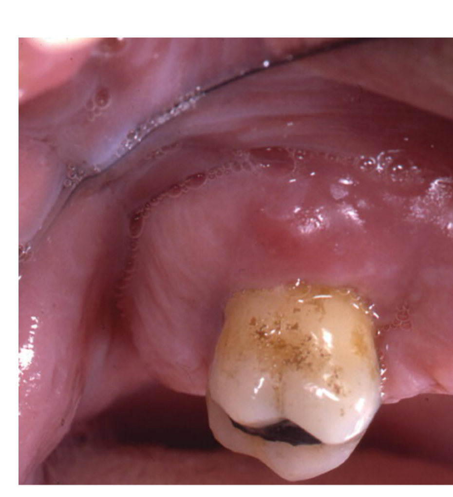
Confined to marginal gingival tissue
-
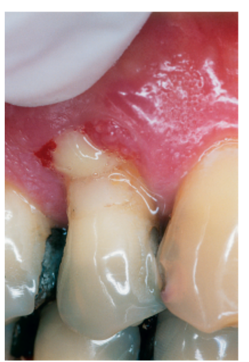
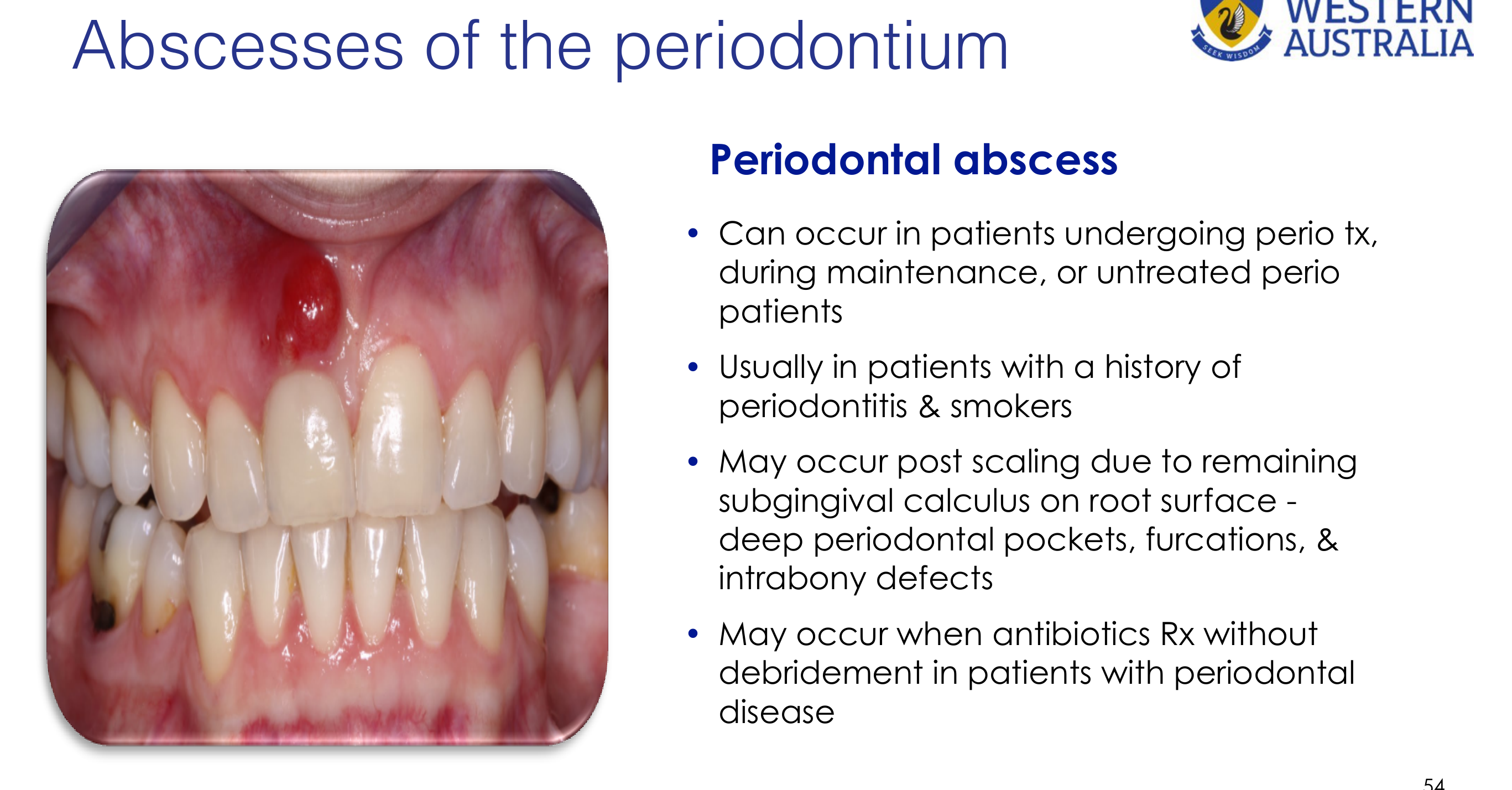
Ovoid swelling in gum
-
Gingiva is oedematous & red, with a smooth, shiny surface
-
Very common - Pus may be expressed from the gingival margin by gentle digital pressure, or by probing
-
Uncommon - Fistula
-
Symptoms vary from a slight discomfort, tender gums, to severe pain & swelling, or tooth “elevation”
-
The tooth may feel “high” or “popped up” in the socket
-
Increased tooth mobility, TTP or soreness with chewing
-
Extra-oral uncommon – swollen face (3.6%)
-
Lymphadenopathy, fever & malaise (7-40%)
Periodontal abscess diagnosis
-
Deep probing depths >5mm (7-9mm on average)
-
Positive response to pulp vitality test
- The tooth is usually vital (responds to cold tests), which differentiates it from an endodontic abscess
-
Radiographs – no apical radiolucency or endodontic lesion, but bone loss observed
-
Can occur in patients undergoing perio tx, during maintenance, or untreated perio patients
-
Usually in patients with a history of periodontitis & smokers
-
May occur post scaling due to remaining subgingival calculus on root surface - deep periodontal pockets, furcations, & intrabony defects
-
May occur when antibiotics Rx without debridement in patients with periodontal disease
 |  |
Management and Complications51
management
- Drain abscess by debriding through the pocket or an incision & removing plaque, calculus, & other irritants
- Occlusal adjustment
- Necessary if the tooth is elevated and causing pain on biting
- CHX mouthwash
- Rx. Analgesics
- Resolution 2-7 days
- Recurring abscess – surgery to access site to find other cause for abscess
management
Rarely needed! Only if spreading locally + mild systemic involvement
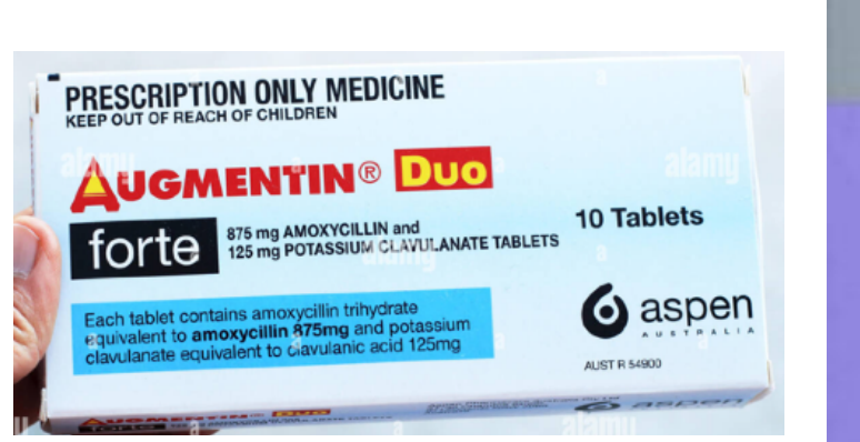
- Phenoxymethylpenicillin 500mg x4 daily for 5 days or Amoxycillin 500mg tds for 5 days (plus metronidazole 400mg bds 5 days) TG
- PENICILLIN ALLERGY: CLINDAMYCIN 300mg tds for 5 days
- If severe systemic + spreading - immediate referral to a hospital!!
without treatment…
- Tooth loss
- Spread of infection to other parts of the body – bacteraemia, lung infection, brain abscess
- Life-threatening systemic spread such as cellulitis
 |  |
Differential Diagnosis and Other Conditions52
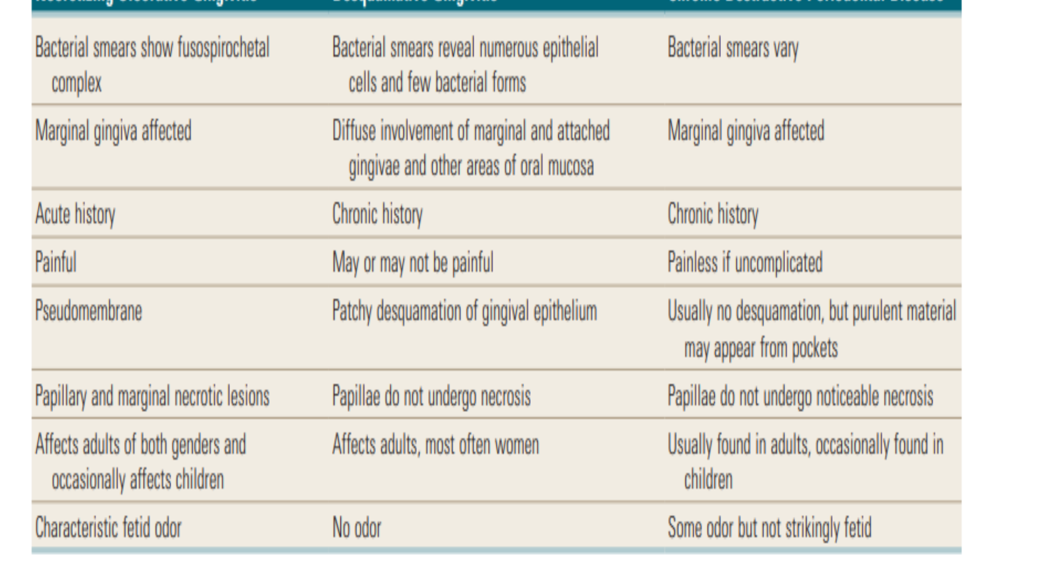
Differentiation of Necrotizing Ulcerative Gingivitis, Chronic Desquamative Gingivitis, and Chronic Periodontal Disease
| Necrotizing Ulcerative Gingivitis | Desquamative Gingivitis | Chronic Destructive Periodontal Disease |
|---|---|---|
| Bacterial smears show fusospirochetal complex | Bacterial smears reveal numerous epithelial cells and few bacterial forms | Bacterial smears vary |
| Marginal gingiva affected | Diffuse involvement of marginal and attached gingivae and other areas of oral mucosa | Marginal gingiva affected |
| Acute history | Chronic history | Chronic history |
| Painful | May or may not be painful | Painless if uncomplicated |
| Pseudomembrane | Patchy desquamation of gingival epithelium | Usually no desquamation, but purulent material may appear from pockets |
| Papillary and marginal necrotic lesions | Papillae do not undergo necrosis | Papillae do not undergo noticeable necrosis |
| Affects adults of both genders and occasionally affects children | Affects adults, most often women | Usually found in adults, occasionally found in children |
| Characteristic fetid odor | No odor | Some odor but not strikingly fetid |
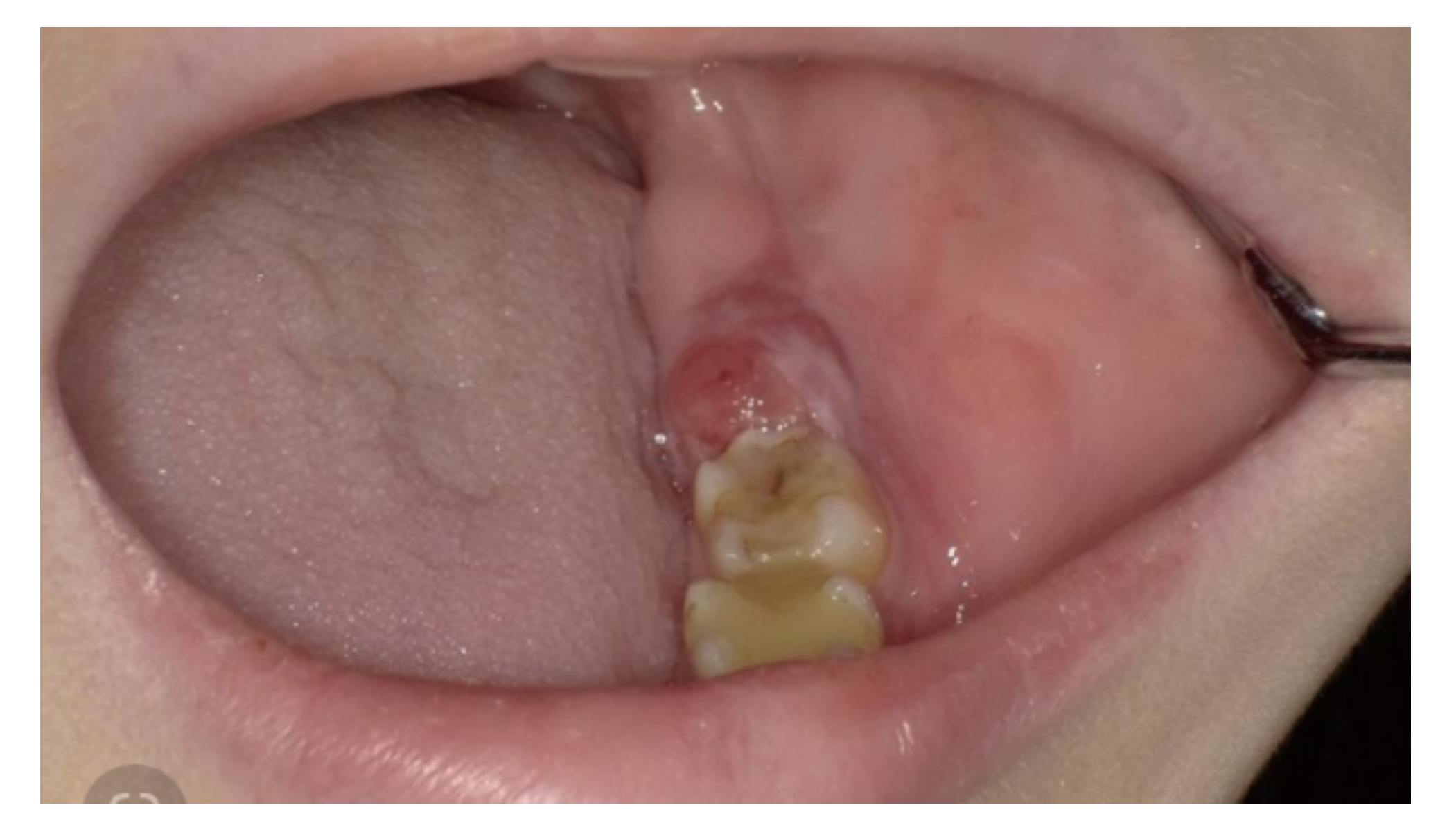
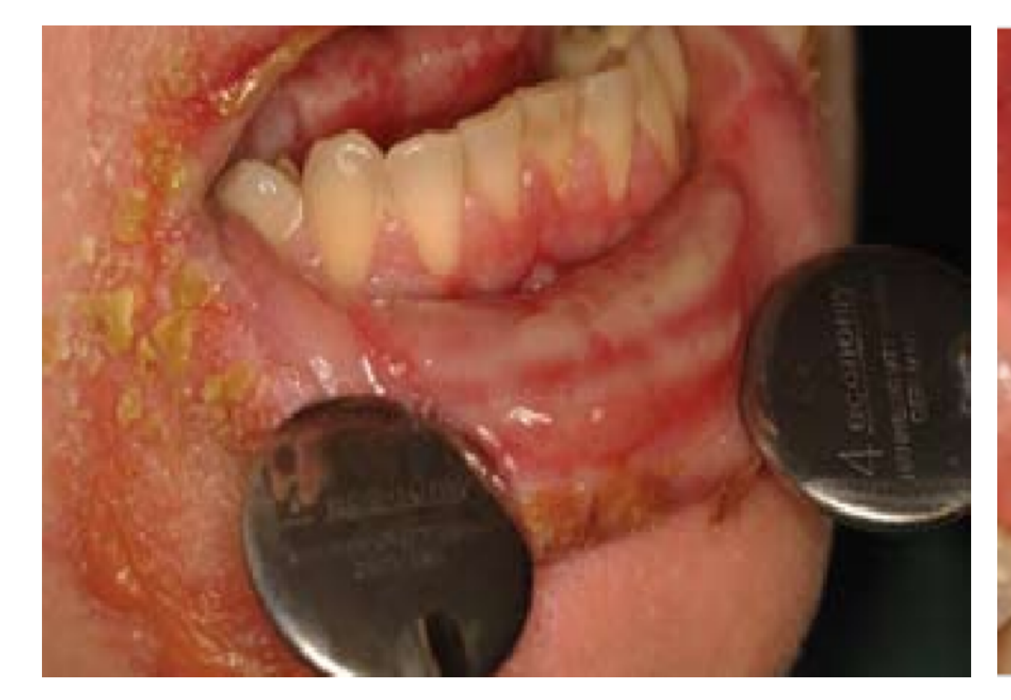
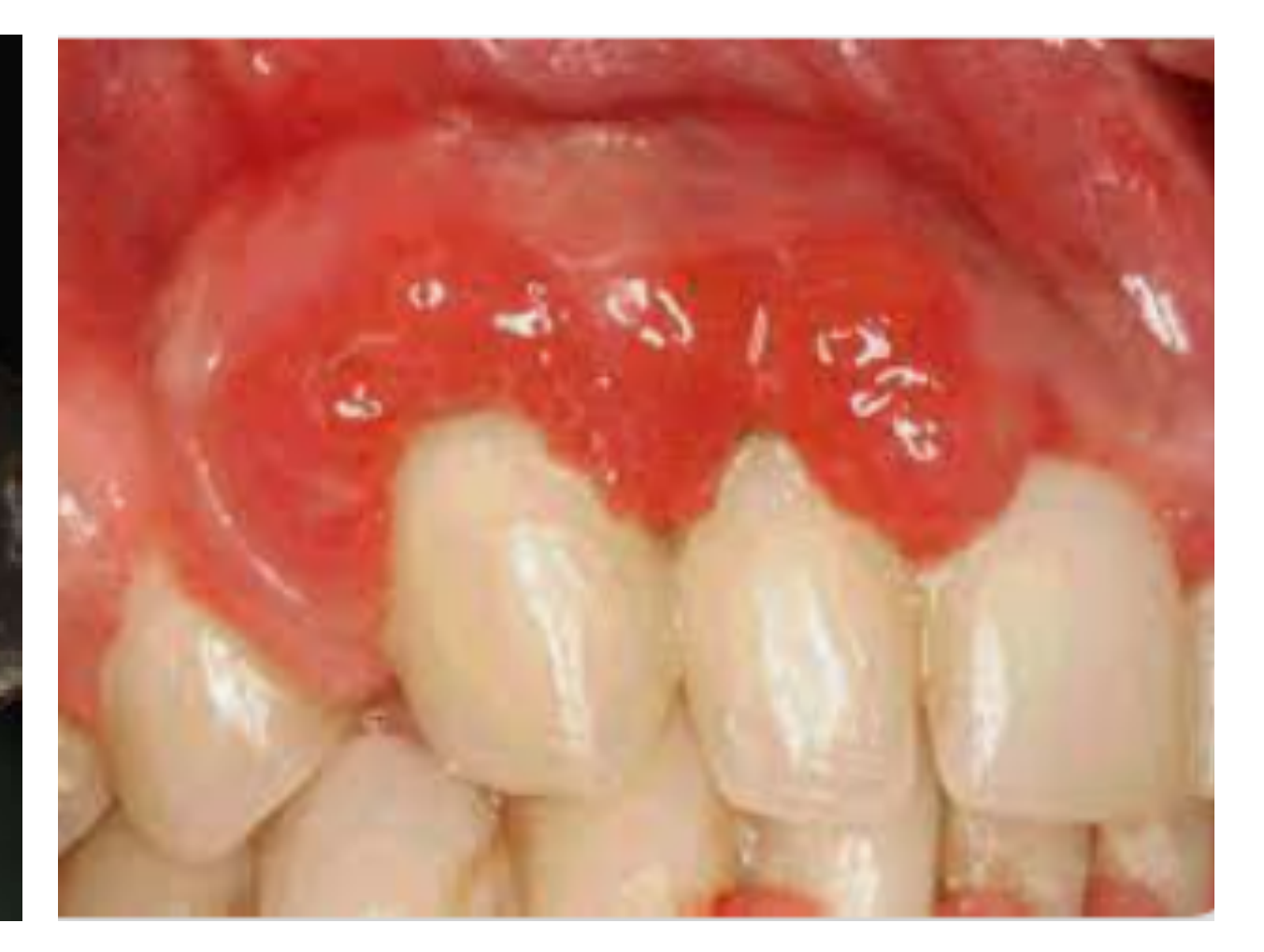
Less Common Acute Conditions53545556
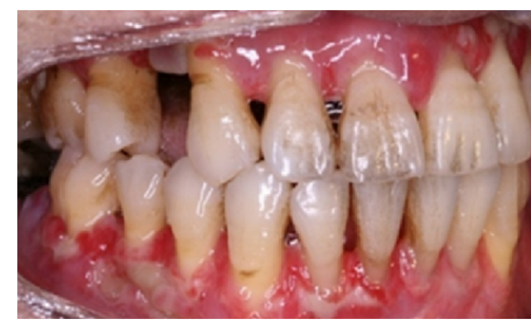
**Acute Candida infection in a cancer patient receiving chemotherapy
- Fungal infection, often seen in immunocompromised patients (e.g., those on chemotherapy).
- Neoplasia: Oral cancers can sometimes mimic the appearance of an abscess or pericoronitis.**
 |  |  |
 |  |  |
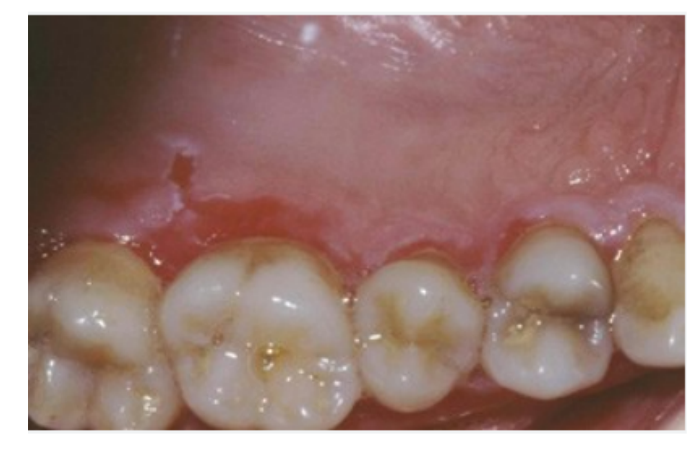
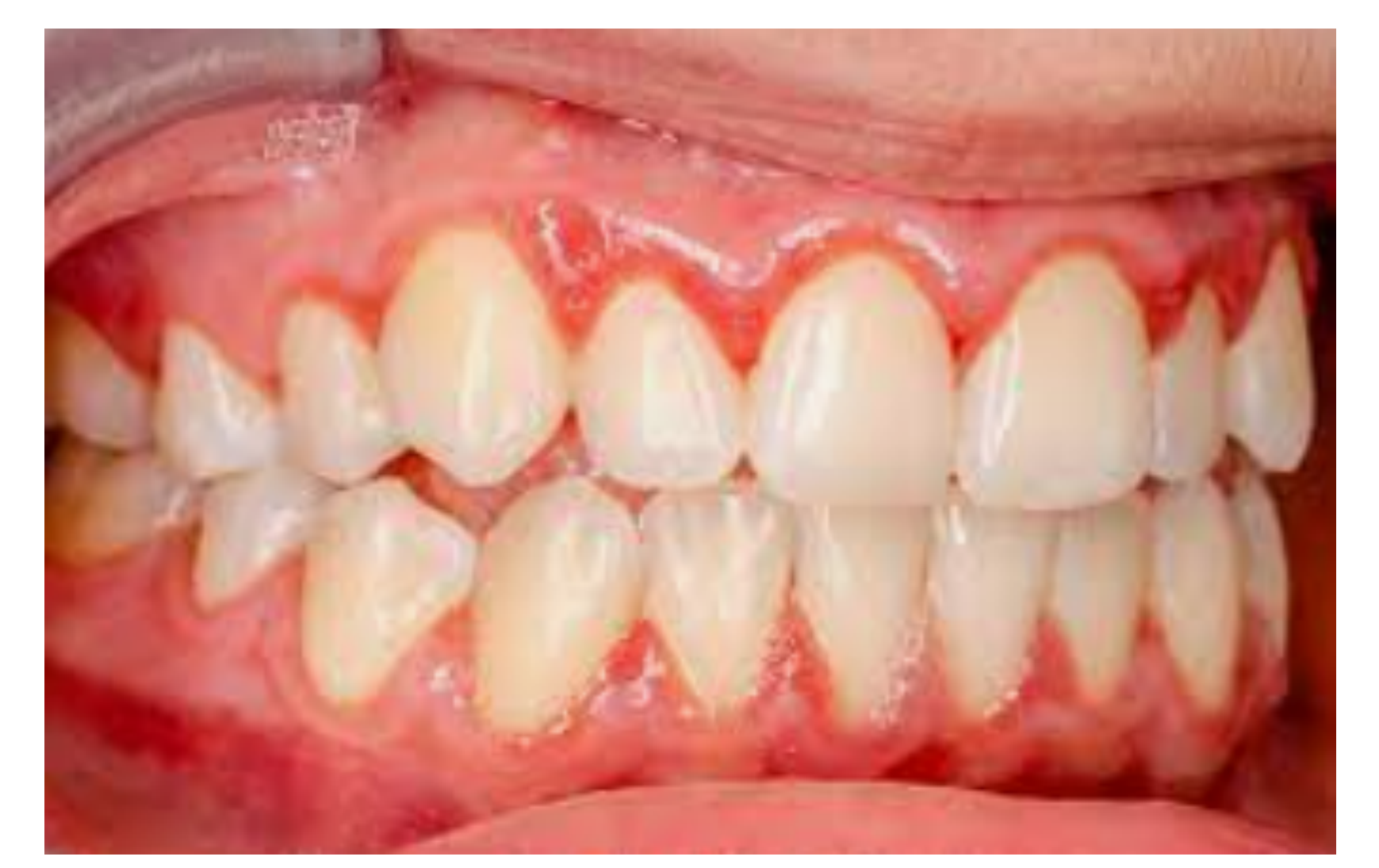
Desquamative gingivitis in a patient with auto-immune disease57
- Characterized by the sloughing off of squamous cells.
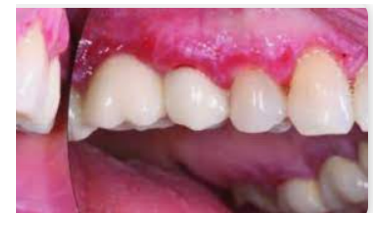
- The gingiva appears severely ulcerated and “nasty,” often linked to systemic autoimmune conditions. Allergic reactions
- Can cause acute gingival swelling.
References and Further Reading5859
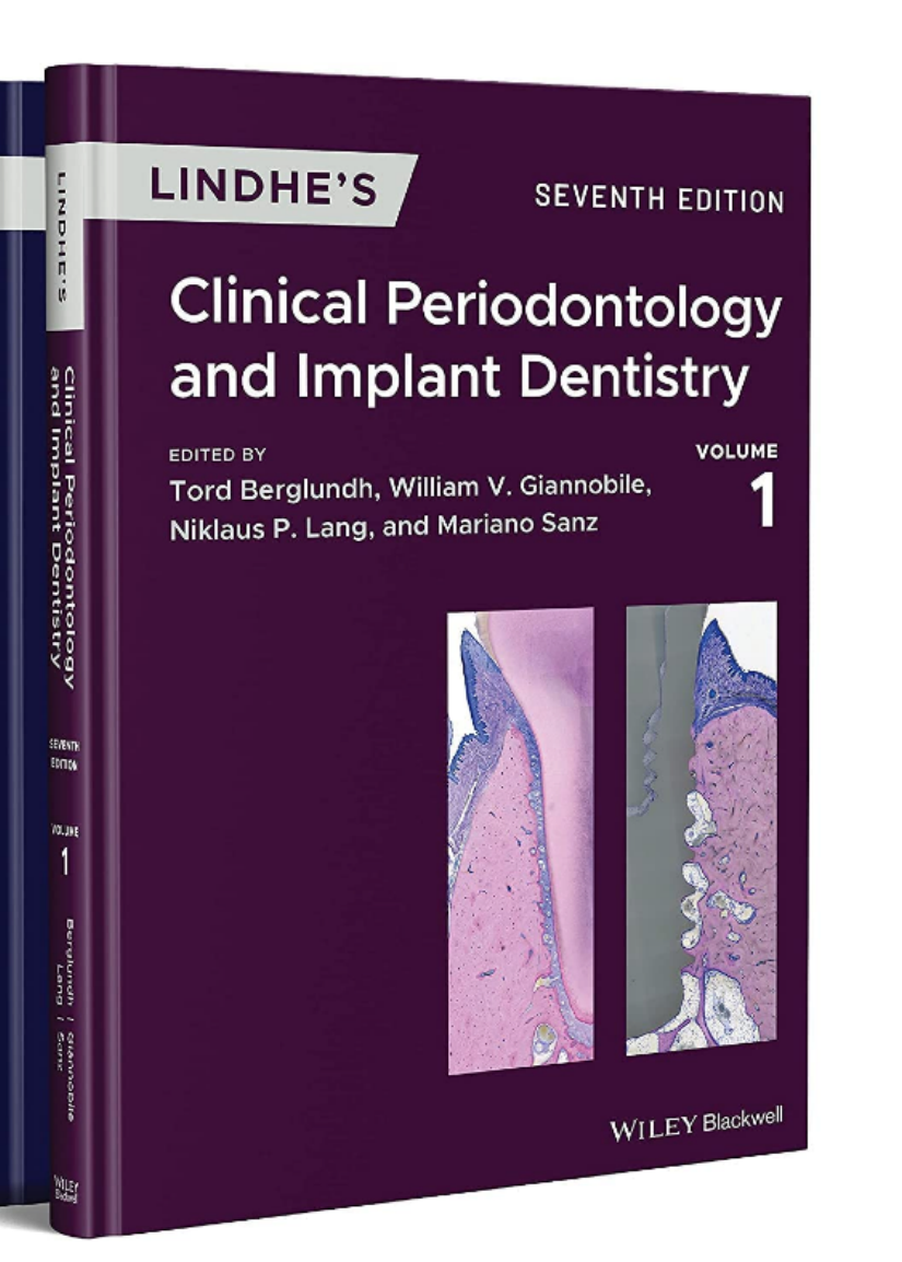
Chapter 14 Non-Plaque-Induced Gingival Diseases Palle Holmstrup¹ and Mats Jontell² ¹ Department of Periodontology, School of Dentistry, University of Copenhagen, Copenhagen, Denmark ² Oral Medicine and Pathology, Institute of Odontology, The Sahlgrenska Academy at University of Gothenburg, Gothenburg, Sweden
Chapter 19 Abscesses, Necrotizing Lesions of the Periodontium, and Endo-Periodontal Lesions David Herrera¹ and Magda Feres² ¹ ETEP (Etiology and Therapy of Periodontal and Peri-Implant Diseases) Research Group, Complutense University of Madrid, Madrid, Spain ² Department of Periodontology, Dental Research Division, Guarulhos University, Guarulhos, São Paulo, Brazil and The Forsyth Institute, Cambridge, MA, USA
The University of Western Australia
Questions?
Images in presentation not otherwise credited are licence-free from https://www.freepik.com/
Recommended Resources
- Therapeutic Guidelines: The primary resource for evidence-based prescribing (accessible on clinic computers).
- eMIMS: Used to check drug formulations and packaging sizes.
- Textbook: Refer to the specific chapter on acute periodontal conditions for further detail.
Audio Appendix
Additional Audio Content
The following sections from the lecture audio did not correspond to any heading in the main document.
Case Study: 18-Month-Old Baby
An 18-month-old baby presented with constant screaming and refusal to eat. The mother had a healing cold sore. The baby was diagnosed with acute herpetic gingivostomatitis after contracting the virus from the mother.
Case Study: Young Male Smoker
The lecturer observed a case of necrotising gingivitis in a young male patient who was “partying a lot” and smoking heavily. The diagnosis was tipped off by the classic “punched-out” appearance of the papilla and the patient’s heavy smoking habit.
 |  |
Footnotes
-
Original PDF page 1: L14AcuteConditions, p.1 ↩
-
Original PDF page 2: L14AcuteConditions, p.2 ↩
-
Original PDF page 3: L14AcuteConditions, p.3 ↩
-
Original PDF page 4: L14AcuteConditions, p.4 ↩
-
Original PDF page 5: L14AcuteConditions, p.5 ↩
-
Original PDF page 6: L14AcuteConditions, p.6 ↩
-
Original PDF page 7: L14AcuteConditions, p.7 ↩
-
Original PDF page 8: L14AcuteConditions, p.8 ↩
-
Original PDF page 9: L14AcuteConditions, p.9 ↩
-
Original PDF page 10: L14AcuteConditions, p.10 ↩
-
Original PDF page 11: L14AcuteConditions, p.11 ↩
-
Original PDF page 12: L14AcuteConditions, p.12 ↩
-
Original PDF page 13: L14AcuteConditions, p.13 ↩
-
Original PDF page 14: L14AcuteConditions, p.14 ↩
-
Original PDF page 15: L14AcuteConditions, p.15 ↩
-
Original PDF page 16: L14AcuteConditions, p.16 ↩
-
Original PDF page 17: L14AcuteConditions, p.17 ↩
-
Original PDF page 18: L14AcuteConditions, p.18 ↩
-
Original PDF page 19: L14AcuteConditions, p.19 ↩
-
Original PDF page 20: L14AcuteConditions, p.20 ↩
-
Original PDF page 21: L14AcuteConditions, p.21 ↩
-
Original PDF page 22: L14AcuteConditions, p.22 ↩
-
Original PDF page 23: L14AcuteConditions, p.23 ↩
-
Original PDF page 24: L14AcuteConditions, p.24 ↩
-
Original PDF page 25: L14AcuteConditions, p.25 ↩
-
Original PDF page 26: L14AcuteConditions, p.26 ↩
-
Original PDF page 27: L14AcuteConditions, p.27 ↩
-
Original PDF page 28: L14AcuteConditions, p.28 ↩
-
Original PDF page 29: L14AcuteConditions, p.29 ↩
-
Original PDF page 30: L14AcuteConditions, p.30 ↩
-
Original PDF page 31: L14AcuteConditions, p.31 ↩
-
Original PDF page 32: L14AcuteConditions, p.32 ↩
-
Original PDF page 33: L14AcuteConditions, p.33 ↩
-
Original PDF page 34: L14AcuteConditions, p.34 ↩
-
Original PDF page 35: L14AcuteConditions, p.35 ↩
-
Original PDF page 36: L14AcuteConditions, p.36 ↩
-
Original PDF page 37: L14AcuteConditions, p.37 ↩
-
Original PDF page 38: L14AcuteConditions, p.38 ↩
-
Original PDF page 39: L14AcuteConditions, p.39 ↩
-
Original PDF page 40: L14AcuteConditions, p.40 ↩
-
Original PDF page 42: L14AcuteConditions, p.42 ↩
-
Original PDF page 41: L14AcuteConditions, p.41 ↩
-
Original PDF page 43: L14AcuteConditions, p.43 ↩
-
Original PDF page 44: L14AcuteConditions, p.44 ↩
-
Original PDF page 45: L14AcuteConditions, p.45 ↩
-
Original PDF page 46: L14AcuteConditions, p.46 ↩
-
Original PDF page 47: L14AcuteConditions, p.47 ↩
-
Original PDF page 48: L14AcuteConditions, p.48 ↩
-
Original PDF page 52: L14AcuteConditions, p.52 ↩
-
Original PDF page 54: L14AcuteConditions, p.54 ↩
-
Original PDF page 56: L14AcuteConditions, p.56 ↩
-
Original PDF page 58: L14AcuteConditions, p.58 ↩
-
Original PDF page 59: L14AcuteConditions, p.59 ↩
-
Original PDF page 61: L14AcuteConditions, p.61 ↩
-
Original PDF page 62: L14AcuteConditions, p.62 ↩
-
Original PDF page 63: L14AcuteConditions, p.63 ↩
-
Original PDF page 60: L14AcuteConditions, p.60 ↩
-
Original PDF page 64: L14AcuteConditions, p.64 ↩
-
Original PDF page 65: L14AcuteConditions, p.65 ↩