Etiopathogenesis Of Periodontal Disease1
Dr. Pradeep Koppolu BDS, MDS (Perio), PhD (Malaysia), FICOI, FPFA, PDCR Discipline Lead & Program Convenor Periodontics and Implantology
Introduction To The Human Microbiota2
-
Is Human foetus inside the uterus sterile?
-
Within 2 weeks, a nearly mature microbiota is established in the gut of the newborn baby.
-
After weaning (>2 years), the entire human microbiota is formed and comprises a very complex collection of hundreds of different types of bacteria with approximately 10¹⁴ microbial cells.
-
From this moment on, our body contains 1.3 to 10 times more bacteria than human cells.
-
It has been estimated that, for a normal, healthy human being, the bacterial population comprises 2 kg of the total body weight. This is fascinating if one realizes that the average human brain weighs only about 1.4 kg.
Development Of The Oral Microbiome
Introduction3
Microbial Colonization In Infants
- Within hours after birth sterile oral cavity is colonized by numbers of mainly facultative and aerobic bacteria.
- From 2nd day, anaerobic bacteria can be detected.
- First and most dominant oral microbes in the oral cavity of newborn infants: Streptococcus salivarius and Streptococcus mitis.
- Veillonella spp., Neisseria spp., Actinomyces spp. and Staphylococcus spp. are also among the first colonizers of the oral cavity.
Adult Oral Microbiome And Dysbiosis4
-
It is estimated that the oral bacterial microbiome of adults encompasses approximately 750 commonly occurring species, roughly half of which can be present at any time in any individual.
-
A shift in microbial community leads to an imbalance, or dysbiosis, in the microbiota.
-
This shift in the microbial community can be largely independent of the acquisition of new members of the microbiota and instead reflects changes in the abundance of either individual organisms or consortia of organism’s resident within the subgingival biofilm (previously referred to as plaque).
Ecological Determinants Of Oral Microbiota
Host Factors Influencing Oral Microbiota5
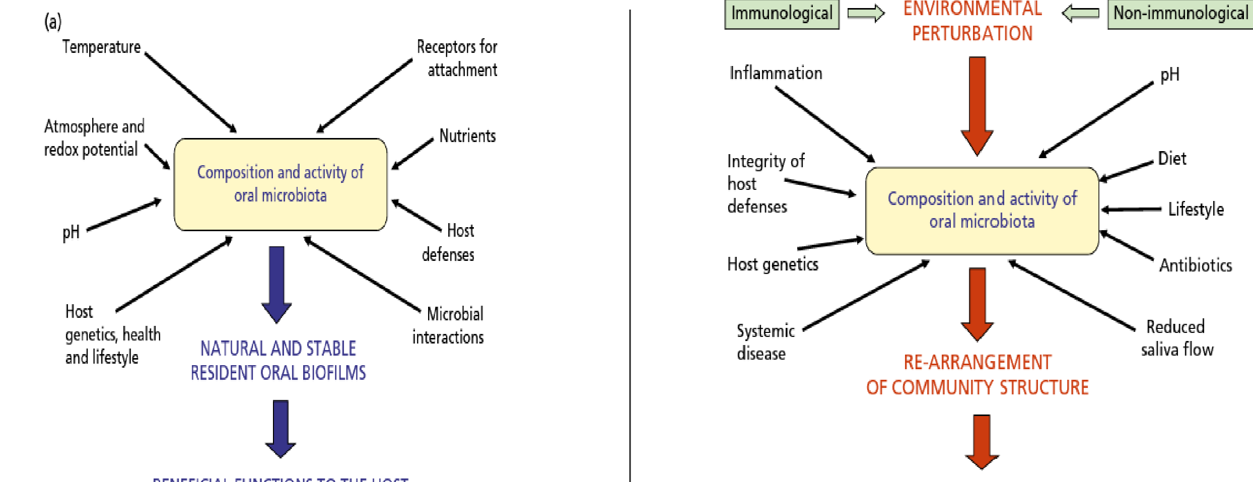
Temperature Atmosphere redox potential pH Host genetics, health and lifestyle Receptors for attachment Nutrients Host defenses Microbial interactions
Composition and activity of oral microbiota
NATURAL AND STABLE RESIDENT ORAL BIOFILMS
BENEFICIAL FUNCTIONS TO THE HOST
(b)
Environmental Factors And Host Interactions67
Immunological ENVIRONMENTAL PERTURBATION Non-immunological
Inflammation Integrity of host defenses Host genetics Systemic disease pH Diet Lifestyle Antibiotics Reduced saliva flow
Composition and activity of oral microbiota
RE-ARRANGEMENT OF COMMUNITY STRUCTURE
INCREASED RISK OF DISEASE
Fig. 8-1 Host factors that influence the microbial composition, activity and stability of the resident oral microbiota. (a) A number of host factors help to determine the composition and activity of the natural and beneficial oral microbiota. (b) A perturbation in a key environmental factor can disrupt the natural stability (microbial homeostasis) of the resident microbiota at a site and result in a re-arrangement of the composition and activity of the resident microbial community; such a change might predispose the site to disease. (Source: Adapted from Marsh et al. 2011.)
From an ecologic viewpoint, the oral cavity, which communicates with the pharynx, should be considered an “open growth system” with an uninterrupted ingestion and removal of microorganisms and their nutrients.
Any microorganisms that are unable to adhere to a surface within the mouth are washed away in the flow of saliva and swallowed.
The composition and diversity of the microbial community is therefore impacted by physical gradients of temperature, humidity, nutrient content, salivary flow, oxygen tension and pH, as well as shear forces due to mastication.
Frequent exposure to dietary sugars can significantly affect the dynamics of bacterial adhesion and accumulation.
 |  |  |
 |  |  |
Saliva As A Transport And Nutrient Medium8
- The constant lubrication of the oral cavity by saliva allows microbes to disperse and reach distant sites within the oral cavity, with saliva effectively acting as a transport medium.
- Although saliva has antimicrobial properties, it has also been reported to promote growth of a health-associated microbiota.
- The primary source of nutrients for the oral microbiome is provided by the host, and include the proteins and glycoproteins present in saliva and gingival crevicular fluid (GCF).
- The mouth is maintained at a temperature of around 35–37 °C, which is suitable for the growth of a broad range of microbes, though temperature does increase at subgingival sites during inflammation, which can favour the growth and metabolism of some putative periodontal pathogens.
Role Of pH In Bacterial Distribution9
- pH is a major determinant of bacterial distribution and metabolism in the mouth.
- The buffering activity of saliva plays a major role in maintaining the intraoral pH at around neutrality, which favours the growth of the resident oral microbiome.
- The pH in dental biofilms falls rapidly to below pH 5.0 following the intake of
The oral microbiome
Sedghi L, DiMassa V, Harrington A, Lynch SV, Kapila YL. The oral microbiome: Role of key organisms and complex networks in oral health and disease. Periodontol 2000. 2021;87:107–131
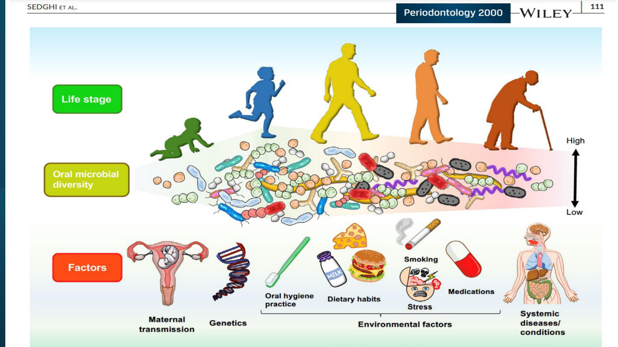
Life stage
Oral microbial diversity
Factors
Maternal transmission
Genetics
Oral hygiene practice
Dietary habits
Smoking
Environmental factors
Stress
Medications
Systemic diseases/conditions
Periodontology 2000
SEDGHI ET AL.
WILEY
111
High
Low
FIGURE 1 The Oral Microbiome: From First Encounters to Lifelong Encounters. Prenatal. The prenatal oral cavity is thought to be sterile until birth, with colonization occurring soon after delivery. The composition of the oral microbiota in infants has been shown to correlate with mode of delivery. However, infants share an oral microbiota similar to that of their mothers, suggesting that the infant oral microbiota may derive from hematogenous or intrauterine transmission from the mother. Detection of oral microbes of maternal origin among several intrauterine locations, as well as associations with adverse pregnancy outcomes, demonstrate the role of the maternal oral microbiome in prenatal health and suggests in utero colonization. Early life. Microbial colonization begins shortly following birth through vertical transmission from the mother, transmission from the diet, and transmission from infant-to-human interactions. Microbial diversity increases upon eruption of primary teeth as this process permits the expansion of microbial niches in the oral cavity. Eruption of primary teeth also results in deviation from the maternal oral microbiota. As children age, their oral microbiotas begin to stabilize. Adult life. The oral microbiota continues to be shaped throughout life by genetic and environmental factors. Environmental factors that influence the composition and function of the oral microbiome include diet, stress, oral hygiene practices, drinking alcohol, and smoking. Genetic factors are linked to conserved phylogenetic and functional microbial signatures related to development of dental caries and heritable predisposition to periodontal disease. Aging and systemic disease. Oral microbiome diversity has been shown to decrease with age. The phylogeny and functional signatures of the oral microbiome are
Microbial Distribution Across Oral Sites10
The Oral Cavity From a Microbe’s Perspective
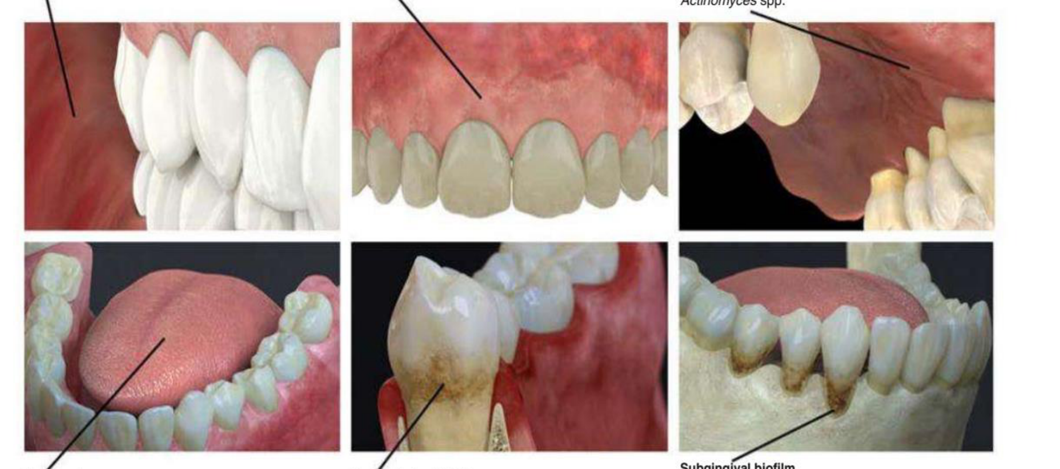
Buccal mucosa Streptococcus spp., Haemophilus spp., Gemella spp.
Gingiva Streptococcus spp. (S. mitis)
Palate Streptococcus spp., Veillonella spp., Prevotella spp., Actinomyces spp.
Tongue dorsum Streptococcus spp. (S. salivarius, S. parasanguinis), Veillonella spp. (V. parvula), Prevotella spp. (P. intermedia), Actinomyces, Granulicatella adiacens
Supragingival biofilm Streptococcus spp. (S. mitis, S. sanguinis, S. gordonii, S. oralis, S. constellatus), Actinomyces spp., Capnocytophaga spp., Corynebacterium spp.
Subgingival biofilm Streptococcus spp., Peptostreptococcus spp., Fusobacterium spp., Capnocytophaga spp., Prevotella spp., Actinomyces spp., Corynebacterium spp., Treponema spp., Porphyromonas spp., Aggregatibacter actinomycetemcomitans
Teeth And Implants As Non-Shedding Surfaces1112
From a microbiological viewpoint, teeth and implants are unique for two reasons:
(1) They provide a hard, non-shedding surface that allows for the development of extensive structured bacterial deposits
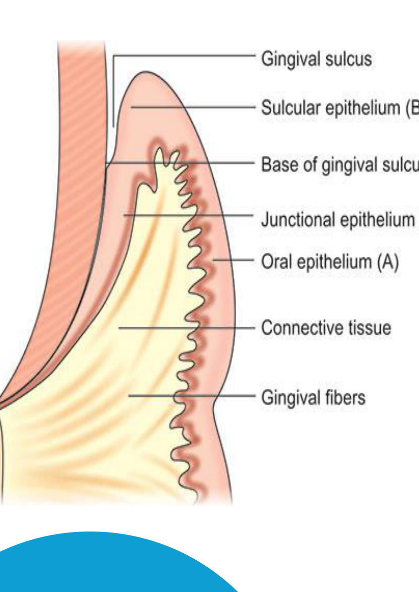
(2) They form a unique ectodermal interruption. A special seal of epithelium (junctional epithelium) and connective tissue is present between the external environment and the internal parts of the body.
-
The accumulation and metabolism of bacteria on these hard surfaces are considered the primary causes of caries, gingivitis, periodontitis, peri-implantitis, and, sometimes, bad breath.
-
In periodontitis patients, periodontal pockets form, creating a subgingival niche that increases the root surface area for microbial colonization, resulting in a subgingival microbial biofilm.
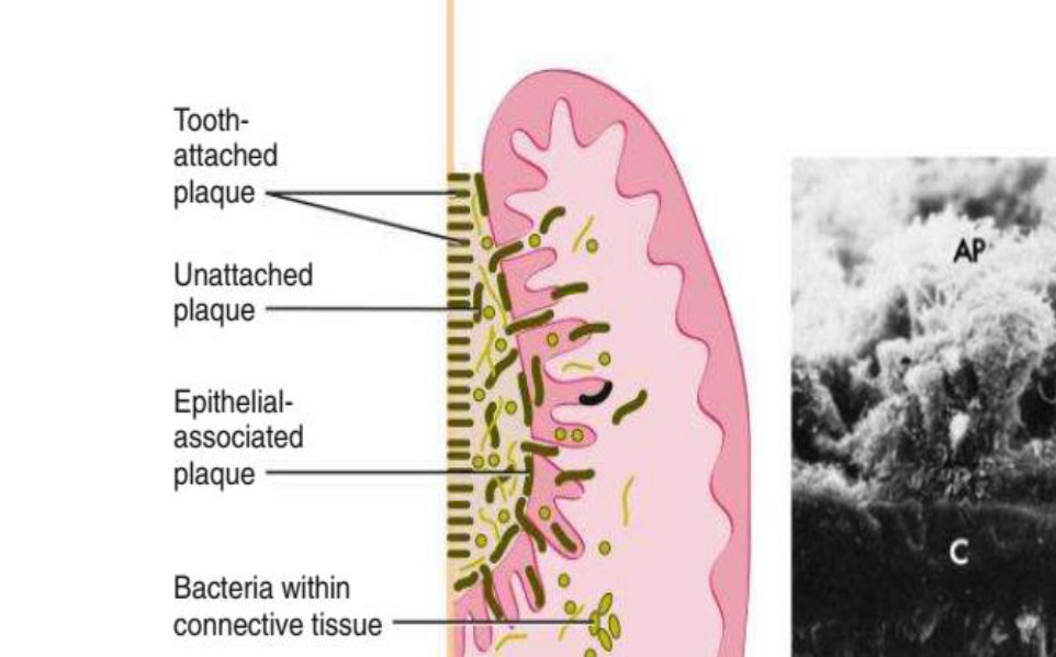
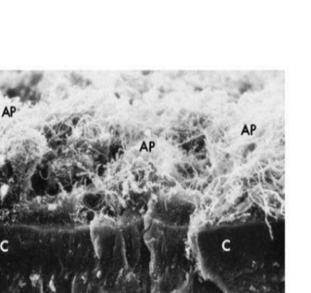
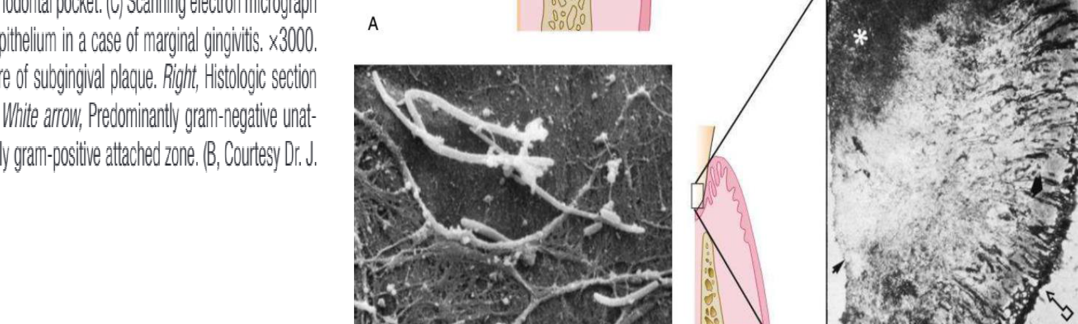
Fig. 10.2 Subgingival plaque. (A) Diagram depicting the plaque–bacteria association between the tooth surface and the periodontal tissues. (B) Scanning electron photomicrograph of a cross-section of cementum (C) with attached subgingival plaque (AP). The area shown is within a periodontal pocket. (C) Scanning electron micrograph of cocci and filaments associated with the surface of pocket epithelium in a case of marginal gingivitis. x3000. (D) Left, Diagrammatic representation of the histologic structure of subgingival plaque. Right, Histologic section of subgingival plaque. Arrow with box, Sulcular epithelium. White arrow, Predominantly gram-negative unattached zone. Black arrow, Tooth surface. Asterisk, Predominantly gram-positive attached zone. (B, Courtesy Dr. J. Sottosanti, La Jolla, California.)
 |  |  |
 |  |
Site-Specific Microbial Composition1314
• Bacterial species identified in all sites investigated by Aas and coworkers belonged to the genera Gemella, Granulicatella, Streptococcus, and Veillonella.
• The most commonly found species in all subjects and all intra-oral sites investigated was S. mitis, which is considered a pioneer species, or primary colonizer.
• Streptococci are thought to be universally present in all sites within the mouth, but the distribution is thought to vary in different ecological niches of the oral cavity.
• It has been suggested that teeth are the primary habitat for periodontal pathogens because soon after a full-mouth tooth extraction in patients with severe periodontitis, key pathogens such as Aggregatibacter actinomycetemcomitans and P. gingivalis disappeared from the oral cavity, as determined by bacterial culturing techniques.
• Prevotella intermedia and other black-pigmented Prevotella spp. remained, but at lower detection frequencies and numbers.
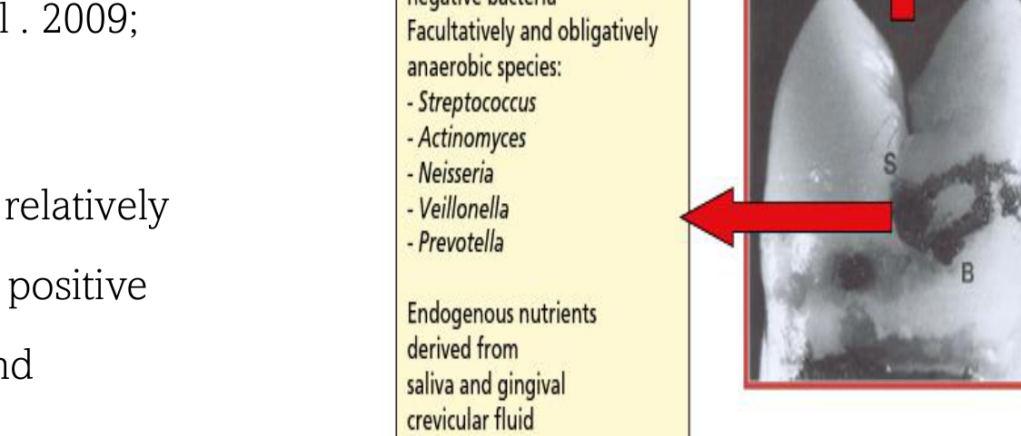
The development and composition of the oral microbiome The mother is the main source of the oral microbiome in the newborn baby. The microbial composition of dental biofilms varies at distinct sites on a tooth (fissures, approximal surfaces, gingival crevice) due to inherent differences in their anatomy and biology (Papaioannou et al . 2009; Marsh et al . 2016b) Fissures are influenced by saliva and the diet and support a relatively sparse microbiota consisting of mainly saccharolytic Gram- positive bacteria, such as streptococci, while obligately anaerobic, and especially Gram- negative species, are rarely recovered.
| Dental X-ray showing a tooth from 3 views with descriptions of bacterial composition and environments at the fissure, approximal, and gingival crevice locations. |
Dental Biofilms
Structure And Architecture Of Biofilms15
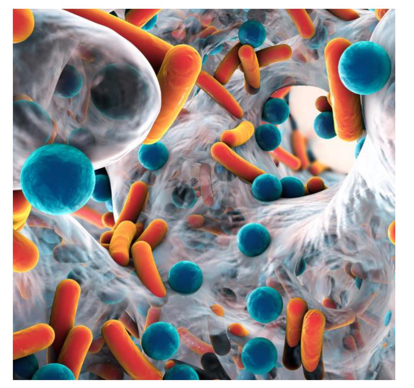
- Microbial populations on the surfaces of teeth are excellent examples of biofilm communities.
- The architecture of a dental biofilm has many features in common with other biofilms.
- It is heterogeneous in structure, with clear evidence of open fluid- filled channels running through the biofilm mass.
- Nutrients reach the sessile (attached) microcolonies by diffusion through water channels to microbial microcolonies.
Phases Of Biofilm Formation16
-
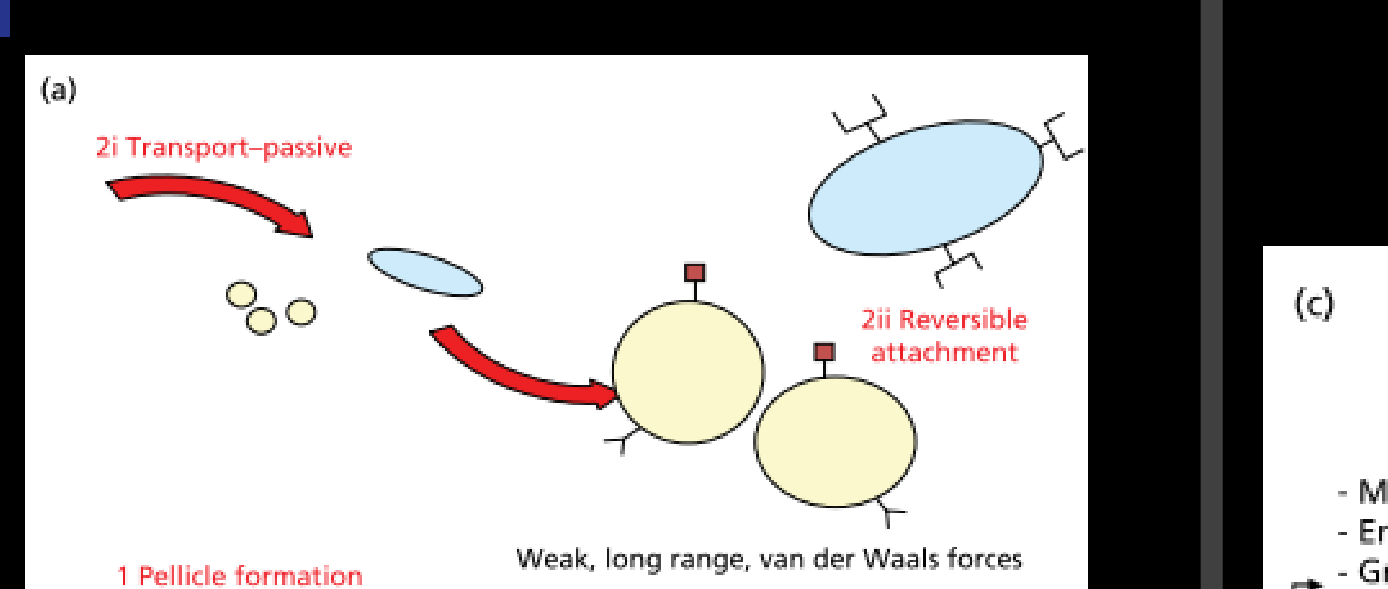
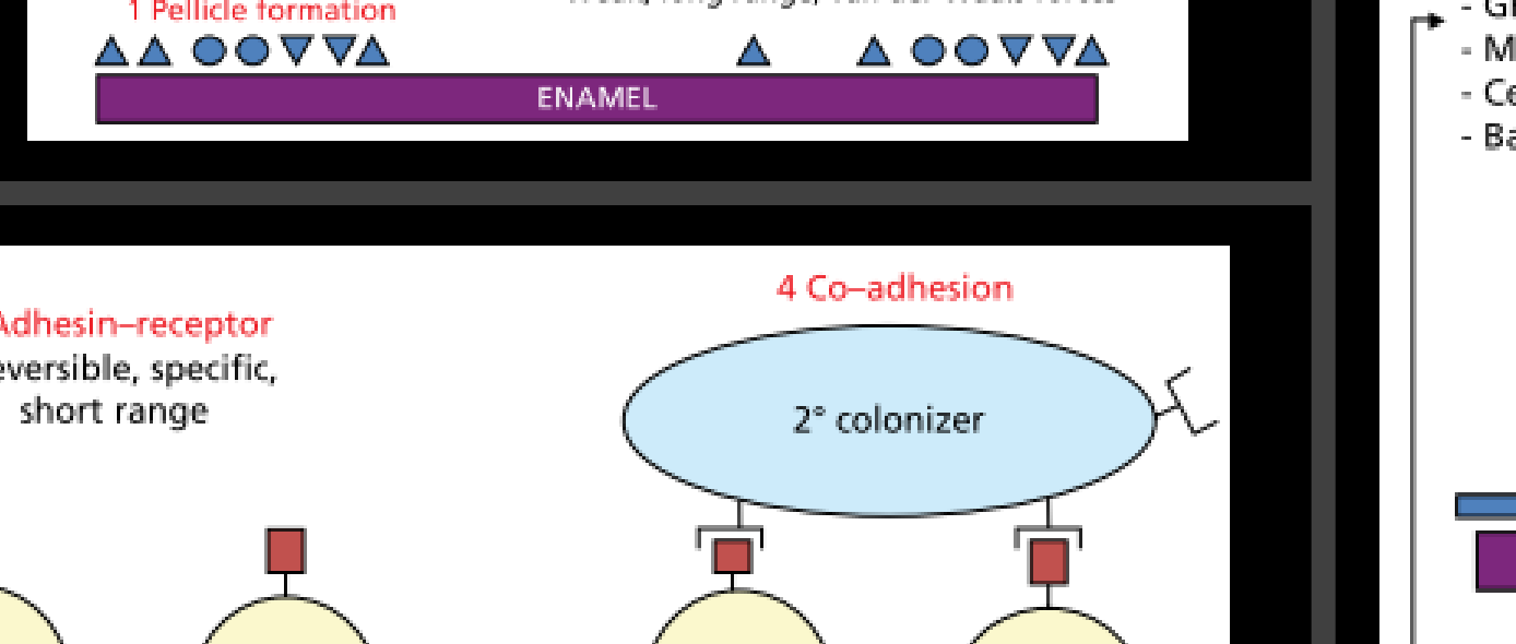
The process of dental biofilm formation can be divided into several phases: (1) The formation of the pellicle on the tooth surface (2) The initial adhesion/attachment of bacteria (3) Colonization/biofilm maturation
-
The development of dental biofilms follows a well-established sequence of events where initial colonizers, predominantly streptococci, act as a foundation to establish an environment suitable for later colonization by potentially more pathogenic species
-
Initially, bacteria can be held reversibly near to the surface by weak, long- range, physicochemical forces between the electrical charge on the molecules on the pellicle- coated surface and those on the microbial cell.
Formation Of The Acquired Pellicle17
(1) The formation of the pellicle on the tooth surface
-
All surfaces in the oral cavity, including hard and soft tissues, are coated with a layer of organic material known as the acquired pellicle.
-
Microorganisms rarely colonize clean enamel.
-
Within seconds of eruption, or following cleaning, tooth surfaces become coated with a conditioning film (acquired pellicle) of molecules (biologically active proteins, phosphoproteins, and glycoproteins) derived mainly from saliva (but also from GCF and bacteria) (Hannig et al . 2005).
-
The pellicle on tooth surfaces consists of more than 180 peptides, proteins, and glycoproteins, including keratins, mucins, proline-rich proteins, and other molecules that can function as adhesion sites (receptors) for bacteria.
Initial Adhesion And Primary Colonizers181920
2-Initial Adhesion/Attachment of Bacteria The University of Western Australia logo is present in the top-left corner.
- The interactions between microbial cell surface “adhesin” molecules and salivary pellicle receptors determine if a bacterial cell will stay on the surface.
- Only a relatively small proportion of oral bacteria possess adhesins that interact with receptors in the host pellicle. These species are considered the “primary colonizers” of tooth surfaces. The primary colonizers provide new binding sites for adhesion by other oral bacteria.
- Initial colonizers such as streptococci produce lactic acid which can be utilized as a carbon and energy source by Veillonella species.
- Veillonella spp. are amongst the most prevalent bacterial species in dental biofilms and are thought to have a similar role to oral fusobacteria (such as Fusobacterium nucleatum in that they are able to act as bridging organisms that are able to support the colonization and growth of later colonizers, by their ability to bind to both early and late colonizers
Colonization of teeth by bacteria occur in three phases
- Phase 1 is transport to the tooth surface—Brownian motion (average displacement, 40 μm/hour)
- Phase 2 is initial reversible adhesion—Van der Waals attractive forces and electrostatic repulsive forces
- Phase 3 is strong attachment - Proteins, Glycoproteins, or polysaccharides
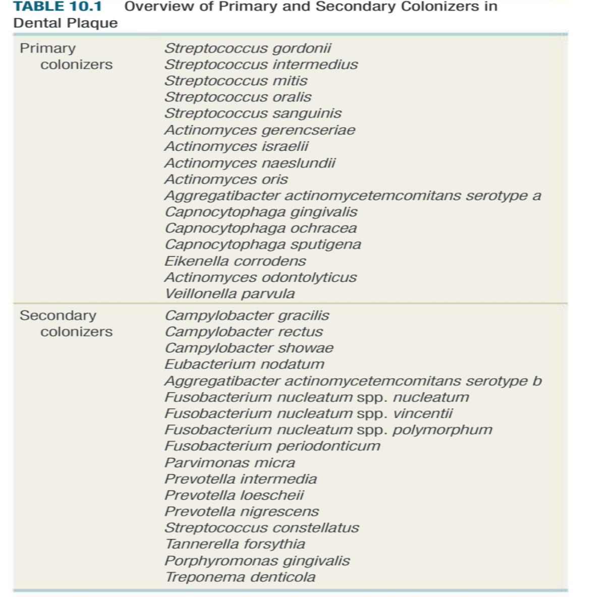
| Primary colonizers |
Streptococcus gordonii Streptococcus intermedius Streptococcus mitis Streptococcus oralis Streptococcus sanguinis Actinomyces gerencseriae Actinomyces israelii Actinomyces naeslundii Actinomyces oris Aggregatibacter actinomycetemcomitans serotype a Capnocytophaga gingivalis Capnocytophaga ochracea Capnocytophaga sputigena Eikenella corrodens Actinomyces odontolyticus Veillonella parvula |
| Secondary colonizers |
Campylobacter gracilis Campylobacter rectus Campylobacter showae Eubacterium nodatum Aggregatibacter actinomycetemcomitans serotype b Fusobacterium nucleatum spp. nucleatum Fusobacterium nucleatum spp. vincentii Fusobacterium nucleatum spp. polymorphum Fusobacterium periodonticum Parvimonas micra Prevotella intermedia Prevotella loescheii Prevotella nigrescens Streptococcus constellatus Tannerella forsythia Porphyromonas gingivalis Treponema denticola |
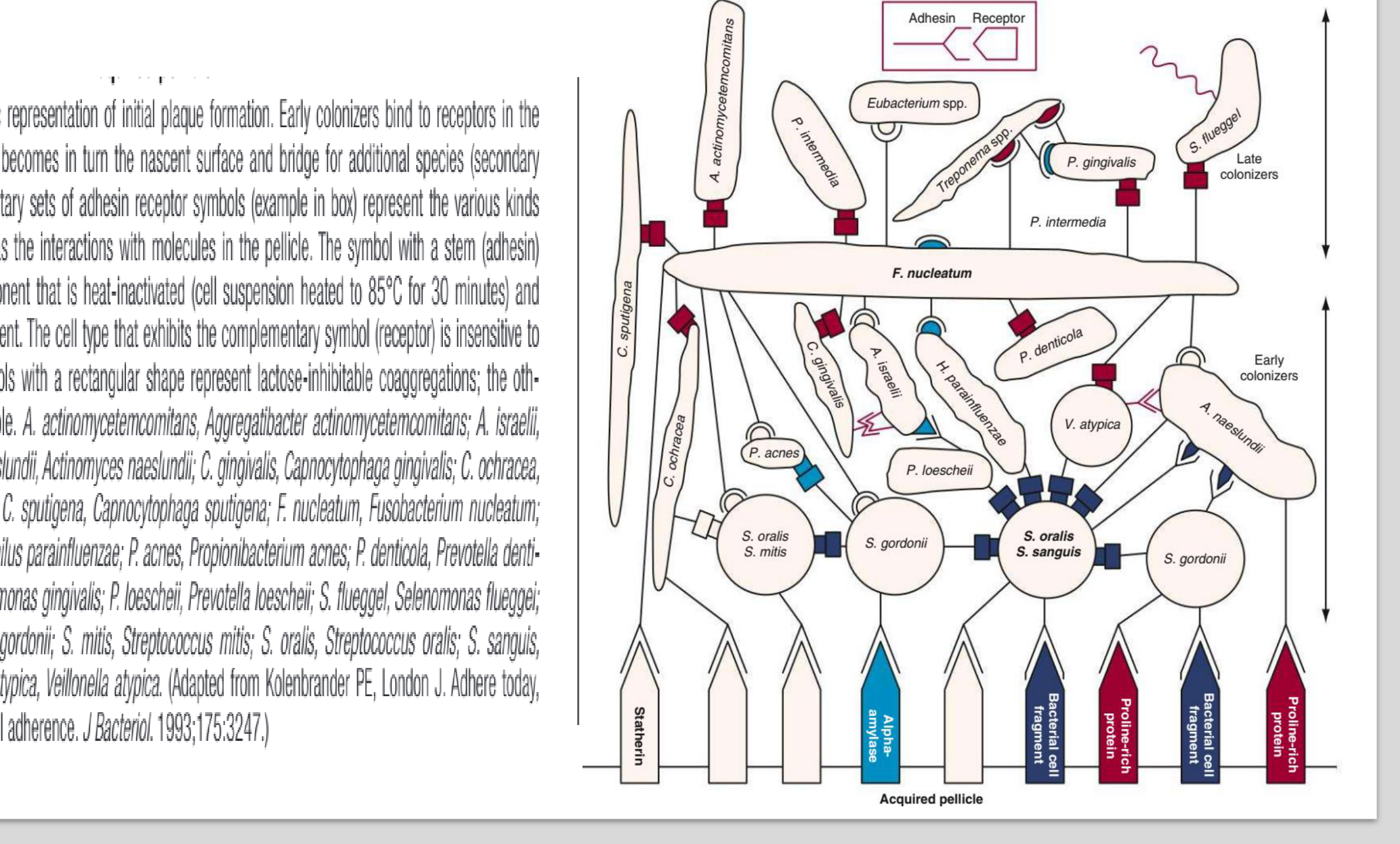
- The primary colonizing bacteria adhered to the tooth surface provide new receptors for attachment by other bacteria, in a process known as “coadhesion” or “coaggregation”.
- The transition from early supragingival dental biofilm to more mature biofilm developing beneath the gingival margin involves a shift in the microbial population from primarily gram-positive organisms to high numbers of gram-negative bacteria.
- A key organism in dental biofilm development is Fusobacterium nucleatum. This species can co-adhere to most oral bacteria and acts as an important bridging organism between early and later colonizing species.
 |  |
 |
Secondary Colonization And Coaggregation2122
Fig. 10.10 Diagrammatic representation of initial plaque formation. Early colonizers bind to receptors in the pellicle. Each adherent cell becomes in turn the nascent surface and bridge for additional species (secondary colonizers). The complementary sets of adhesin receptor symbols (example in box) represent the various kinds of coaggregations as well as the interactions with molecules in the pellicle. The symbol with a stem (adhesin) represents a cellular component that is heat-inactivated (cell suspension heated to 85°C for 30 minutes) and sensitive to protease treatment. The cell type that exhibits the complementary symbol (receptor) is insensitive to either treatment. The symbols with a rectangular shape represent lactose-inhibitable coaggregations; the others are lactose noninhibitable.
A. actinomycetemcomitans, Aggregatibacter actinomycetemcomitans; A. israelii, Actinomyces israelii; A. naeslundii, Actinomyces naeslundii; C. gingivalis, Capnocytophaga gingivalis; C. ochracea, Capnocytophaga ochracea; C. sputigena, Capnocytophaga sputigena; F. nucleatum, Fusobacterium nucleatum; H. parainfluenzae, Haemophilus parainfluenzae; P. acnes, Propionibacterium acnes; P. denticola, Prevotella denticola; P. gingivalis, Porphyromonas gingivalis; P. loescheii, Prevotella loescheii; S. flueggei, Selenomonas flueggei; S. gordonii, Streptococcus gordonii; S. mitis, Streptococcus mitis; S. oralis, Streptococcus oralis; S. sanguis, Streptococcus sanguis; V. atypica, Veillonella atypica. (Adapted from Kolenbrander PE, London J. Adhere today, here tomorrow: oral bacterial adherence. J Bacteriol. 1993;175:3247.)
 |  |
Communication Between Biofilm Bacteria23
• Bacterial cells do not exist in isolation. In a biofilm, capacity to communicate with each other.
• One example of this is “quorum sensing”, in which signaling molecule that accumulates in the local environment triggers a response such as a change in the expression genes once they reach a critical threshold concentration.
There are 2 types of signaling molecules of plaque bacteria:
• Peptides released by gram-positive organisms during pump…
• In Streptococcus mutans , quorum sensing is mediated by competence stimulating peptide (CSP) (Li et al . 2017).
• “Universal” signal molecule autoinducer 2 (AI-2).
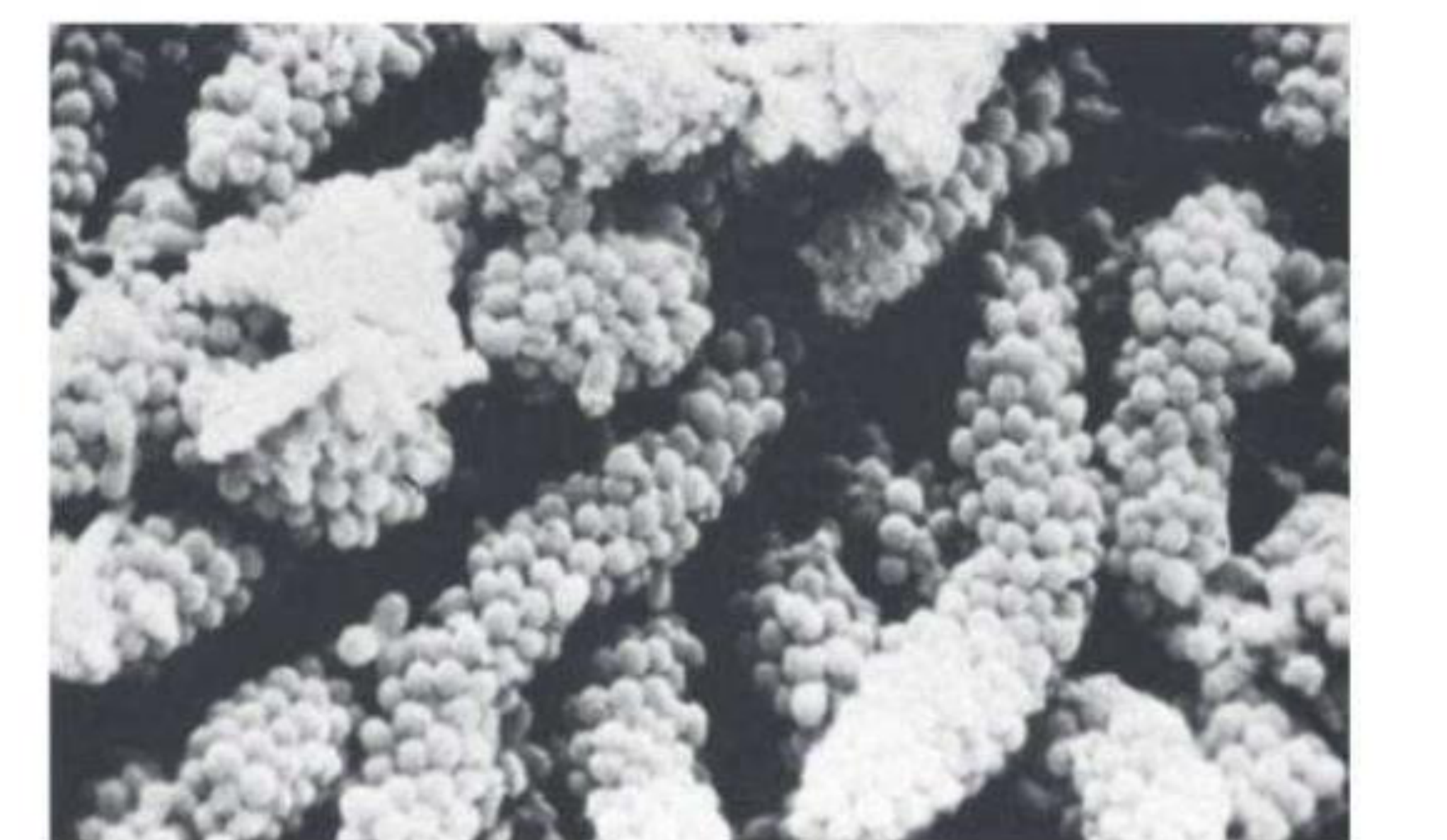
Fig. 10.11 Long-standing supragingival plaque near the gingival margin demonstrating a “corn cob” arrangement. A central gram-negative filamentous core supports the outer coccal cells, which are firmly attached by inter-bacterial adherence or coaggregation.
Hypotheses Of Periodontal Disease Etiology
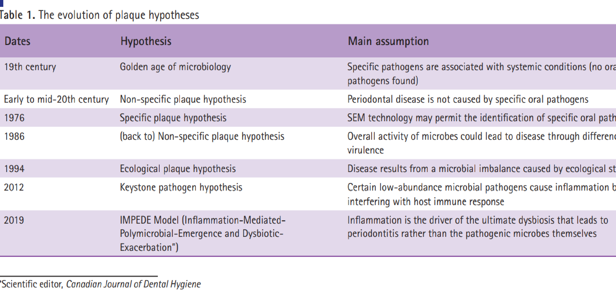
| |\input| | --- |\input| | Dates |\input| | Hypothesis |\input| | Main assumption |\input| | 19th century |\input| | Golden age of microbiology |\input| | Specific pathogens are associated with systemic conditions (no oral pathogens found) ||\input| | Early to mid-20th century |\input| | Non-specific plaque hypothesis |\input| | Periodontal disease is not caused by specific oral pathogens ||\input| | 1976 |\input| | Specific plaque hypothesis |\input| | SEM technology may permit the identification of specific oral pathogens ||\input| | 1986 |\input| | (back to) Non-specific plaque hypothesis |\input| | Overall activity of microbes could lead to disease through differences in virulence ||\input| | 1994 |\input| | Ecological plaque hypothesis |\input| | Disease results from a microbial imbalance caused by ecological stress ||\input| | 2012 |\input| | Keystone pathogen hypothesis |\input| | Certain low-abundance microbial pathogens cause inflammation by interfering with host immune response ||\input| | 2019 |\input| | IMPEDE Model (Inflammation-Mediated-Polymicrobial-Emergence and Dysbiotic-Exacerbation”) | |\input| | Inflammation is the driver of the ultimate dysbiosis that leads to periodontitis rather than the pathogenic microbes themselves ||\input|
Scientific editor, Canadian Journal of Dental Hygiene
Specific Versus Non-Specific Plaque Hypotheses242526
Correspondence: Dr Salme E Lavigne; scientificeditor@cdha.ca
©2023 Canadian Dental Hygienists Association
Can J Dent Hyg 2023;57(2): 75–77 | 75
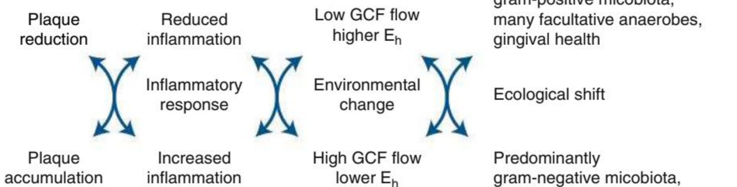
| Plaque reduction | Reduced inflammation | Low GCF flow | Predominantly |
| higher Eh | gram-positive | ||
| Plaque accumulation | micobiota, | ||
| many facultative | |||
| Increased inflammation | anaerobes, | ||
| Environmental change | gingival health | ||
| High GCF flow | Ecological shift | ||
| lower Eh | |||
| Predominantly | |||
| gram-negative | |||
| micobiota, | |||
| many obligate | |||
| anaerobes, | |||
| periodontal disease |
Fig. 10.25 Ecologic plaque hypothesis in relation to periodontal diseases: gingivitis and periodontitis. The accumulation of plaque causes the inflammation of adjacent tissues (gingivitis) and other environmental changes that favor the growth of gram-negative anaerobes and proteolytic species, including periodontal pathogens. The increased proportions of such species result in the destruction of periodontal tissues (i.e., periodontitis). Eh, Redox potential; GCF, gingival crevicular fluid. (Adapted from Marsh PD. Microbial ecology of dental plaque and its significance in health and disease. Adv Dent Res. 1994;8:263.)
The University of Western Australia
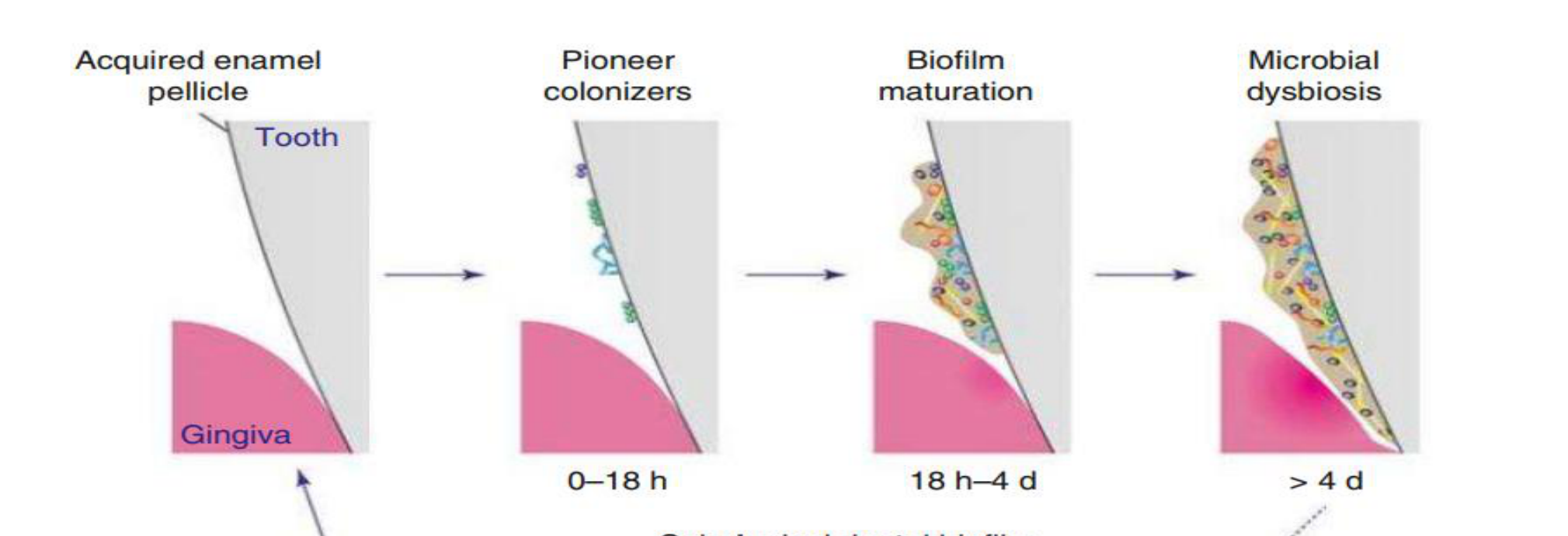
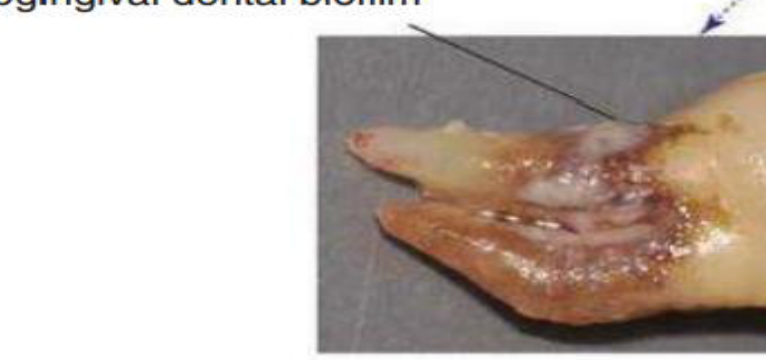
Acquired enamel pellicle Tooth Gingiva 0–18 h Pioneer colonizers 18 h–4 d Biofilm maturation > 4 d Microbial dysbiosis Subgingival dental biofilm
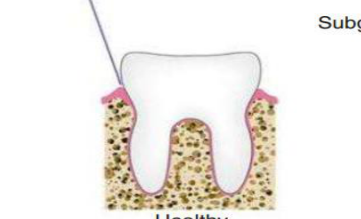
Healthy tooth Periodontal disease
Fig. 10.7 Accumulation of dental biofilm and progression to disease. Good oral hygiene maintains low levels of dental biofilm at the gum margin. The acquired enamel pellicle, a layer of protein and glycoprotein largely derived from saliva, is not removed during tooth cleaning and forms attachment sites for pioneer colonizing bacteria. Initial attachment starts within minutes. Without further oral hygiene, the dental biofilm thickens and an extracellular matrix accumulates (“biofilm maturation”) after around 18 h. This mature biofilm reorganizes over the next few days, without dramatically increasing in thickness. Interactions between the biofilm and gingival tissue triggers inflammation, indicated by reddening of the gingiva. Ultimately, this can lead to microbial dysbiosis and the accumulation of significant biofilm below the gingival margin which leads to chronic inflammation and loss of tissue surrounding the tooth. Ultimately, the biofilm-laden tooth becomes mobile and requires extraction. A tooth extracted for periodontal disease is shown.
 |  |  |
 |  |
Microbial Complexes In Subgingival Plaque2728
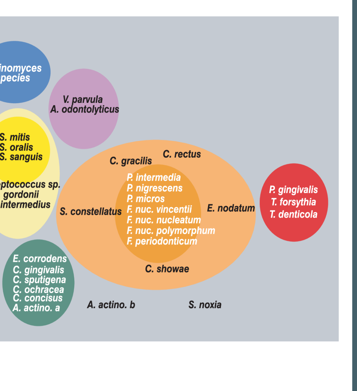
Bold Text: Fig. 10.24
Main Caption and Body Text: Associations among subgingival species. The data were derived from 13,261 subgingival plaque samples taken from the mesial aspect of each tooth in 185 adult subjects. Each sample was individually analyzed for the presence of 40 subgingival species with the use of checkerboard DNA-DNA hybridization. Associations were sought among species via cluster analysis and community ordination techniques. The complexes to the left consist of species that are thought to colonize the tooth surface and to proliferate at an early stage. The orange complex becomes numerically dominant later; it is thought to bridge the early colonizers and the red complex species, which become numerically more dominant during the later stages of plaque development. A. actinomyces comitans, Aggregatibacter actinomycetemcomitans; A. odontolytica, Actinomyces odontolytica; C. concisus, Campylobacter concisus; C. gingivalis, Capnocytophaga gingivalis; C. gracilis, Campylobacter gracilis; C. ochracea, Capnocytophaga ochracea; C. rectus, Campylobacter rectus; C. showae, Campylobacter showae; C. sputigena, Capnocytophaga sputigena; E. corrodens, Eikenella corrodens; F. nuc., Fusobacterium nucleatum; F. periodonticum, Fusobacterium periodonticum; P. gingivalis, Porphyromonas gingivalis; P. micra, Parvimonas micra; P. nigrescens, Prevotella nigrescens; S. constellatus, Streptococcus constellatus; S. mitis, Streptococcus mitis; S. noxia, Selenomonas noxia; S. oralis, Streptococcus oralis; S. sanguis, Streptococcus sanguis; T. denticola, Treponema denticola; T. forsythia, Tannerella forsythia; V. parvula, Veillonella parvula. (Adapted from Socransky SS, Haffajee AD, Cugini MA, et al. Microbial complexes in subgingival plaque. J Clin Periodontol. 1998;25:134.)
Figure Content (Bulleted List of Species Groups):
-
Blue Oval (Top Left):
- Actinomyces species
-
Purple Oval (Top Right):
- V. parvula
- A. odontolyticus
-
Yellow Oval (Left):
- S. mitis
- S. oralis
- S. sanguis
- Streptococcus sp.
- S. gordonii
- S. intermedius
-
Large Orange Oval (Center):
- C. gracilis
- C. rectus
- S. constellatus
- E. nodatum
- P. intermedia
- P. nigrescens
- P. micros
- F. nuc. vincentii
- F. nuc. nucleatum
- F. nuc. polymorphum
- F. periodonticum
- C. showae
- A. actino. b
- S. noxia
-
Small Green Oval (Bottom Left):
- E. corrodens
- C. gingivalis
- C. sputigena
- C. ochracea
- C. concisus
- A. actino. a
-
Red Oval (Far Right):
- P. gingivalis
- T. forsythia
- T. denticola
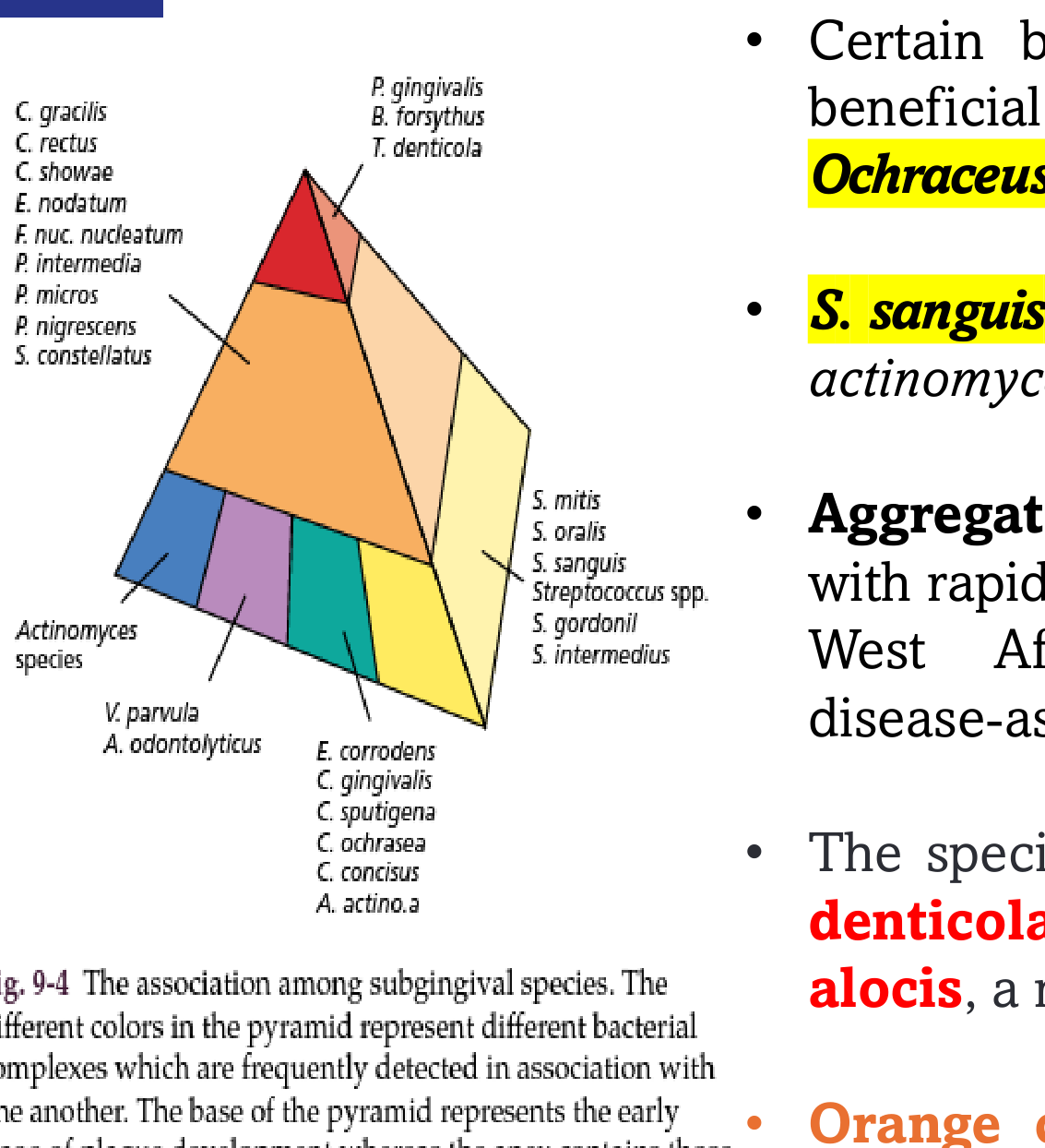
The presentation text is transcripted below. The pyramid on the left represents a classification of oral bacteria.
V. parvula A. odontolyticus E. corrodens C. gingivalis C. sputigena C. ochracea C. concius A. actino.a
The base of the pyramid represents the early stage of plaque development whereas the apex contains those organisms thought to be the last species to become established in the microbiota. The red complex of bacteria are those organisms frequently associated with sites of periodontal disease.
(Source: Reprinted from Socransky & Haffajee 2002, with permission.)
 |  |
Clinical Deposits And Calculus
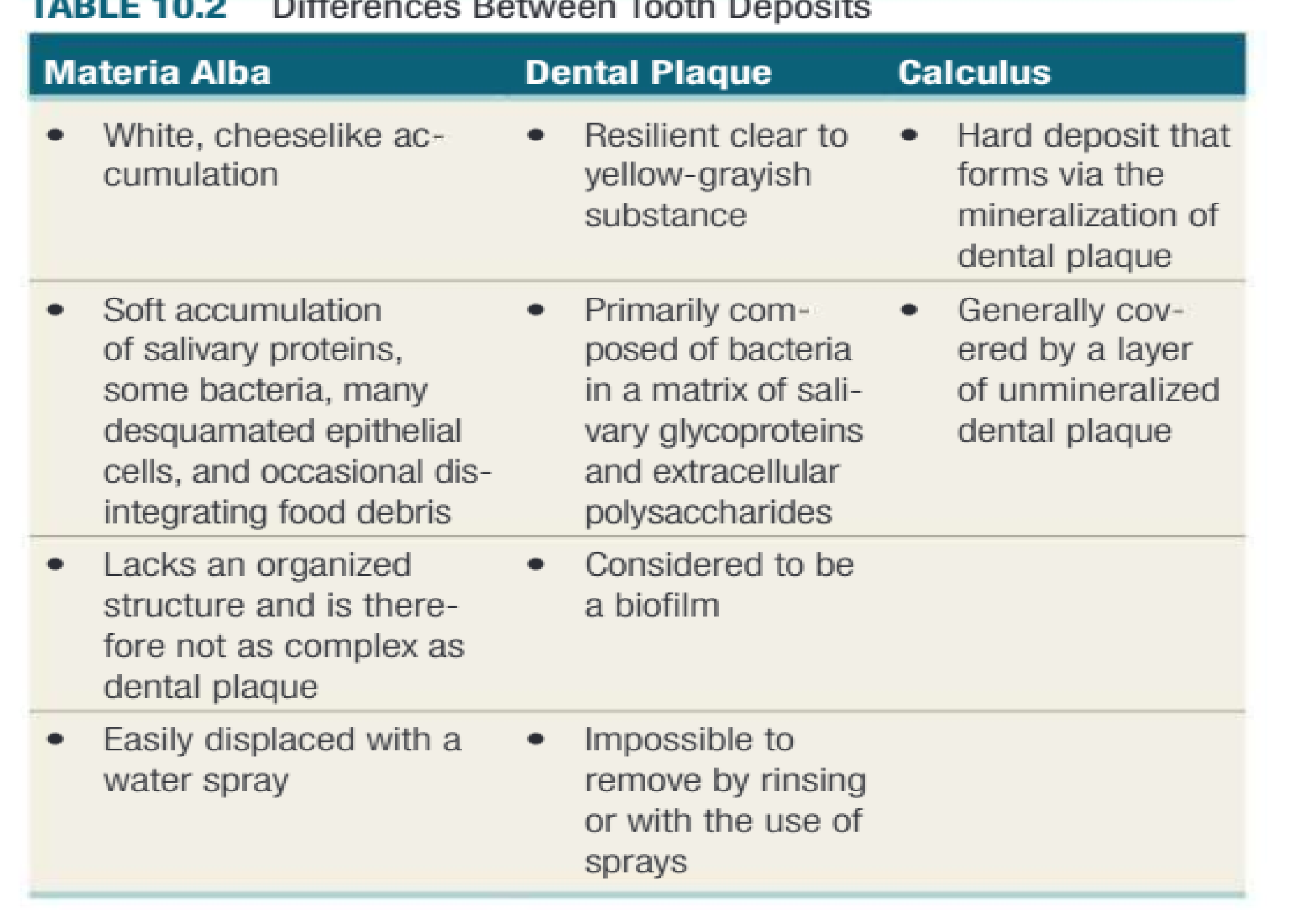
| Materia Alba | Dental Plaque | Calculus |
|---|---|---|
|
White, cheeseslike ac- cumulation | Resilient clear to
yellow-grayish substance | Hard deposit that
forms via the mineralization of dental plaque |
|
Soft accumulation of salivary proteins, some bacteria, many desquamated epithelial cells, and occasional dis- integrating food debris Materia Alba Versus Dental Plaque29 | Primarily com-
posed of bacteria in a matrix of sali- vary glycoproteins and extracellular polysaccharides | Generally covered by a layer of unmineralized dental plaque |
| Lacks an organized
structure and is there- fore not as complex as dental plaque | Considered to be a biofilm | |
| Easily displaced with a
water spray | Impossible to
remove by rinsing or with the use of sprays |
Dental Calculus Characteristics303132
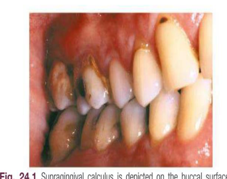
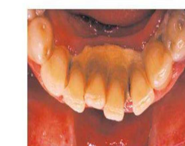
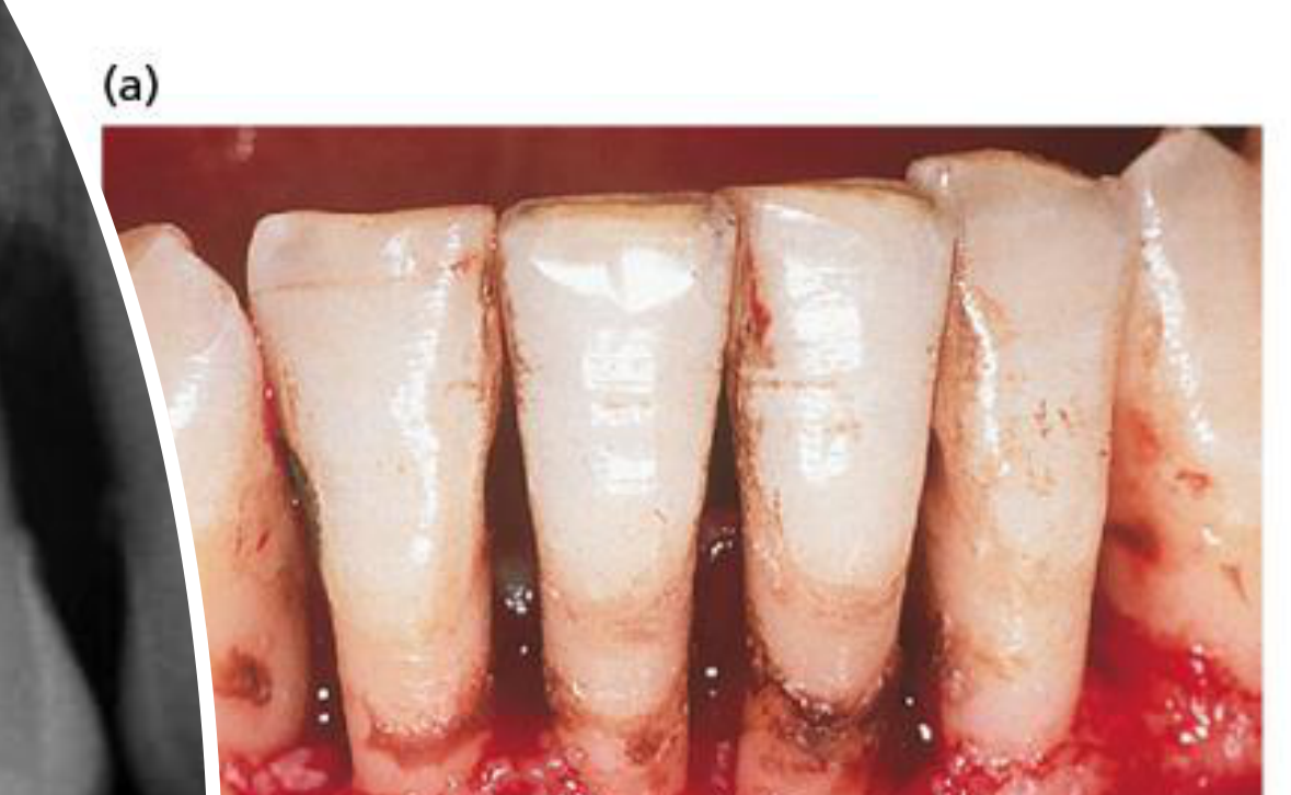
Dental calculus
- Dental calculus or tartar represents mineralized bacterial biofilm, although calculus formation can be induced in germ- free animals because of precipitation of mineral salts originating from saliva.
- As the mineral content of dental biofilms increases, the biofilm mass becomes calcified to form calculus Calculus is frequently found in areas of the dentition adjacent to salivary ducts (e.g., the lingual surface of the mandibular incisors and canines, the buccal surface of the maxillary first molars), and this reflects.
- The high concentration of minerals available from saliva in those regions.
- The inorganic components of subgingival dental biofilms are derived from crevicular fluid (a serum transudate).
- It should be noted that calculus continually harbors a viable bacterial biofilm.
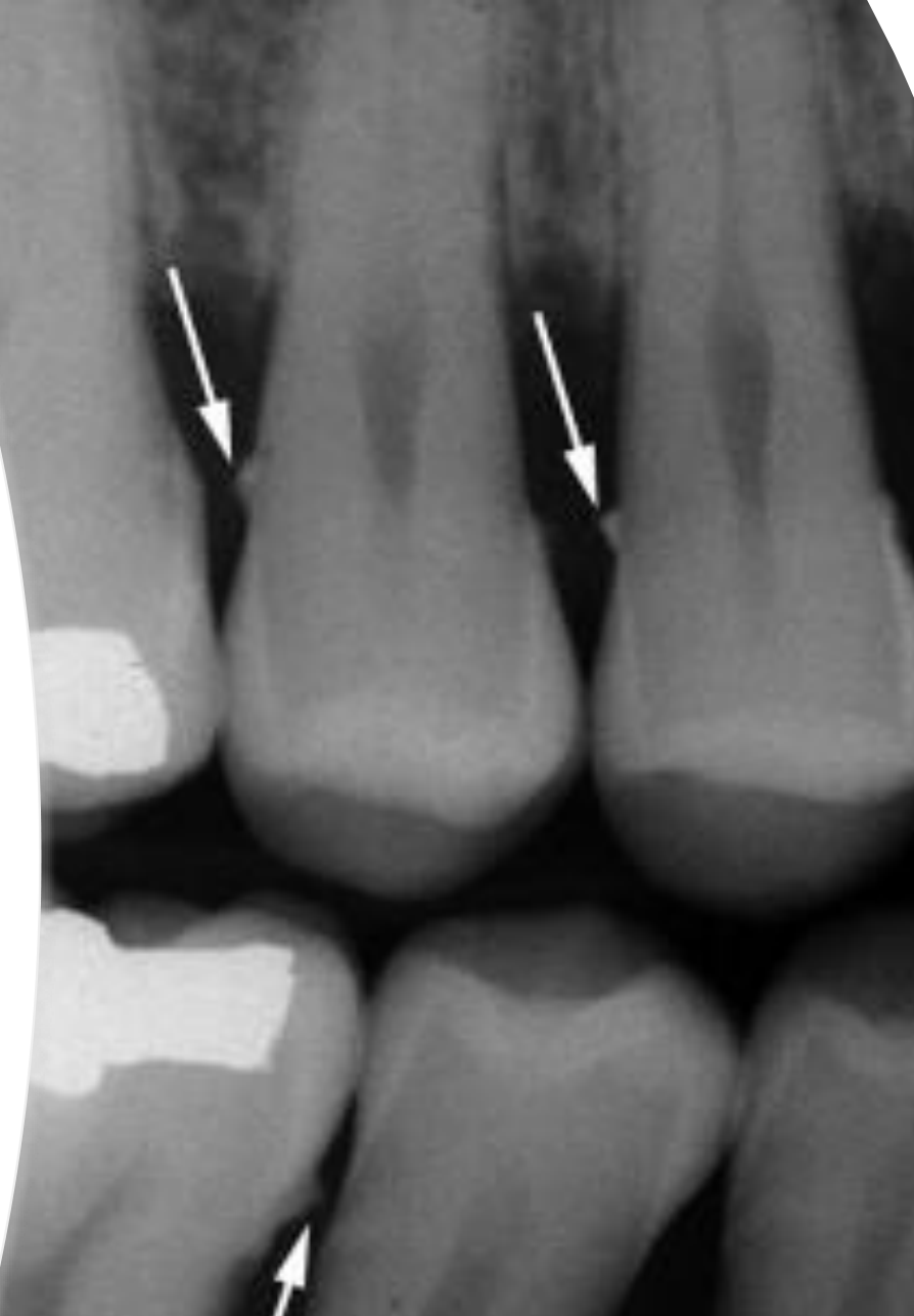
- Subgingivally, calculus may be found by tactile exploration only, since it is usually not visible to the naked eye.
- Subgingival calculus is found in most periodontal pockets, usually extending from the cementoenamel junction to close to the bottom of the pocket.
 |  |  |
 | 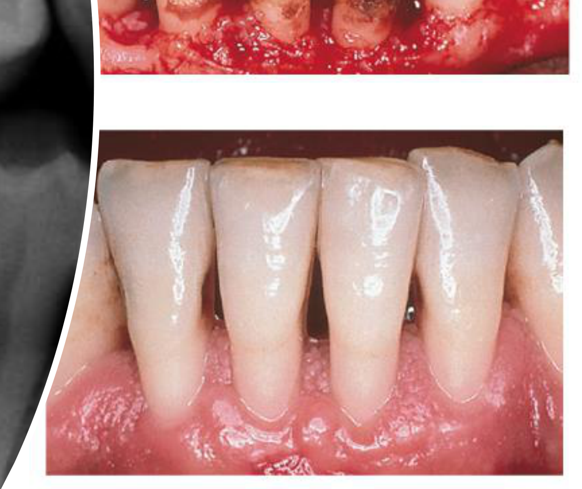 |
Iatrogenic And Predisposing Factors333435
Saudi Dental Journal (2020) 32, 80–85
King Saud University Saudi Dental Journal www.ksu.edu.sa www.sciencedirect.com
المستند King Saud University
الجمعية السعودية لطب الأسنان SAUDI DENTAL SOCIETY
Effect of iatrogenic factors on periodontal health: An epidemiological study
Kankara Vinathi Reddy a,*, Chembolu Nirupama a, Pathakota Krishnajaneya Reddy a, Pradeep Koppolu b, Dalal H. Alotaibi c
Conclusion: This study clearly identified a higher prevalence, 50.8% of sub-gingival restorations causing gingivitis and has shown significant influence on periodontal status of the tooth.
 |  |
Composition And Inorganic Content36
Composition
Inorganic Content
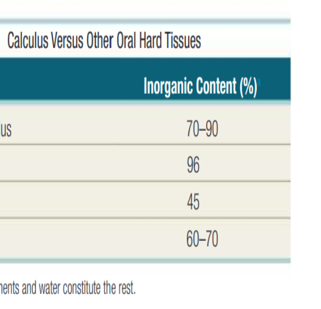
- Dental calculus is primarily composed of inorganic components (70% to 90%) and the organic components constitute the rest.
- The major inorganic proportions of calculus are approximately
- 76% calcium phosphate (Ca3[PO4]2)
- 3% calcium carbonate (CaCO3)
- 4% magnesium phosphate (Mg3[PO4]2)
- 2% carbon dioxide
- Traces of other elements such as sodium, zinc, strontium, bromine, copper, manganese, tungsten, gold, aluminum, silicon, iron, and fluorine.
- The calcium concentration or content in plaque is 2 to 20 times higher than in saliva.
- The time required to reach the maximal level has been reported between 10 weeks & 6 months
| Structure | Inorganic Content (%) |
|---|---|
| Dental calculus | 70–90 |
| Enamel | 96 |
| Dentin | 45 |
| Bone | 60–70 |
| *Organic components and water constitute the rest. | |
Formation And Mineralization Process37
• Formation • Calculus is mineralized dental plaque. • The soft plaque is hardened by the precipitation of mineral salts, which usually starts between the 1st and 14th day of plaque formation. • Calcification has been reported to occur within as little as 4 to 8 hours. • Calcifying plaques may become 50% mineralized in 2 days and 60% to 90% mineralized in 12 days.
KEY FACT Calculus by itself does not contribute directly to gingival inflammation. Like other retentive factors, such as open crown margin or an overhanging restoration, calculus retains dental plaque, which contributes to gingival inflammation.
Modes Of Calculus Attachment38
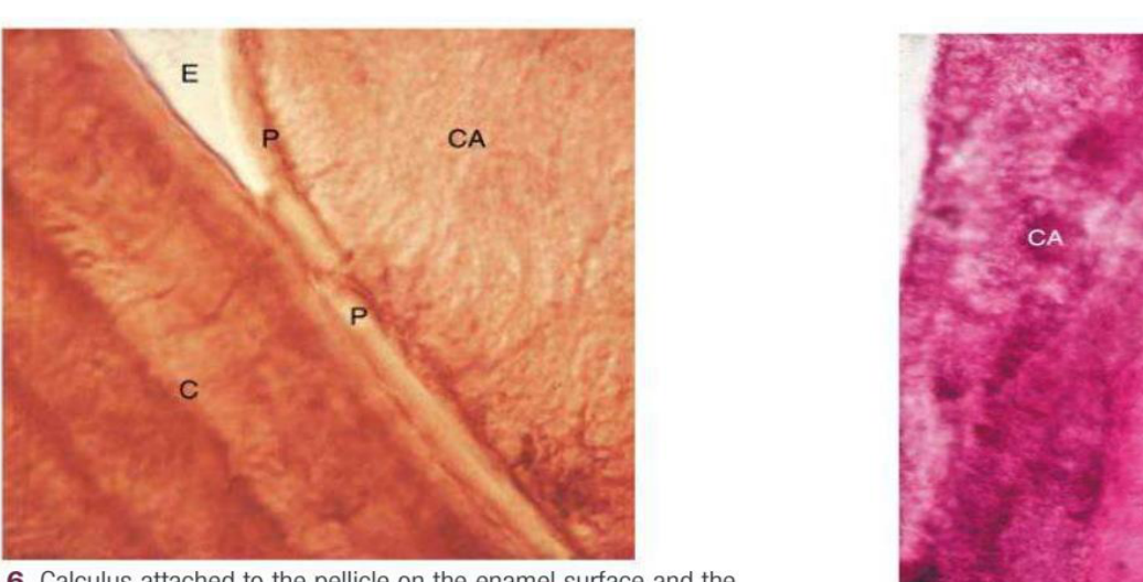
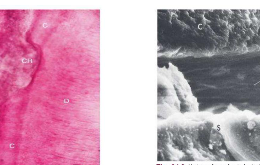
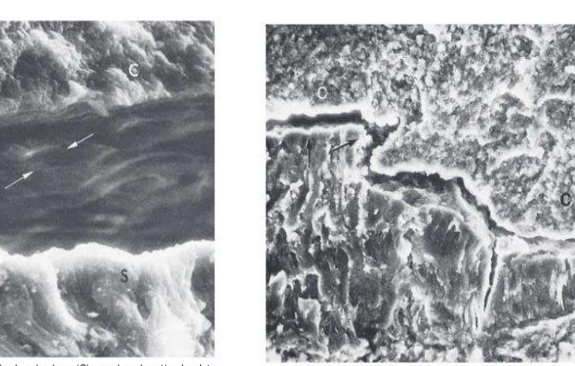
Four modes of attachment have been described.
(1) Attachment by means of an organic pellicle on cementum is depicted in Fig. 24.6
(2) Mechanical locking into surface irregularities, such as caries lesions or resorption lacunae, is illustrated in Fig. 24.8.
(3) Close adaptation of the undersurface of calculus to depressions or gently sloping mounds of the unaltered cementum surface is shown in Fig. 24.9.
(4) Penetration of bacterial calculus into cementum
The reason for firm attachment to the tooth surface is the fact that the pellicle beneath the bacterial biofilm also calcifies. This, in turn, results in an intimate contact with enamel, cementum or dentin crystals
The University of Western Australia
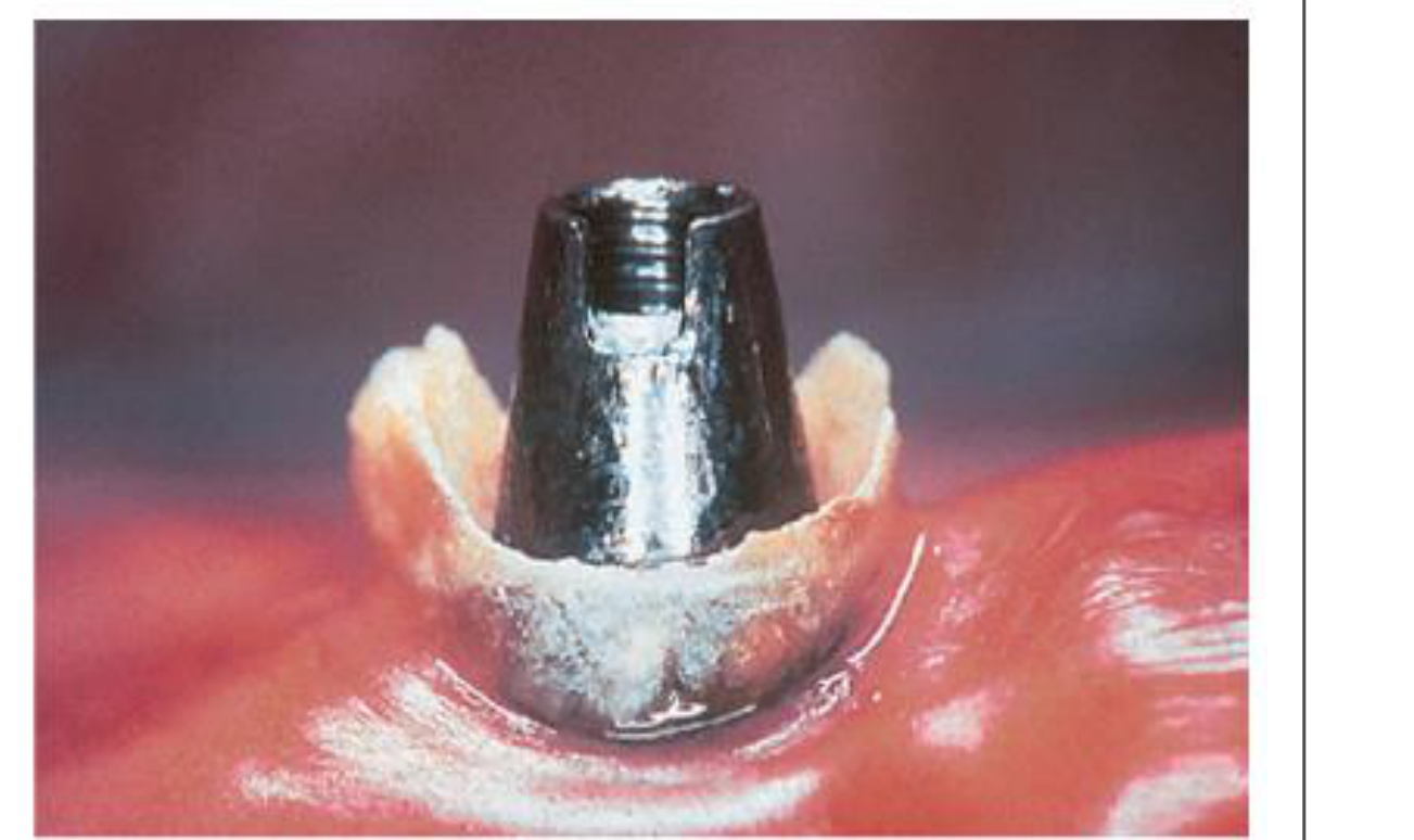
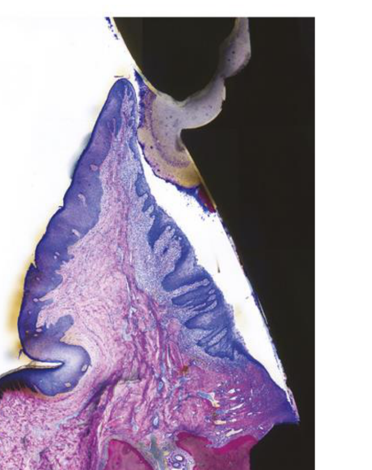
Fig. 8-23 Excess cement at the abutment-crown interface provides an ideal substrate for biofilm and calculus deposition and retention. Bacterial biofilm covers the entire surface of the cement, whereas calculus is present apical to the cement overhang. Detachment of the epithelium indicates pocket formation. The detachment of the apical-most portion of the epithelium, however, may represent an artifact due to histologic processing. Underdecalfied ground section.
 |  |
 |
References And Chapter Outline
References
Chapter 24: The Role of Dental Calculus and Other Local Predisposing Factors39
- Authors: Vivek Thumbrigere-Math, James E. Hinrichs
Chapter Outline
- Calculus
- Supragingival and Subgingival Calculus
- Prevalence
- Composition
- Attachment to the Tooth Surface
- Formation
- Etiologic Significance
- Materia Alba, Food Debris, and Dental Stains
- Other Predisposing Factors
- Iatrogenic Factors
- Margins of Restorations
- Malocclusion
- Periodontal Complications Associated With Orthodontic Therapy
- Extraction of Impacted Third Molars
- Habits and Self-inflicted Injuries
- Smokeless Tobacco
- Radiation Therapy
Chapter 8: Dental Biofilms and Calculus
- Authors: Philip D. Marshall, Mariano Sanz, Nikolaus P. Lang, Dieter D. Bosshardt
- Affiliations:
- Department of Oral Biology, School of Dentistry, University of Leeds, UK
- Faculty of Odontology, ETEP (Etiology and Therapy of Periodontal and Peri-Implant Diseases) Research Group, Complutense University of Madrid, Madrid, Spain; and Department of Periodontology, Faculty of Dentistry, Institute of Clinical Dentistry, University of Oslo, Oslo, Norway
- Department of Periodontology, School of Dental Medicine, University of Bern, Bern, Switzerland
Chapter 10: Biofilm and Periodontal Microbiology
- Authors: Wirm Teughels, Magda Feres, Sukirth M. Ganesan, Mark David Gidley, Yvonne L. Hernandez-Kapila, Nicholas S. Jakubovics
Source Texts
- Newman and Carranza’s Clinical Periodontology and Implantology, 14th Edition
- Lindhe’s Clinical Periodontology and Implant Dentistry, Seventh Edition, Vol 1, Edited by Torleif Lindhe, William F. Giannobile, Niklaus P. Lang, and Mariano Sanz, John Wiley & Sons
Reference
- Sedghi L, DiMassa V, Harrington A, Lynch SV, Kapila YL. The oral microbiome: Role of key organisms and complex networks in health and disease. Periodontol 2000. 2021;87:107–131
Footnotes
-
Original PDF page 1: L4 Etiopathogenesis part 1, p.1 ↩
-
Original PDF page 2: L4 Etiopathogenesis part 1, p.2 ↩
-
Original PDF page 3: L4 Etiopathogenesis part 1, p.3 ↩
-
Original PDF page 4: L4 Etiopathogenesis part 1, p.4 ↩
-
Original PDF page 5: L4 Etiopathogenesis part 1, p.5 ↩
-
Original PDF page 6: L4 Etiopathogenesis part 1, p.6 ↩
-
Original PDF page 9: L4 Etiopathogenesis part 1, p.9 ↩
-
Original PDF page 7: L4 Etiopathogenesis part 1, p.7 ↩
-
Original PDF page 8: L4 Etiopathogenesis part 1, p.8 ↩
-
Original PDF page 10: L4 Etiopathogenesis part 1, p.10 ↩
-
Original PDF page 11: L4 Etiopathogenesis part 1, p.11 ↩
-
Original PDF page 12: L4 Etiopathogenesis part 1, p.12 ↩
-
Original PDF page 13: L4 Etiopathogenesis part 1, p.13 ↩
-
Original PDF page 14: L4 Etiopathogenesis part 1, p.14 ↩
-
Original PDF page 15: L4 Etiopathogenesis part 1, p.15 ↩
-
Original PDF page 16: L4 Etiopathogenesis part 1, p.16 ↩
-
Original PDF page 17: L4 Etiopathogenesis part 1, p.17 ↩
-
Original PDF page 18: L4 Etiopathogenesis part 1, p.18 ↩
-
Original PDF page 19: L4 Etiopathogenesis part 1, p.19 ↩
-
Original PDF page 20: L4 Etiopathogenesis part 1, p.20 ↩
-
Original PDF page 21: L4 Etiopathogenesis part 1, p.21 ↩
-
Original PDF page 23: L4 Etiopathogenesis part 1, p.23 ↩
-
Original PDF page 22: L4 Etiopathogenesis part 1, p.22 ↩
-
Original PDF page 24: L4 Etiopathogenesis part 1, p.24 ↩
-
Original PDF page 25: L4 Etiopathogenesis part 1, p.25 ↩
-
Original PDF page 26: L4 Etiopathogenesis part 1, p.26 ↩
-
Original PDF page 27: L4 Etiopathogenesis part 1, p.27 ↩
-
Original PDF page 28: L4 Etiopathogenesis part 1, p.28 ↩
-
Original PDF page 29: L4 Etiopathogenesis part 1, p.29 ↩
-
Original PDF page 30: L4 Etiopathogenesis part 1, p.30 ↩
-
Original PDF page 31: L4 Etiopathogenesis part 1, p.31 ↩
-
Original PDF page 32: L4 Etiopathogenesis part 1, p.32 ↩
-
Original PDF page 33: L4 Etiopathogenesis part 1, p.33 ↩
-
Original PDF page 37: L4 Etiopathogenesis part 1, p.37 ↩
-
Original PDF page 38: L4 Etiopathogenesis part 1, p.38 ↩
-
Original PDF page 34: L4 Etiopathogenesis part 1, p.34 ↩
-
Original PDF page 35: L4 Etiopathogenesis part 1, p.35 ↩
-
Original PDF page 36: L4 Etiopathogenesis part 1, p.36 ↩
-
Original PDF page 39: L4 Etiopathogenesis part 1, p.39 ↩