Etiopathogenesis: Host Responses to Oral Biofilms1
Etiopathogenesis -2 Host responses to oral biofilms
Associate Professor Leticia A Miranda* DDS, Specialist (Perio), MSc (Perio), PhD (Perio, Medicine)
Dr. Pradeep Koppolu* BDS, MDS (Perio), PhD (Malaysia), FICOI, FPFA, PDCR Discipline Lead & Program Convenor Periodontics and Implantology
*notes
Reading Resources2
BOOK Periodontology at a Glance Clerehugh, Valerie. ; Tugnait, Aradhna. ; Genco, Robert J. ; Somerset : Wiley; 2013 [Available Online] →
Clinical Periodontology and Implant Dentistry, 2 Volume Set by Niklaus P. Lang, Jan Lindhe, and Niklaus P Lang
PUBLISHER John Wiley & Sons, Incorporated
DATE 2015-03-25
NEWMAN AND CARRANZA’S CLINICAL PERIODONTOLOGY AND IMPLANTOLOGY 14th EDITION
CHAPTER 11 Host-Microbe Interactions and the Inflammatory Response Keith L. Kirkwood | Carlos Rossa Jr. | George Hajishengallis | Ann Decker | Yvonne L. Hernandez-Kapla
 |  |
 |  |
Learning Outcomes3
- Recognize periodontal health and disease;
- Understand the peculiarities of the periodontal niche;
- Identify the key components of the host response in the dento-gingival area;
- Understand the histopathology of periodontal diseases.
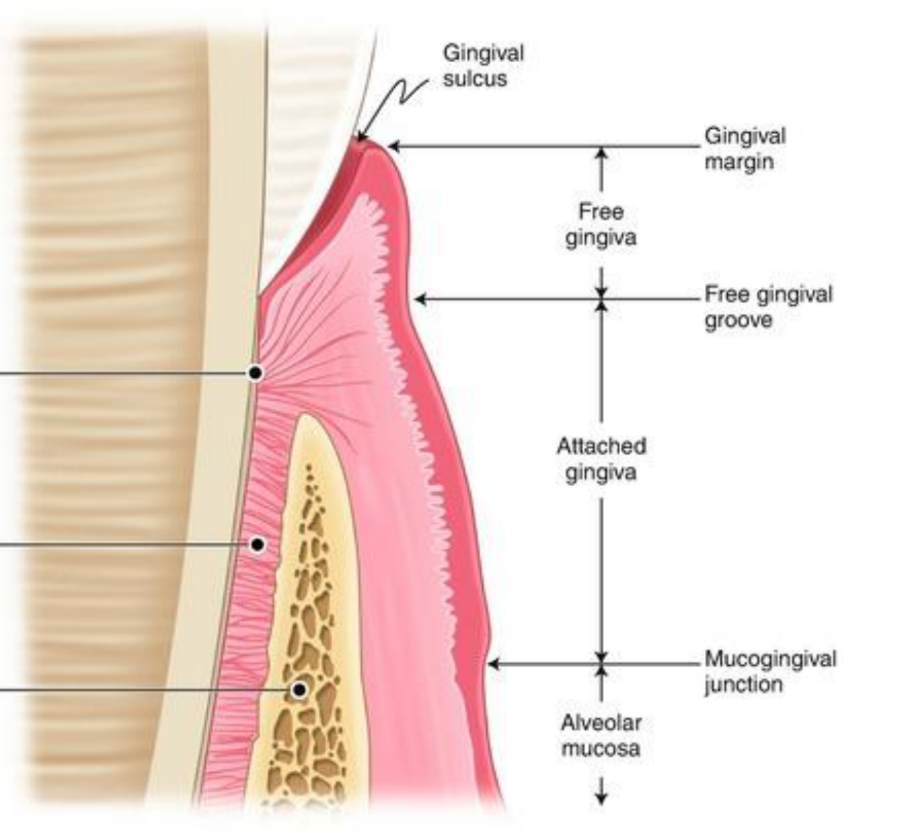
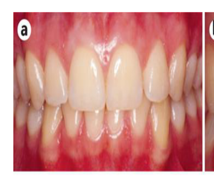
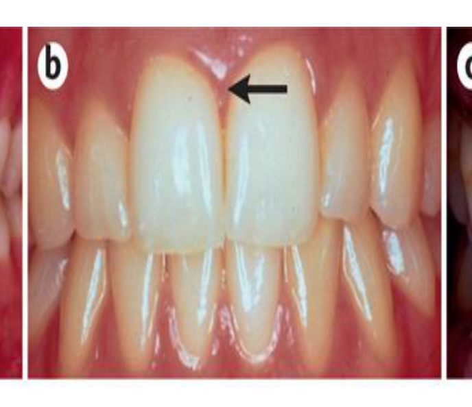
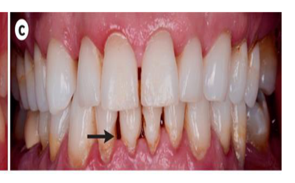
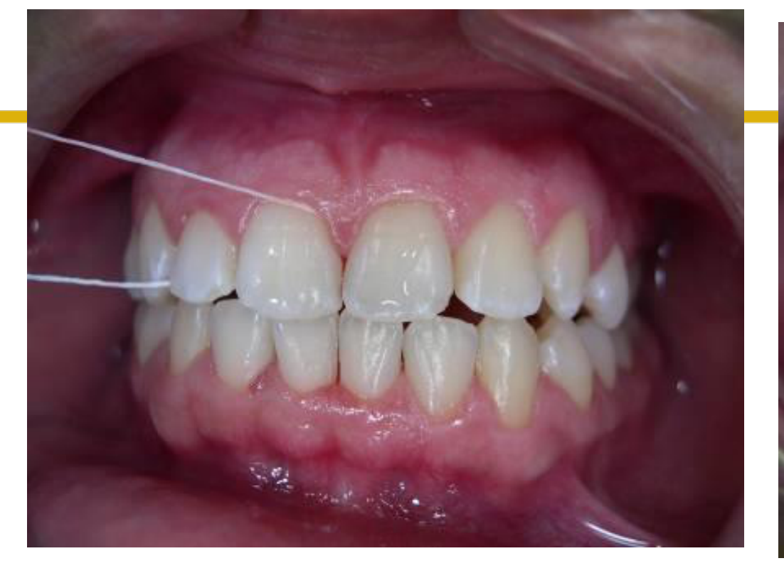
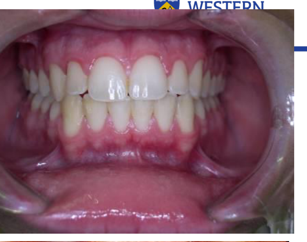
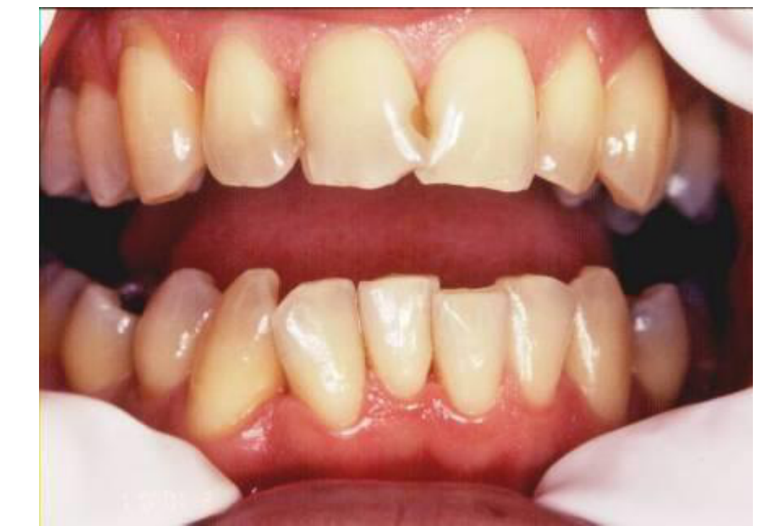
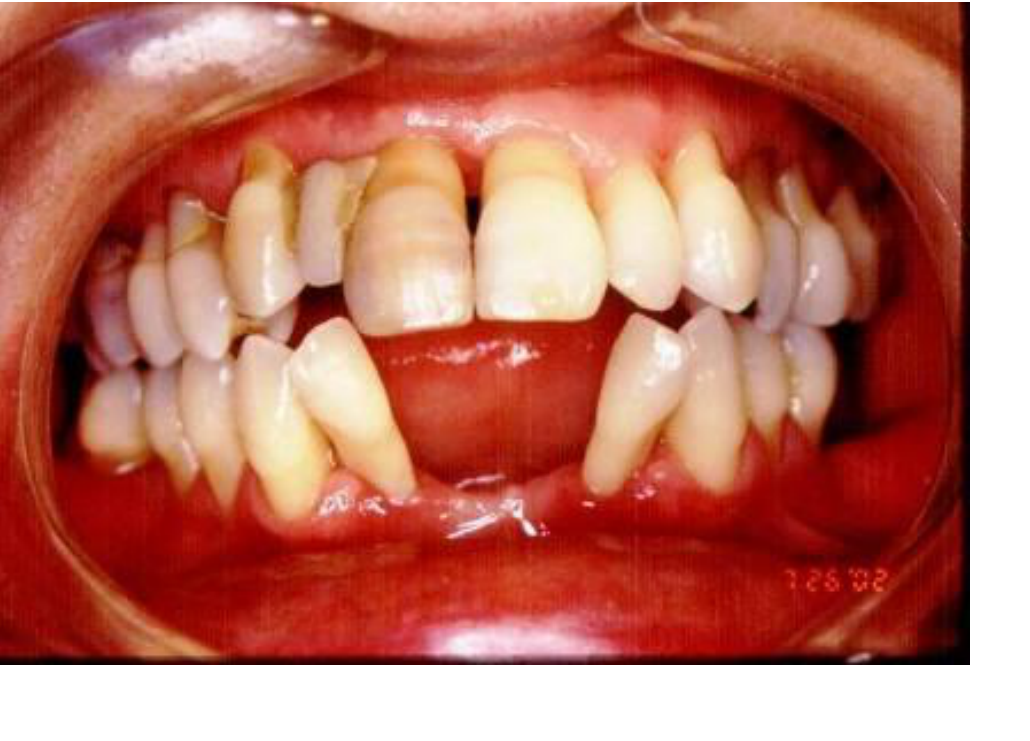
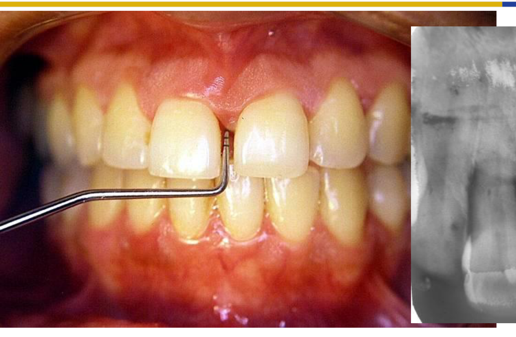
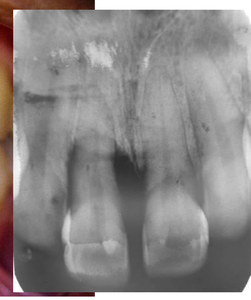
Periodontal Health and Disease Overview4567
Periodontium
Periodontal health and diseases
THE UNIVERSITY OF WESTERN AUSTRALIA
 |  |  |
 |  |  |
 |  |  |
 |
Periodontal Area Peculiarities8
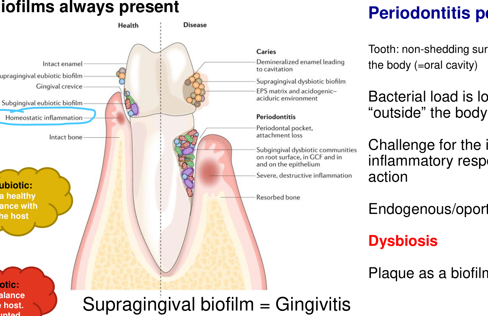
Biofilms always present
Health Disease
-
Intact enamel
-
Supragingival eubiotic biofilm
-
Gingival crevice
-
Subgingival eubiotic biofilm
-
Homeostatic inflammation
-
Intact bone
-
Supragingival dysbiotic biofilm
-
EPS matrix and acidogenic–aciduric environment
Periodontitis
- Periodontal pocket, attachment loss
- Subgingival dysbiotic communities on root surface, in GCF and in and on the epithelium
- Severe, destructive inflammation
- Resorbed bone
Caries
- Demineralized enamel leading to cavitation
Eubiotic: in a healthy balance with the host. Dysbiotic: When balance with the host is disrupted
- Supragingival biofilm = Gingivitis
- Subgingival biofilm = Periodontitis
Periodontitis peculiarities
- Tooth: non-shedding surface, partially outside the body (=oral cavity)
- Bacterial load is located generally “outside” the body
- Challenge for the immune-inflammatory response to take action
- Endogenous/opportunistic infection
- Dysbiosis
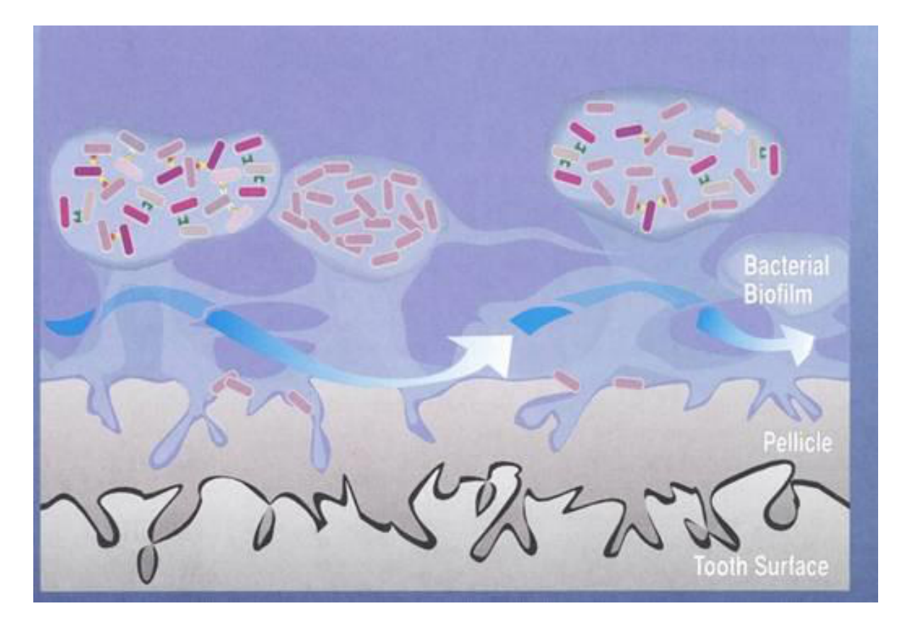
- Plaque as a biofilm
Hajishengallis et al 2015
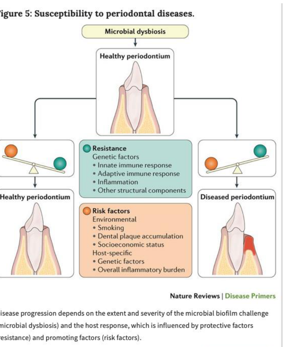
Microbial Dysbiosis and Host Defense Balance910
Dysbiosis
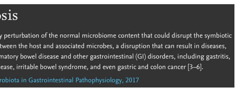
Dysbiosis is any perturbation of the normal microbiome content that could disrupt the symbiotic relationship between the host and associated microbes, a disruption that can result in diseases, such as inflammatory bowel disease and other gastrointestinal (GI) disorders, including gastritis, peptic ulcer disease, irritable bowel syndrome, and even gastric and colon cancer [3–6].
From: The Microbiota in Gastrointestinal Pathophysiology, 2017
Kinane, D. F. et al. (2017) Periodontal diseases Nat. Rev. Dis. Primers doi:10.1038/nrdp.2017.38
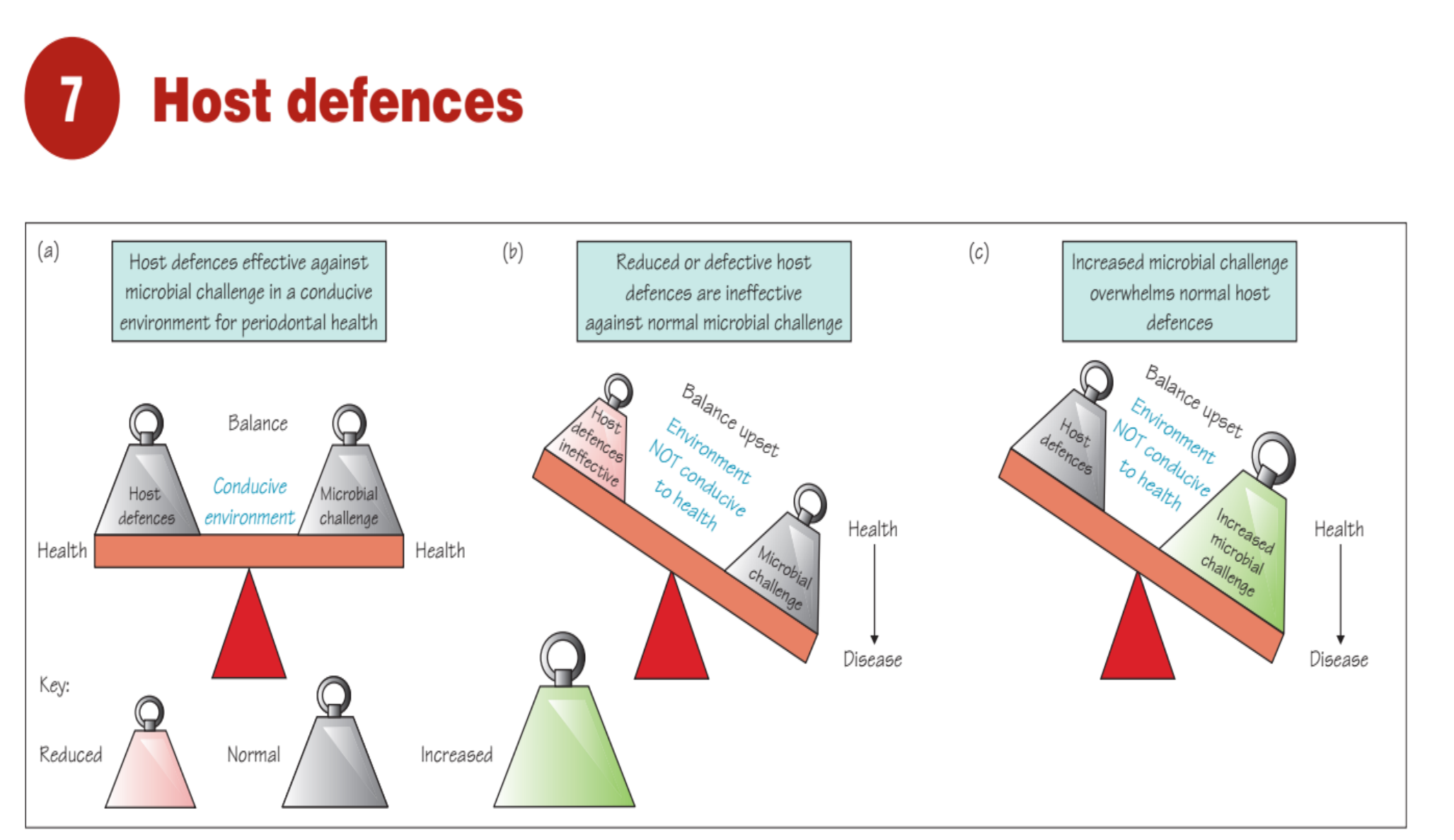
Host defences
(a) Host defences effective against microbial challenge in a conducive environment for periodontal health
Conducive environment
(b) Reduced or defective host defences are ineffective against normal microbial challenge
Host defences ineffective
Balance upset
Environment NOT conducive to health
(c) Increased microbial challenge overwhelms normal host defences
Host defences
Balance upset
Environment NOT conducive to health
Key:
- Reduced
- Normal
- Increased
Health -> Disease
Microbial challenge
Health -> Disease
Increased microbial challenge
Figure 7.1 Concept of balance between host defences, microbial challenge and environment. (a) Balance and periodontal health. (b) Host defences have a defect or are ineffective against microbial challenge, tipping the balance to periodontal destruction. (c) Microbial challenge overwhelms the host defences leading to an upset balance and periodontal destruction - this may relate to the environment not being conducive and/or changes in quality, quantity or virulence of microorganisms.
 |  |
 |
Current Etiopathogenesis Model11
Current PD etiopathogenesis model
The University of Western Australia
-
Behavourial risk factors absent
-
Environmental risk factors absent
-
Genetic risk factors absent
-
Epipenetic effects not evident
-
Behavourial risk factors present
-
Environmental risk factors evident
-
Genetic risk factors present
-
Enzigenetic effects evident
Clinical Health Gingivitis Periodontitis
Health Promoting biofilm = Symbiosis
Incipient Dysbiosis (Quorum Sensing Bacteria)
Frank Dysbiosis (Pathogenic Biofilm)
- Low biomass
- High biomass
- High biomass
Complement PMNs
Proportionate Host response
Antibody PMNs ++ T & B cells
Proportionate Host response
Antibody PMNs +++ Plasma cells
Acute Resolution of inflammation
Chronic Resolution of inflammation
Faled Resolution of inflammation
Antigens Bact’1 DNA fMLP
High biomass
Antigens Virulence Factors LPS
Gingivapinas LPS
Connective Tissue & Zone Damage
Dis-proportionate Host response (hyper-inflammatory)
DAMPs Haem GCF
Chronic non-Resolving inflammation
Cytokines Prostanoids MMPs Oxidative Stress
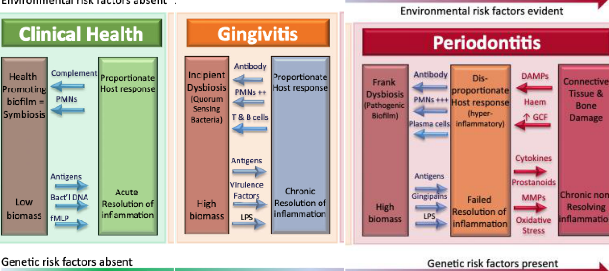
FIGURE 1 Contemporary model of host–microbe interactions in the pathogenesis of periodontitis, in which the host response drives an incipient dysbiosis (gingivitis). If the biofilm is not disrupted/removed, frank dysbiosis results and perpetuates a chronic nonresolving and destructive inflammation. DAMPs, damage-associated molecular patterns; fMLP, N- formylmethionyl- leucyl- phenylalanine; GCF, gingival crevicular fluid; LPS, lipopolysaccharide; MMPs, matrix metalloproteinases; PMNs, polymorphonuclear neutrophils. This figure is referred fromref. 106.
Murakami S, Mealey BL, Mariotti A,Chapple ILC. Dental plaque–induced gingival conditions.J C i n Periodontol. 2018;45(Suppl 20):S17–S2
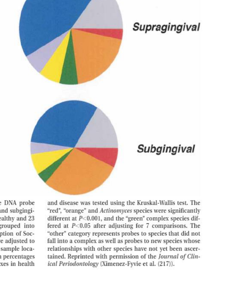
Periodontal Microbiota and Virulence Factors1213
Periodontal microbiota
- P. gingivalis
- B. forsythus
- T. denticola
- C. gracilis
- C. rectus
- C. showae
- E. nodatum
- F. nuc. nucleatum
- F. nuc. polymorphum
- P. intermedia
- P. micros
- P. nigrescens
- S. constellatus
- Actinomyces species
- V. parvula
- A. odontolyticus
- E. corrodens
- C. gingivalis
- C. sputigena
- C. ochracea
- C. concisus
- A. actino. a
- S. mitis
- S. oralis
- S. sanguis
- Streptococcus sp.
- S. gordonii
- S. intermedius
Periodontal Health
- Actinomyces
- other
Periodontitis
- Supragingival
- Subgingival
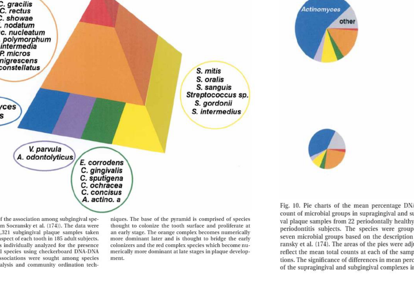
SOCRANSKY, S.S. and HAFFAJEE, A.D. (2002), Dental biofilms: difficult therapeutic targets. Periodontology 2000, 28: 12-55.
Virulence factors – perio microbiota
- Enzymes
- Waste products
- Proteinases
- Leukotoxins
- Lipopolysaccharides
EVADE HOST RESPONSES
Bacterial Biofilm on tooth surface with pellicle
 |  |
 |
Dento-Gingival Host Defenses
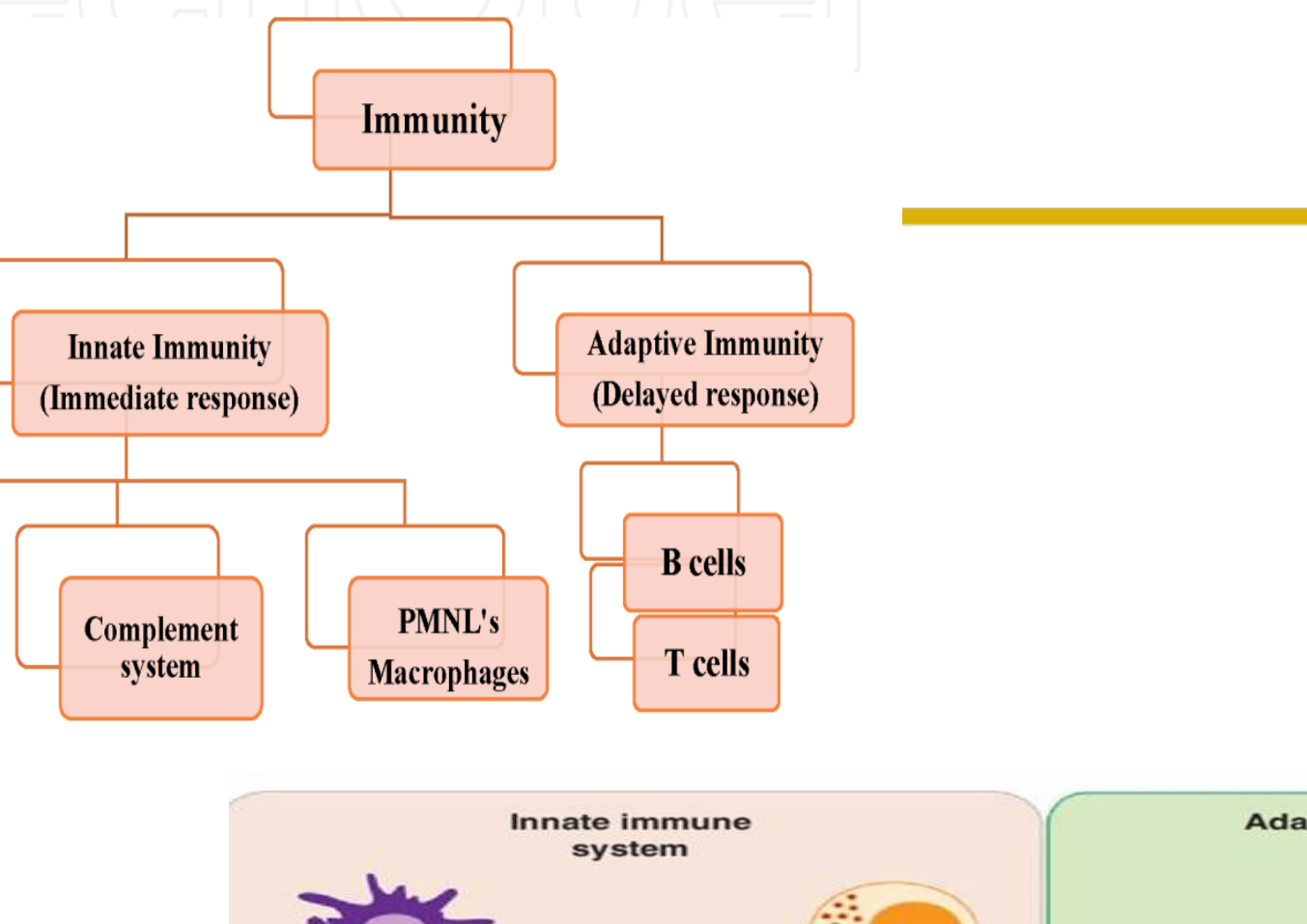
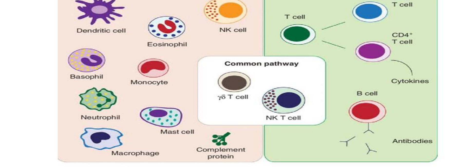
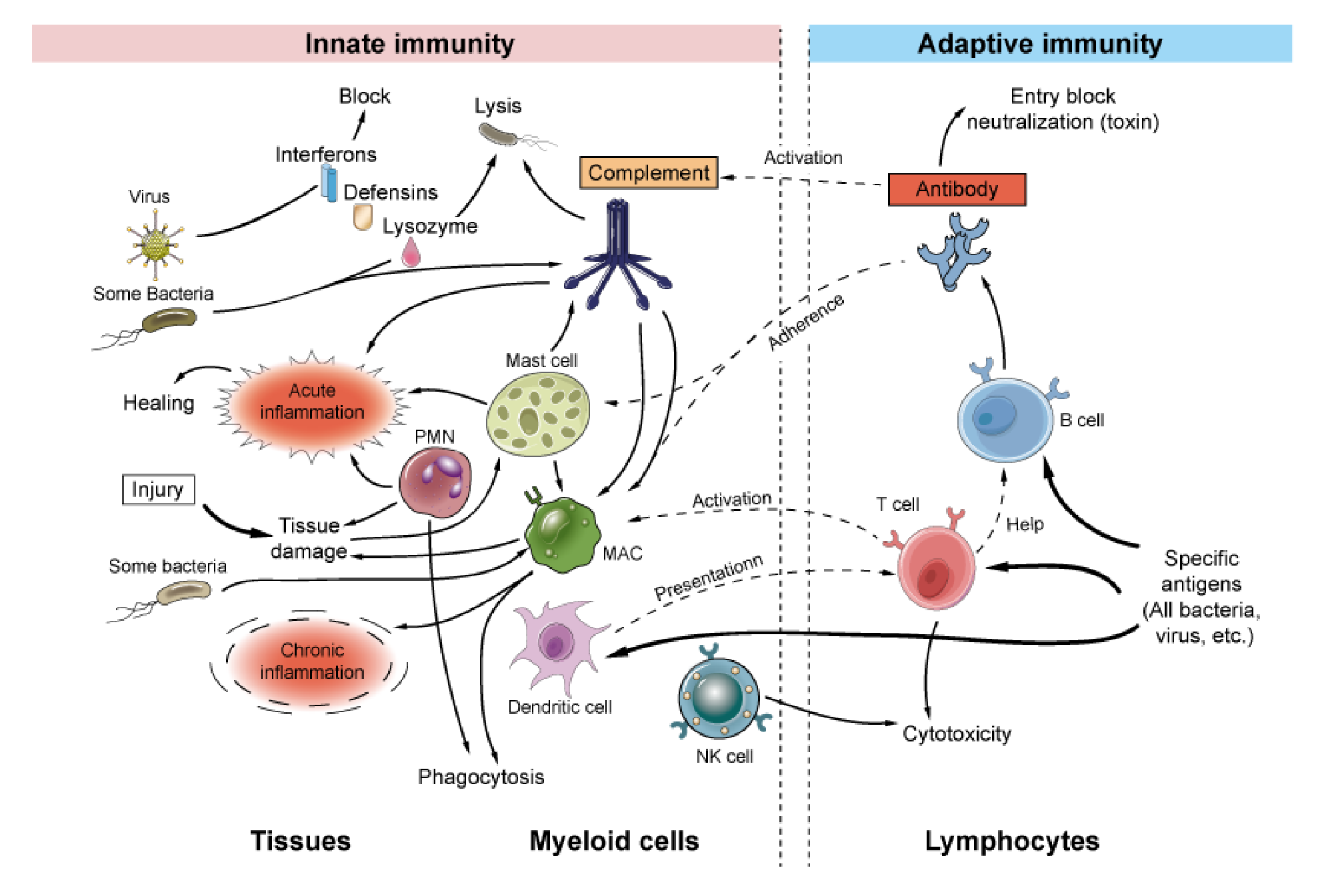
Innate and Adaptive Immunity Systems1415
Immunity
| Innate Immunity (Immediate response) | Adaptive Immunity (Delayed response) |
|---|---|
| Barriers, Complement system, PMNL’s, Macrophages | B cells, T cells |
Innate immune system
- Dendritic cell
- Eosinophil
- NK cell
- Basophil
- Monocyte
- Neutrophil
- Mast cell
- Macrophage
- Complement protein
- Common pathway
- γδ T cell
- NK T cell
Adaptive immune system
- T cell
- CD8+ T cell
- CD4+ T cell
- B cell
- Antibodies
- Cytokines
Innate immunity
- Virus -> Interferons -> Block
- Some Bacteria -> Defensins -> Lysis
- Some Bacteria -> Lysozyme -> Lysis
- Injury -> Tissue_damage -> Acute_inflammation -> Healing
- Some bacteria -> Tissue_damage -> Chronic_inflammation
- Complement -> Mast_cell -> Acute_inflammation
- Complement -> MAC -> Phagocytosis
- Complement -> Acute_inflammation
- PMN -> Phagocytosis
- Dendritic_cell -> Phagocytosis
- NK_cell -> Cytotoxicity
Adaptive immunity
- Specific antigens (All bacteria, virus, etc.) -> B_cell -> Antibody -> Entry_block/neutralization toxin
- Specific antigens -> T_cell
Interactions
- Antibody -> Adherence -> MAC
- Antibody -> Activation -> Complement
- MAC -> Activation -> T_cell
- Dendritic_cell -> Presentation -> T_cell
- T_cell -> Help -> B_cell
 |  |
 |
Saliva and Complement Pathways1617
Dento–gingival host defenses
THE UNIVERSITY OF WESTERN AUSTRALIA
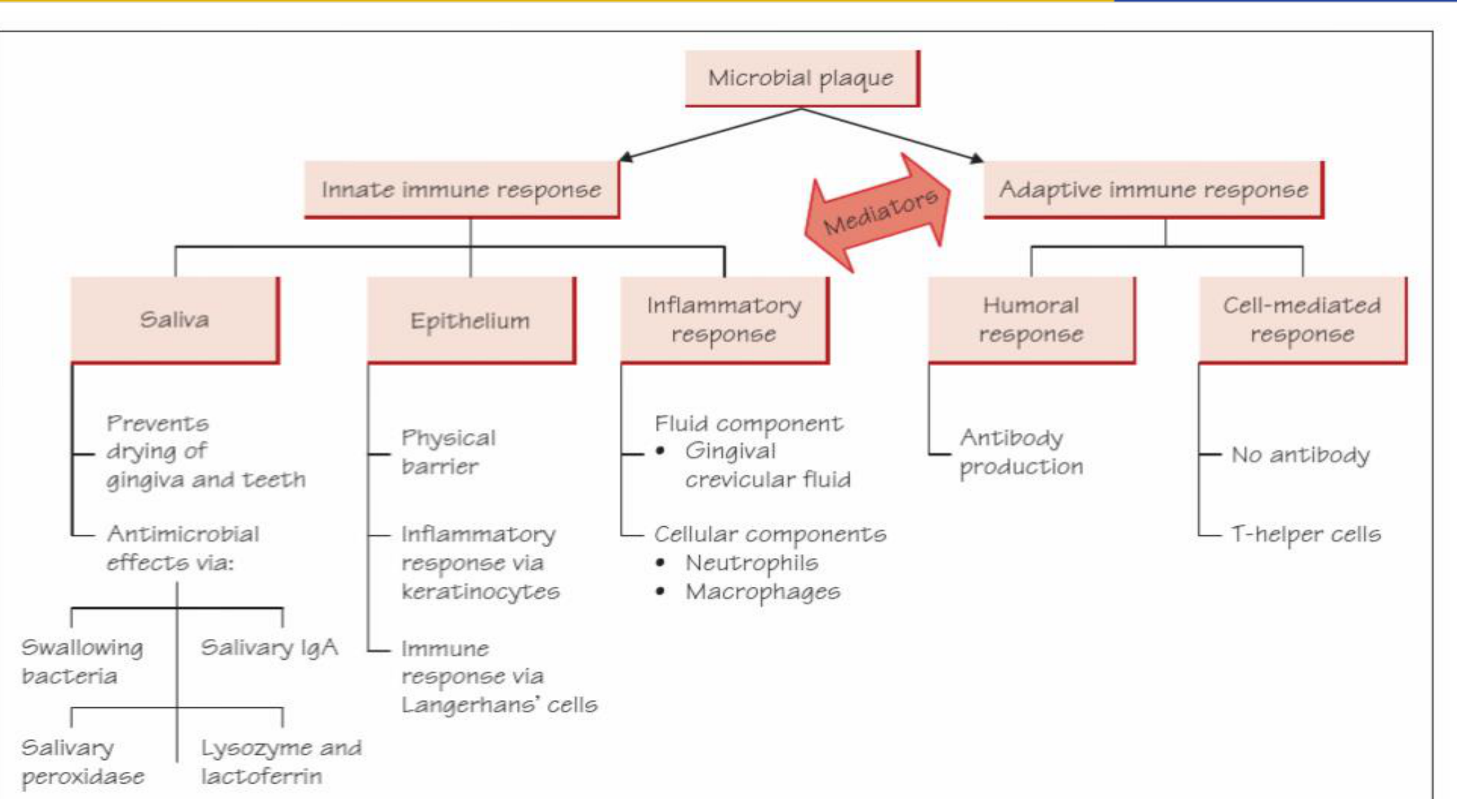
Fig 7.2 Host defences against microbial plaque.
| Innate immune response & Adaptive immune response | Mediators | |
|---|---|---|
| Saliva | Epithelium | Inflammatory response |
| Prevents drying of gingiva and teeth | Physical barrier | Fluid component |
| Antimicrobial effects via: | Inflammatory response via keratinocytes | Gingival crevicular fluid |
| Swallowing bacteria | Immune response via Langerhans’ cells | Cellular components |
| Salivary IgA | Neutrophils | |
| Salivary peroxidase | Macrophages | |
| Lysozyme and lactoferrin |
Clerehugh et al 2013
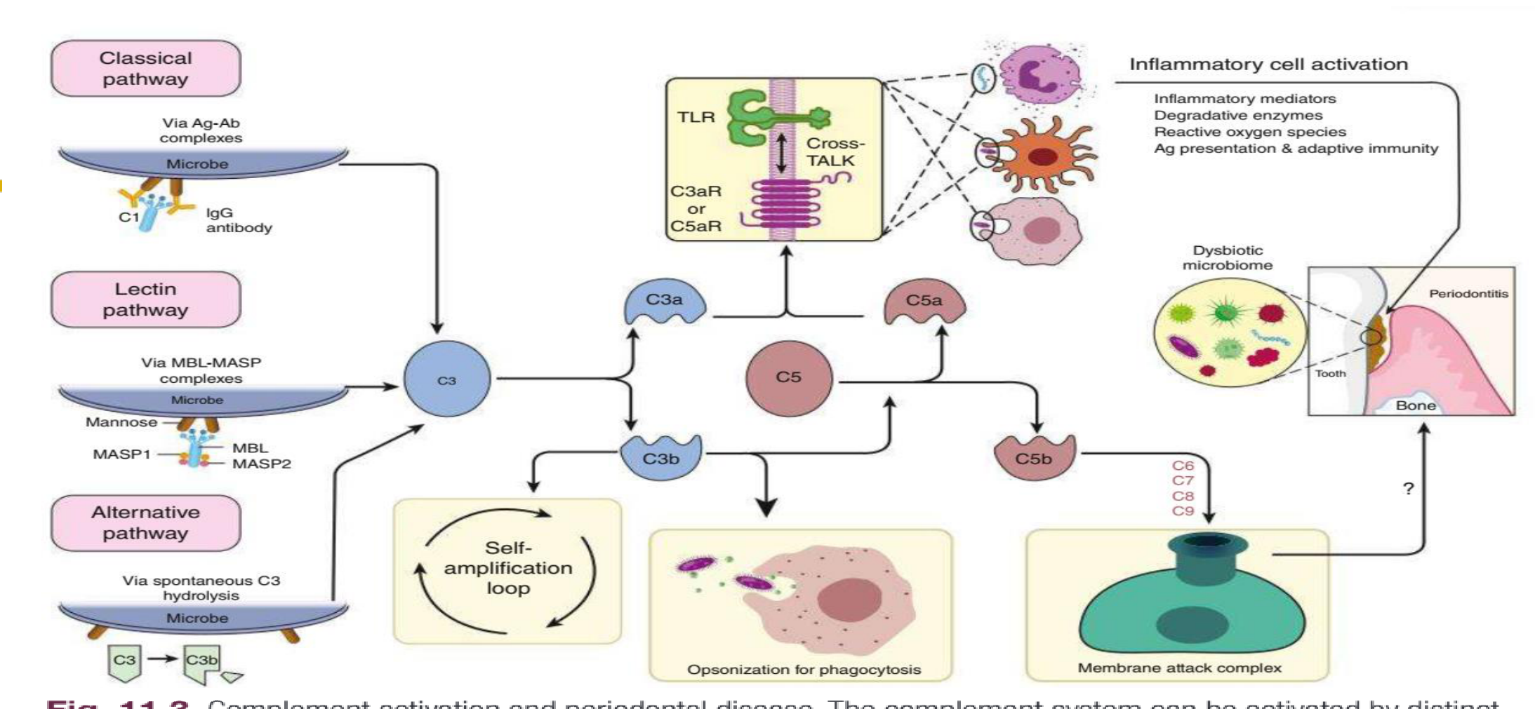
Classical pathway Via Ag-Ab complexes Microbe -> C1 -> IgG antibody
Lectin pathway Via MBL-MASP complexes Microbe -> Mannose -> MASP1 -> MBL -> MASP2
Alternative pathway Via spontaneous C3 hydrolysis Microbe -> C3 >> C3b
C3 Self-amplification loop
- C3a
- C3b -> Opsonization for phagocytosis
- C5 -> C5a -> C5b -> C6 -> C7 -> C8 -> C9 -> Membrane attack complex
TLR C3aR or C5aR -> Cross-TALK -> Inflammatory cell activation -> Inflammatory mediators, Degradative enzymes, Reactive oxygen species, Ag presentation & adaptive immunity
Dysbiotic microbiome -> Periodontitis -> Tooth -> Bone
Fig. 11.3 Complement activation and periodontal disease. The complement system can be activated by distinct initiation mechanisms: The classical pathway is triggered by antigen-antibody complex-mediated activation of the C1 complex. The lectin pathway is initiated when complexes of mannose-binding lectin (MBL) and MBL-associated serine proteases (MASPs) recognize and adhere to microbial surfaces. The alternative pathway is triggered by a “tick-over” mechanism that involves spontaneous C3 hydrolysis, which occurs in the absence of complement regulatory molecules (as is typically the case with foreign surfaces such as microbial cells). In the so-called alternative pathway-amplification loop, additional C3 is cleaved into even more C3b which further fuels the loop, thereby amplifying complement activation irrespective of the initiating mechanism. All three mechanisms of complement initiation and amplification converge at C3. The downstream effects of C3 activation include the generation of effectors that promote inflammation (C3a and C5a), opsonization for phagocytosis (C3b) and the generation C5b-C9 membrane attack complex (MAC). MAC can lyse susceptible targeted bacteria but has also been implicated in destructive inflammation. Whereas the role of MAC in periodontitis is uncertain, C3a and C5a activate specific G-protein-coupled receptors (C3aR and C5aR1), which cross-talk with Toll-like receptors (TLRs). This cross-talk interaction between complement and TLRs activates synergistically inflammatory leukocytes, which directly or indirectly mediate destructive inflammation that leads to periodontal tissue breakdown and alveolar bone loss in periodontitis.
 |  |
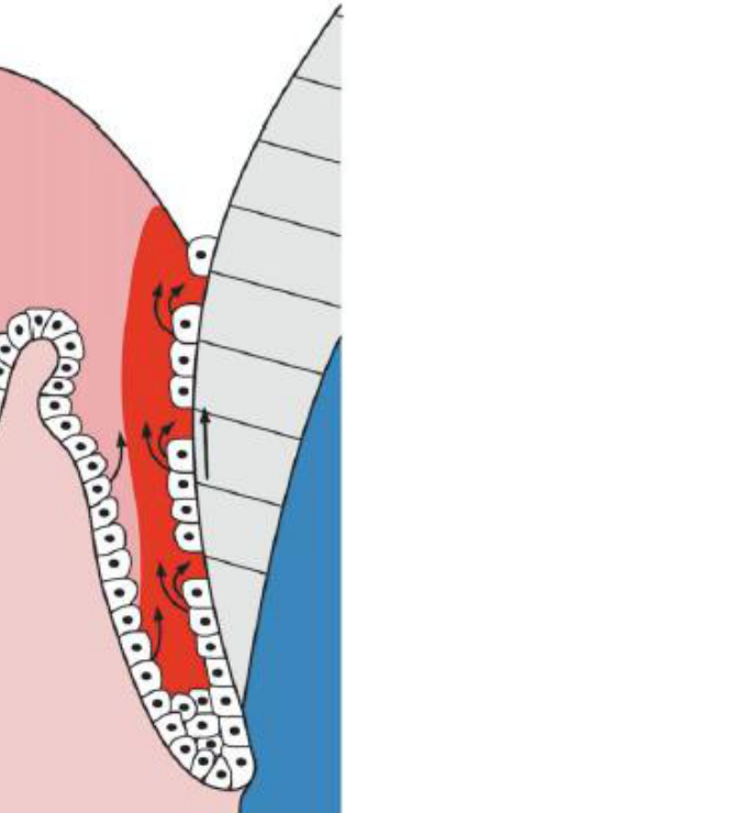
Epithelial Barriers and Gingival Crevicular Fluid1819
Epithelium
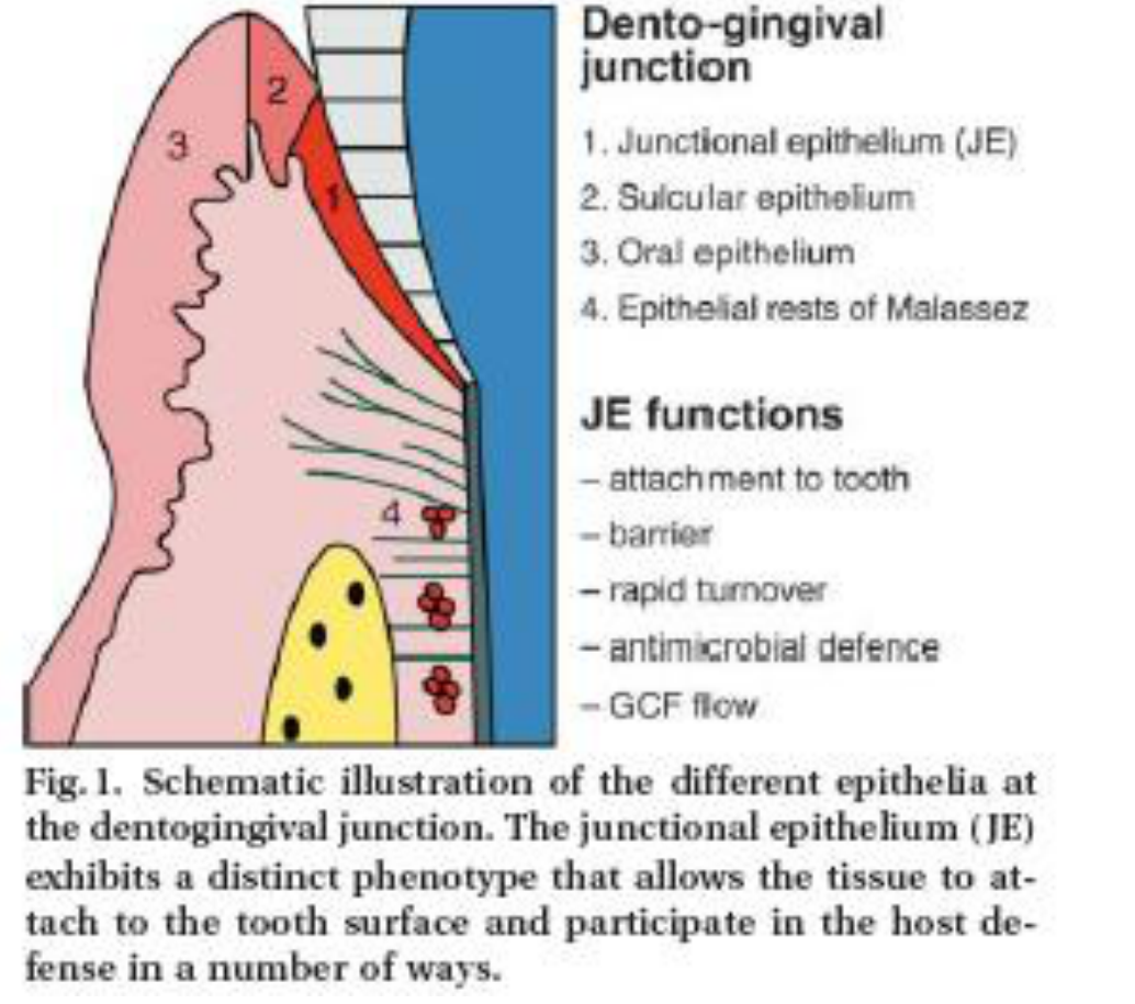
Dento-gingival junction
- Junctional epithelium (JE)
- Sulcular epithelium
- Oral epithelium
- Epithelial rests of Malassez
JE functions
- attachment to tooth
- barrier
- rapid turnover
- antimicrobial defence
- GCF flow
Physical barrier: turnover + peeling
Biological barrier: defensins, IL-8, PMN
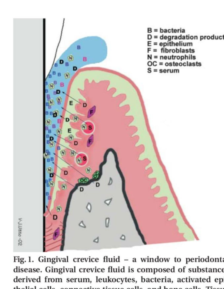
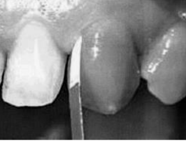
Gingival crevicular fluid (GCF)
Inflammatory exudate released through the crevice, resulting from the increase in permeability of vessels next to the JE and SE.
Composed of: plasma derived substances (antibodies, cytokines, enzymes), epithelium and immune cells, bacteria.
GCF volume and flow increase with increasing inflammation.
 |  |
 |  |
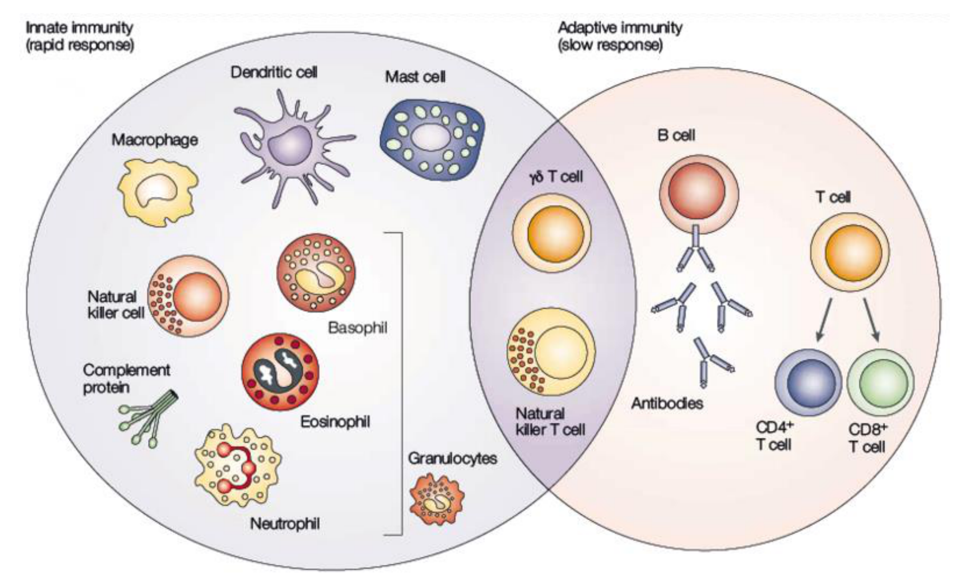
Cellular Components of the Immune Response20
THE UNIVERSITY OF WESTERN AUSTRALIA
Innate immunity (rapid response)
- Dendritic cell
- Mast cell
- Macrophage
- Natural killer cell
- Complement protein
- Basophil
- Eosinophil
- Neutrophil
- Granulocytes
Adaptive immunity (slow response)
- γδ T cell
- Natural killer T cell
- B cell
- Antibodies
- T cell
- CD4+ T cell
- CD8+ T cell
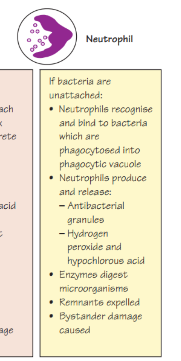
Neutrophil Function and Phagocytosis2122
First defense cells (innate immunity)
Figure 7.3 The role of neutrophils in phagocytosis or the killing of microorganisms.
If bacteria are in plaque matrix:
- Neutrophils attach to plaque matrix
- Neutrophils secrete externally:
- Enzymes
- Hydrogen peroxide and hypochlorous acid
- Neutrophils kill bacteria without phagocytosis
- Plaque matrix dissolved
- Washed away by gingival crevicular fluid
- Bystander damage caused
If bacteria are unattached:
- Neutrophils recognise and bind to bacteria which are phagocytosed into phagocytic vacuole
- Neutrophils produce and release:
- Antibacterial granules
- Hydrogen peroxide and hypochlorous acid
- Enzymes digest microorganisms
- Remnants expelled
- Bystander damage caused
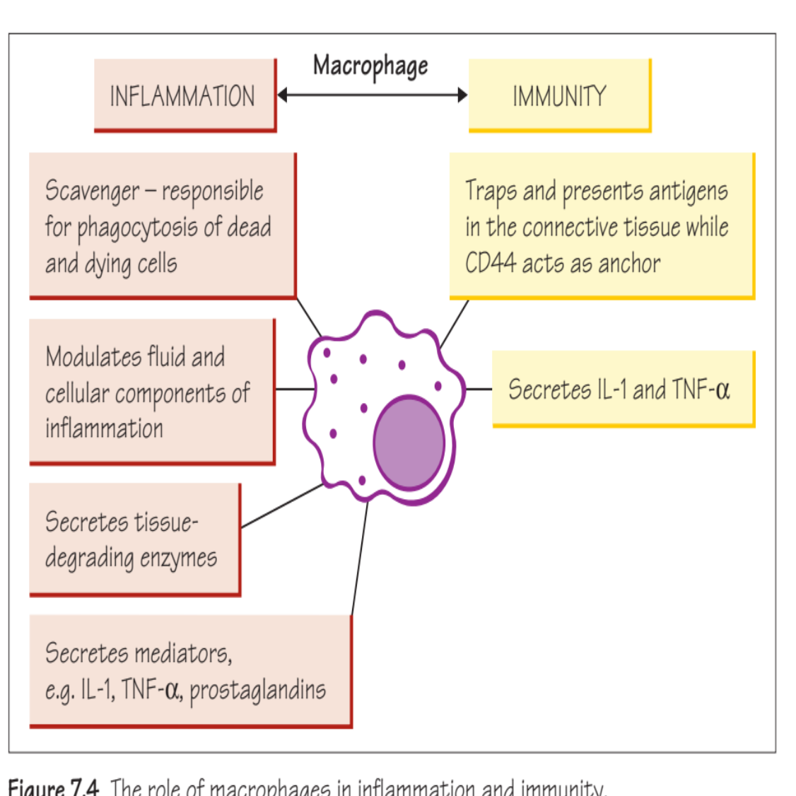
Figure 7.4 The role of macrophages in inflammation and immunity.
INFLAMMATION
- Scavenger - responsible for phagocytosis of dead and dying cells
- Modulates fluid and cellular components of inflammation
- Secretes tissue-degrading enzymes
- Secretes mediators, e.g. IL-1, TNF-α, prostaglandins
IMMUNITY
- Traps and presents antigens in the connective tissue while CD44 acts as anchor
- Secretes IL-1 and TNF-α
Clerehugh et al 2013
The text on the page only makes sense in the context of the diagram (labels such as “Complement,” “Antibody,” “T-cell,” “MMP-8,” etc.), and its meaning is entirely dependent on the visual elements. Per the instructions, the entire content should be treated as a single figure.
 |  |
 |  |
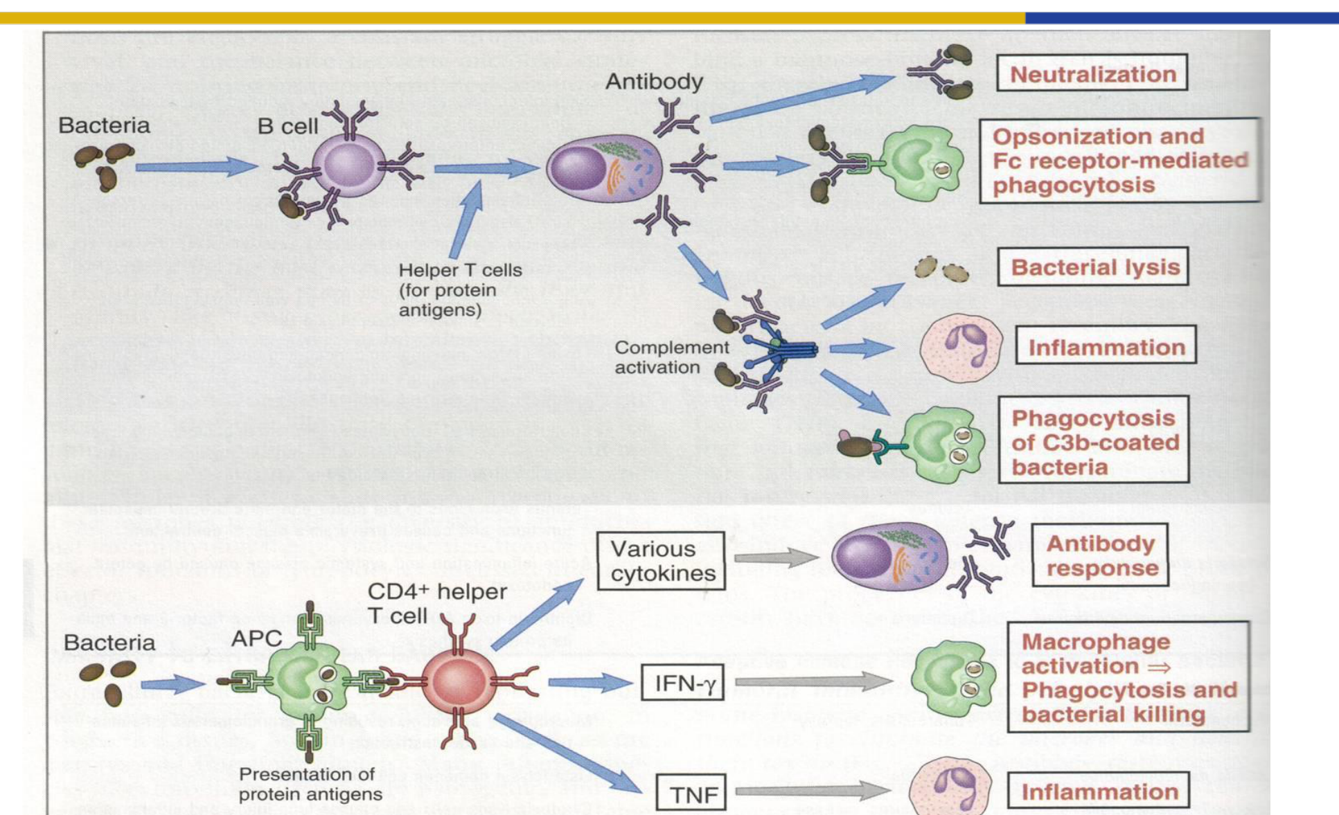
Humoral and Cell-Mediated Responses23242526
Adaptive immunity
Neutralization
Opsonization and Fc receptor-mediated phagocytosis
Bacterial lysis
Inflammation
Phagocytosis of C3b-coated bacteria
Antibody response
Macrophage activation ⇒ Phagocytosis and bacterial killing
Inflammation
Figure 15–1 Adaptive immune responses to extracellular microbes. Adaptive immune responses to extracellular microbes, such as bacteria, and their toxins consist of antibody production and the activation of CD4+ helper T cells. Antibodies neutralize and eliminate microbes and toxins by several mechanisms. Helper T cells produce cytokines that stimulate B cell responses, macrophage activation, and inflammation. APC, antigen-presenting cell; INF-γ, interferon-γ; TNF, tumor necrosis factor.
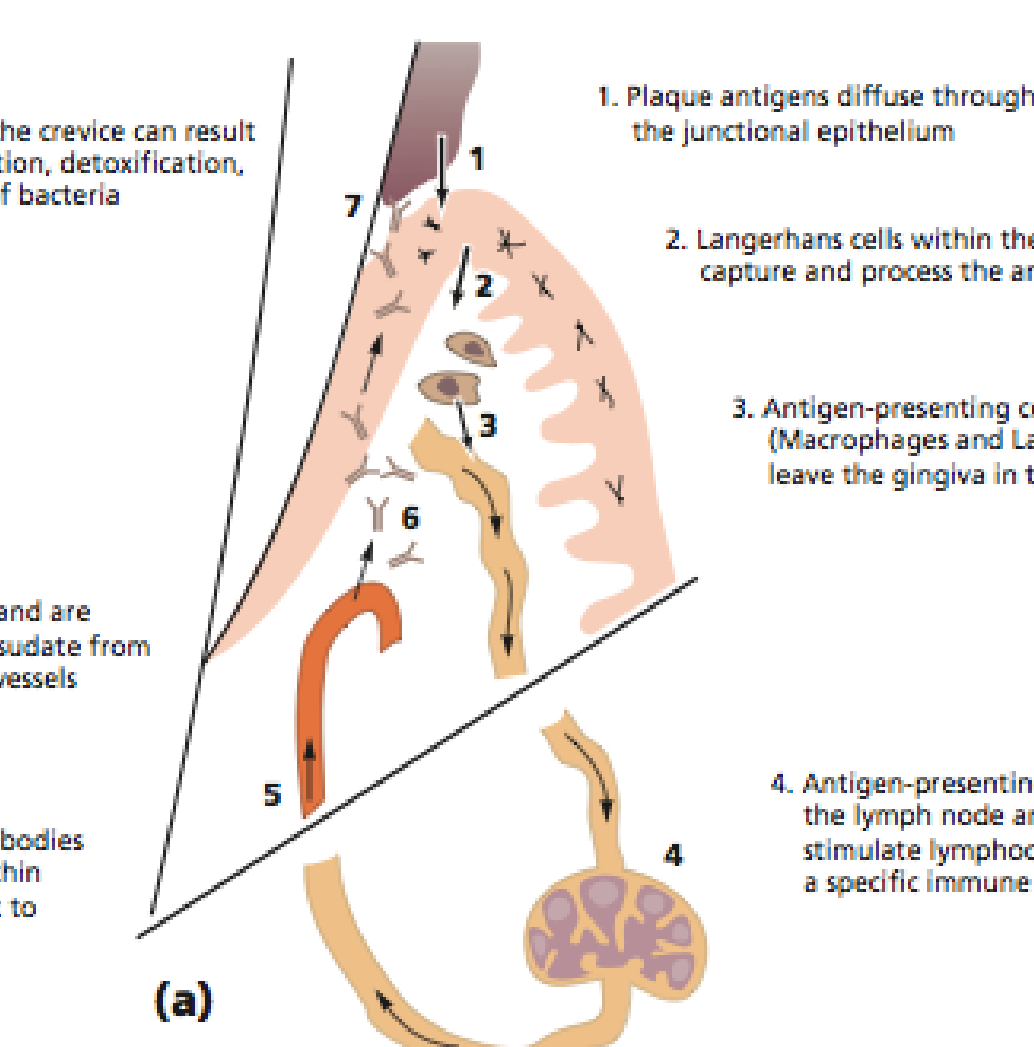
INNATE & ADAPTIVE IMMUNITY
HUMORAL RESPONSE
- Plaque antigens diffuse through the junctional epithelium
- Langerhans cells within the epithelium capture and process the antigens
- Antigen-presenting cells (Macrophages and Langerhans cells) leave the gingiva in the lymphatics
- Antigen-presenting cells reach the lymph node and begin to stimulate lymphocytes to produce a specific immune response
- Periodontal microbe specific antibodies are produced by plasma cells within the lymph nodes and travel back to the gingiva via blood vessels
- Antibodies leave the circulation and are carried to the crevice in the transudate from the inflamed and dilated blood vessels
- Antibody action on microbes in the crevice can result in killing, aggregation, precipitation, detoxification, opsonization and phagocytosis of bacteria
The University of Western Australia
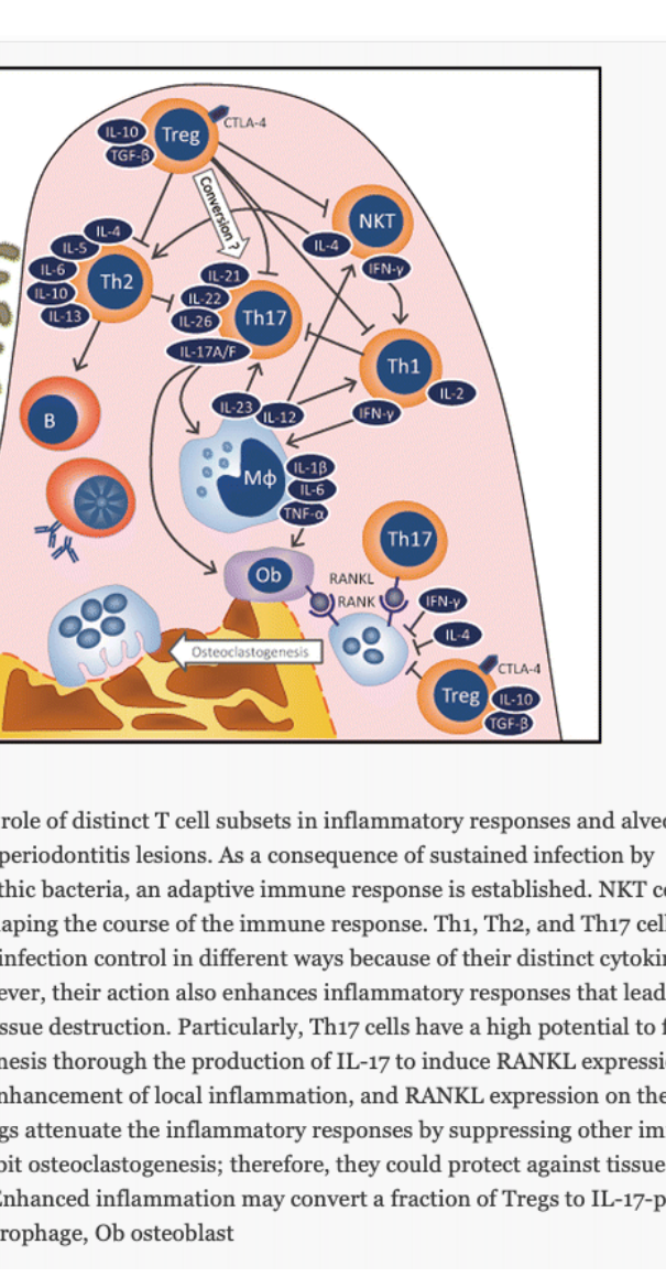
Adaptive - Cell-mediated response
Antigen penetration of JE => APC + T cell naïve =>
Different subsets of T-helper cells (Th1, Th2, Th17, T reg) proliferate and release different cytokines profiles
- Th1: IL-2, IFN-g
- Th2: IL-4, IL-5
- Th17: IL-17
- T reg: IL-10, TGF-b
Cytokines act on other cells (PMN, macrophages, B or T cells) to stimulate, inhibit or kill
T-helper cells on re-exposure proliferate and produce cytokines (memory)
Fig. 1 The potential role of distinct T cell subsets in inflammatory responses and alveolar bone resorption in periodontitis lesions. As a consequence of sustained infection by periodontopathic bacteria, an adaptive immune response is established. NKT cells are involved in shaping the course of the immune response. Th1, Th2, and Th17 cells contribute to infection control in different ways because of their distinct cytokine profiles. However, their action also enhances inflammatory responses that lead to periodontal tissue destruction. Particularly, Th17 cells have a high potential to facilitate osteoclastogenesis through the production of IL-17 to induce RANKL expression on osteoblasts, enhancement of local inflammation, and RANKL expression on themselves. However, Tregs attenuate the inflammatory responses by suppressing other immune cells and inhibit osteoclastogenesis; therefore, they could protect against tissue destruction. Enhanced inflammation may convert a fraction of Tregs to IL-17-producing cells. Mφ macrophage, Ob osteoblast
 |  |
 |  |
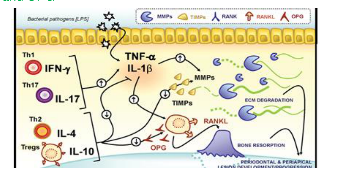
Mechanisms of Tissue Destruction27
Immune response = cells+mediators
- Main aim is to control infection in different ways
- Connective tissue destruction in periodontitis occurs as a “side-effect” of the immune-inflammatory response
- Perio ligament and bone destruction in periodontitis:
- Biofilm essential, but not sufficient
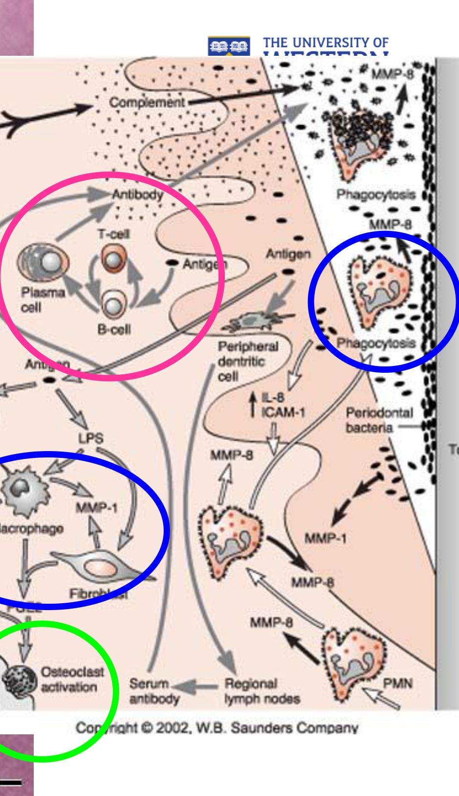
- High levels of pro-inflammatory cytokines (IL1b, TNFa), prostaglandins, MMP’s, RANKL
- Low levels of anti-inflammatory mediators (IL-10, TGF-b, TIMPs and OPG
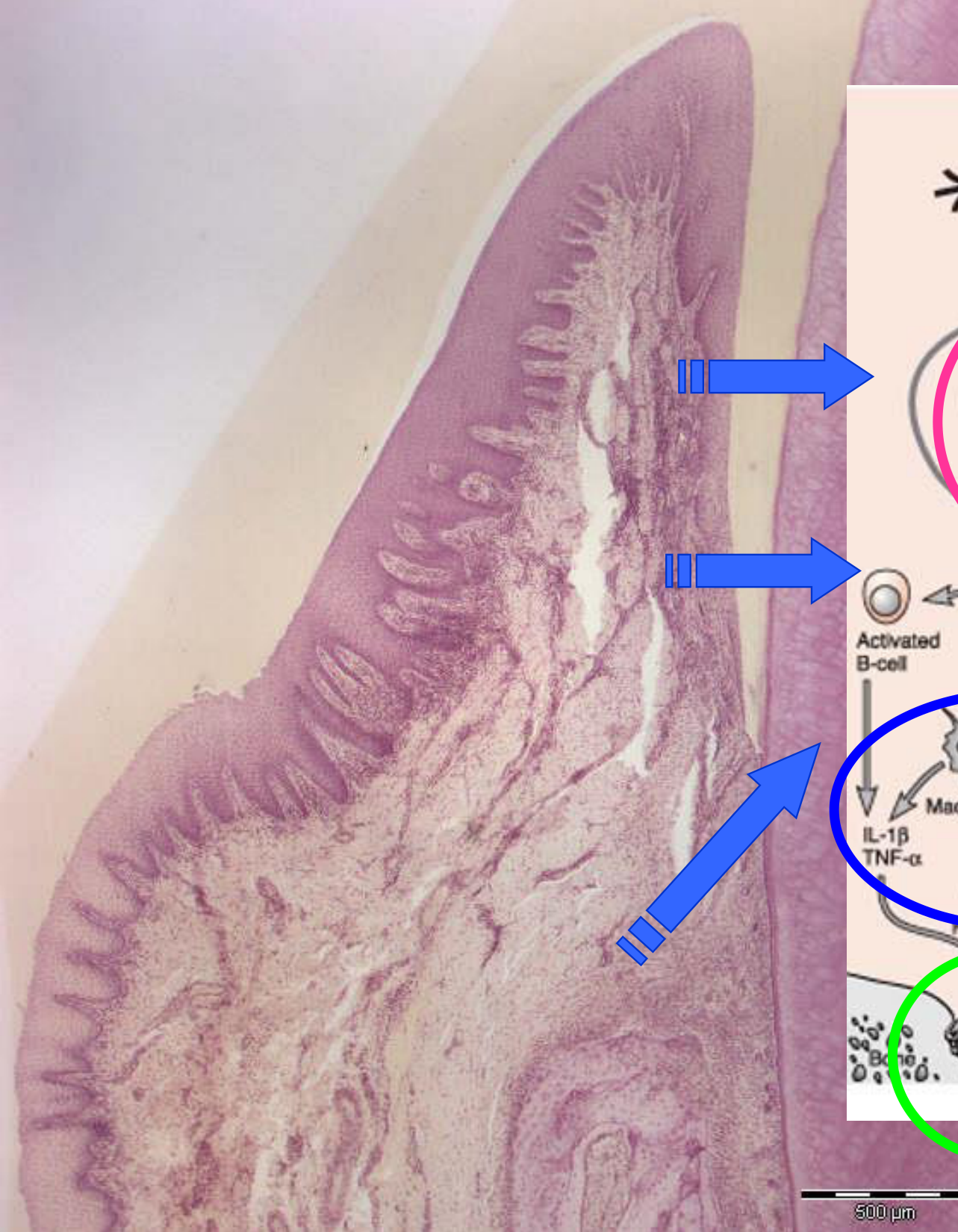
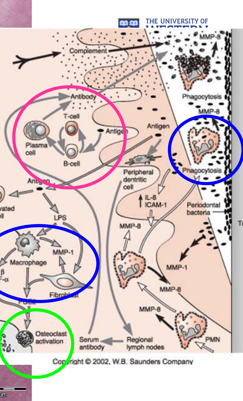
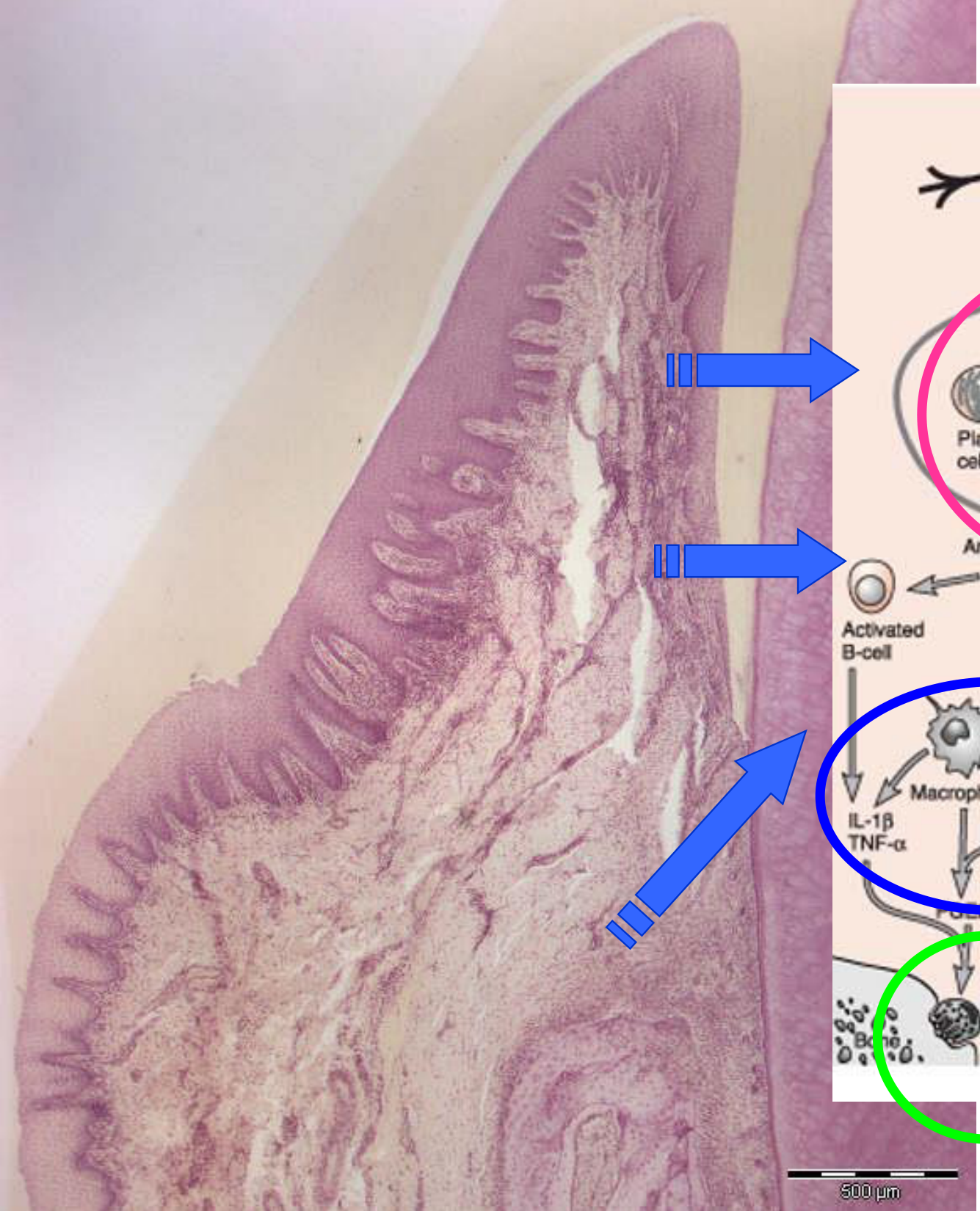
Pro-inflammatory Mediators and Bone Resorption282930
Periodontal bacteria -> LPS -> Activated B-cell Periodontal bacteria -> Peripheral dentitic cell -> T-cell -> B-cell -> Plasma cell -> Antibody -> Complement Periodontal bacteria -> Phagocytosis -> MMP-8 -> PMN
LPS -> Macrophage -> IL-1β TNF-α -> Fibroblast -> MMP-1 -> PMN LPS -> Macrophage -> IL-1β TNF-α -> Osteoclast activation -> Bone
T-cell -> B-cell B-cell -> T-cell
IL-8 ICAM-1 -> PMN
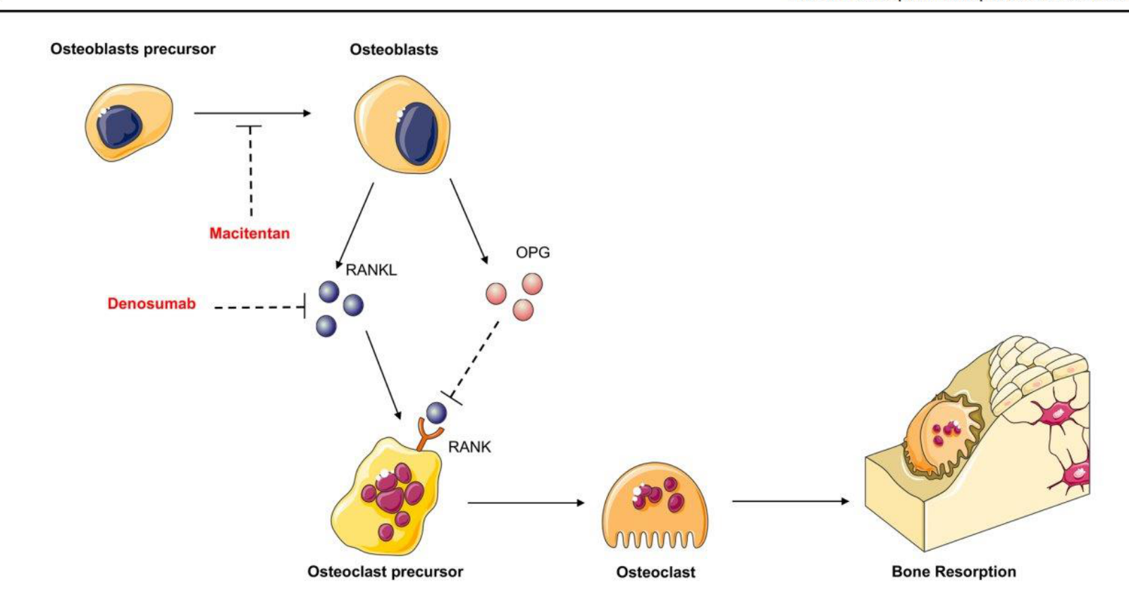
332 Current Osteoporosis Reports (2023) 21:330–343
Macitentan
Denosumab
 |  |
 |  |
Osteoclast Activation and RANKL Regulation3132
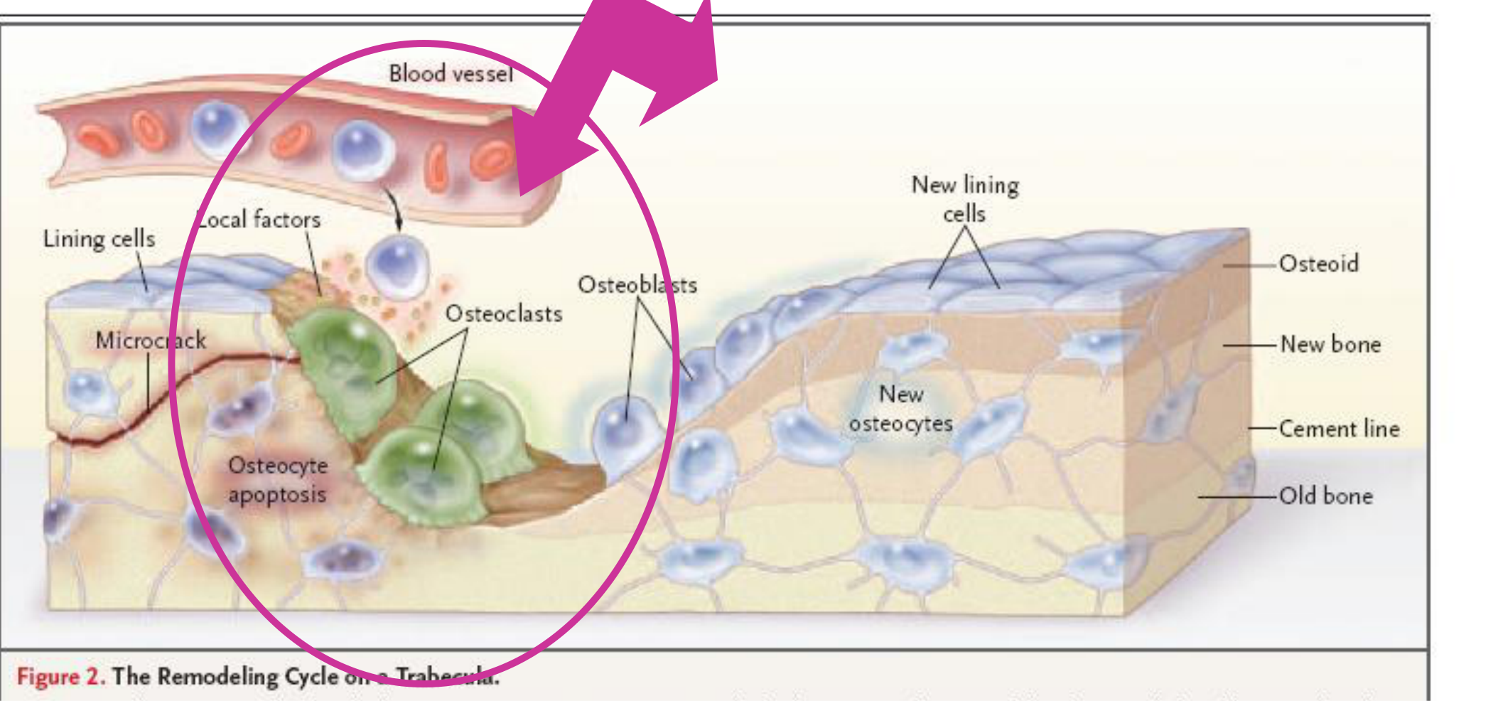
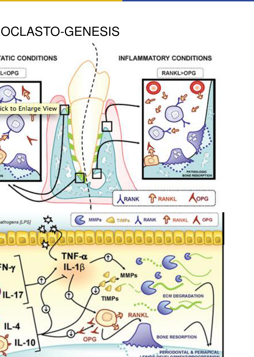
OSTEOCLASTS ACTIVATION
RANKL & OSTEOPROTEGERIN OPG STIMULATE OSTEOCLASTOGENESIS AND BONE RESORPTION.
RANKL /OPG HIGH RATIO PRORESORPTIVE.
RANKL /OPG LOW RATIO ANTIRESORPTIVE.
ACTIVATED T AND B CELLS CELLULAR SOURCE FOR RANKL
IFN-γ AND IL-17 INCREASE RANKL
IL-4 & IL-10 REDUCE RANKL/OPG RATIO
LYMPHOCYTES SUBSETS TH1 & TH17 PRO RESORPTIVE
TH2 & Tregs (Regulatory T) antiresorptive
 |  |
 |  |
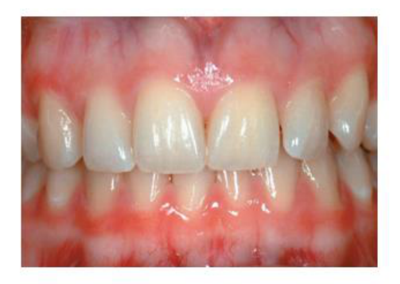
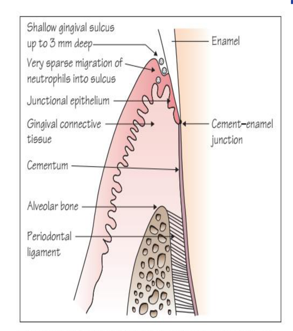
Histopathology of Periodontal Diseases33
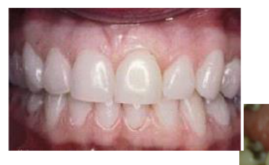
PRISTINE GINGIVA x Histologic Perfection
 |  |
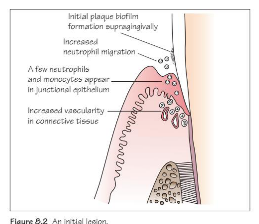
The Initial Lesion and Gingival Health3435
- Clinically healthy gingiva with a limited coronal infiltrate (5-10% of the connective tissue area)
- Neutrophils and monocytes in JE
- Lymphocytes in the connective tissue
- Increase in vascular structures near JE
- Exudative fluid from vessels to tissues = GCF
INITIAL LESION histologically = gingival health clinically
- 24 HOURS PLAQUE ACCUMULATION
- CHANGE IN MICROVASCULAR PLEXUS JE
- ARTERIOLAS, CAPILLARIES AND VENULES DILATION
- HYDROSTATIC PRESSURE INCREASE
- INCREASED PERMEABILITY
- EXUDATE OF FLUIDS & PROTEINS
- INCREASED GCF
- ENHANCED PMNS MIGRATION
- PMNS ACCUMULATE IN JE AND SULCUS
 |  |
The Early Lesion and Early Gingivitis36
EARLY LESION x Early Gingivitis (The University of Western Australia logo)
- One week after plaque accumulation
- JE blood vessels remain dilated
- Increased in number and size of vasculature
- Infiltrate predominant PMNs and lymphocytes = now 15-20% volume
- Very few plasma cells
- Initial fibroblast degeneration and collagen destruction
- Proliferation of basal and JE cells
- Coronal rete pegs
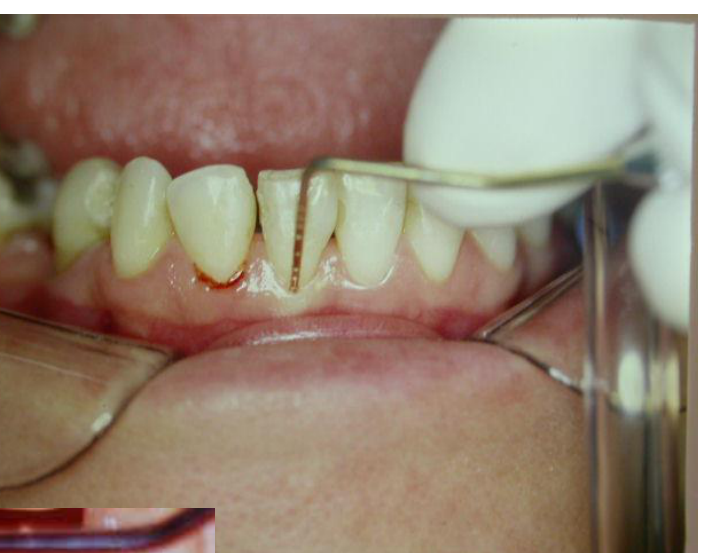
- Inflammation clinically detected – bleeding – due to pocket epithelium ulceration
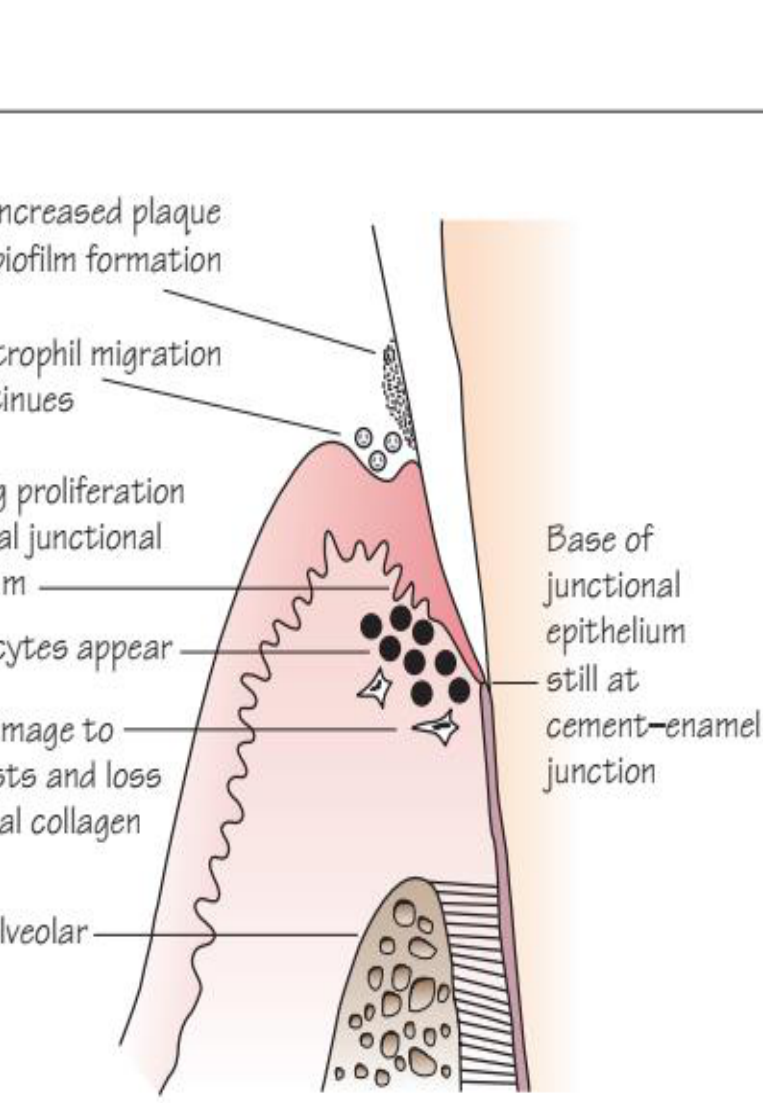
Figure 8.4 An early lesion. (Labels pointing to diagram text):
- Increased plaque biofilm formation
- Neutrophil migration continues
- Rete peg proliferation in coronal junctional epithelium
- Lymphocytes appear
- Early damage to fibroblasts and loss of gingival collagen
- Intact alveolar bone
- Base of junctional epithelium still at cement-enamel junction
pocket epithelium ulceration
The Established Lesion and Chronic Gingivitis373839
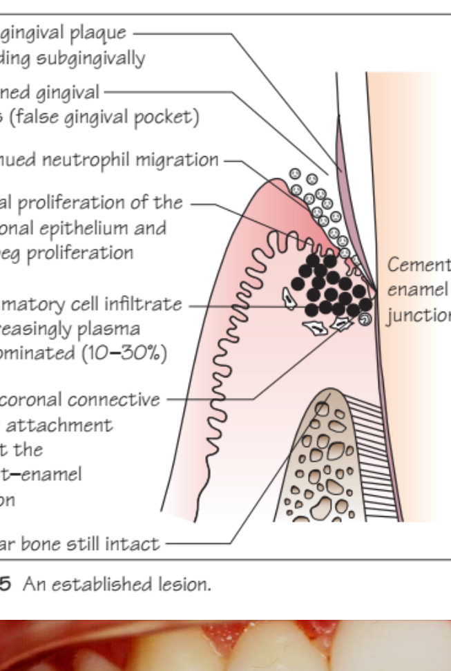
- Increased fluid and leukocyte migration
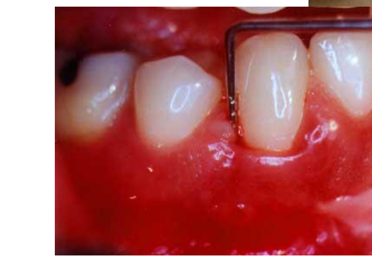
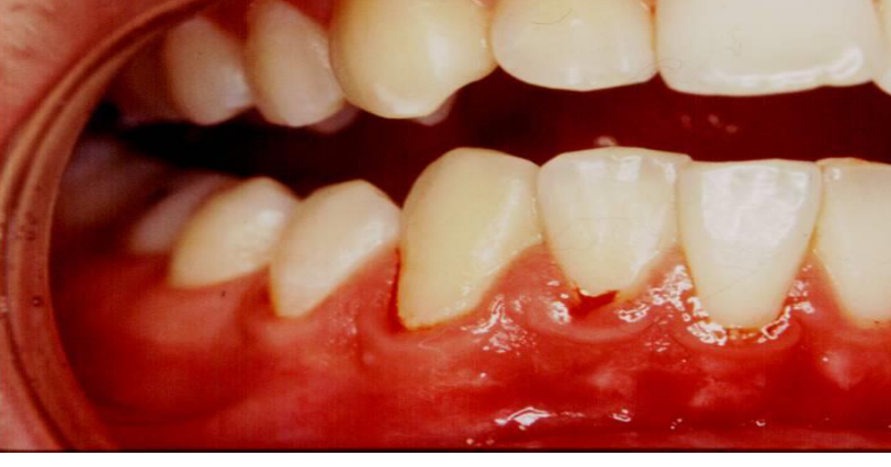
- More edematous swelling clinically
- Plasma cells 10-30 % on coronal connective tissue
- Collagen loss in apical and lateral directions
- Inflammatory cell infiltrate expands
- Extension of rete pegs into connective tissue
- JE deattached to tooth surface
- Pocket epithelium with heavy cell infiltrate PMNs
- Permeable and ulcerated pocket epithelium
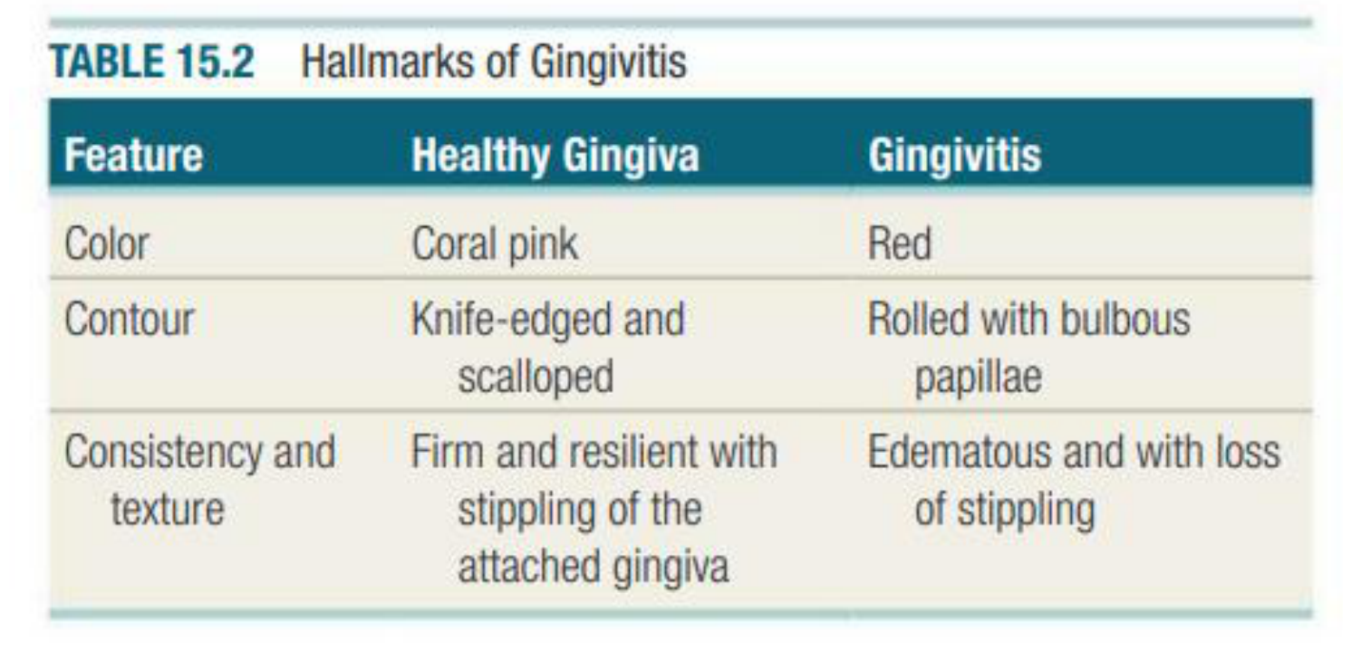
TABLE 15.2 Hallmarks of Gingivitis
| Feature | Healthy Gingiva | Gingivitis |
|---|---|---|
| Color | Coral pink | Red |
| Contour | Knife-edged and scalloped | Rolled with bulbous papillae |
| Consistency and texture | Firm and resilient with stippling of the attached gingiva | Edematous and with loss of stippling |
CHAPTER 15 Dental Biofilm-Induced Gingivitis and Its Management 205
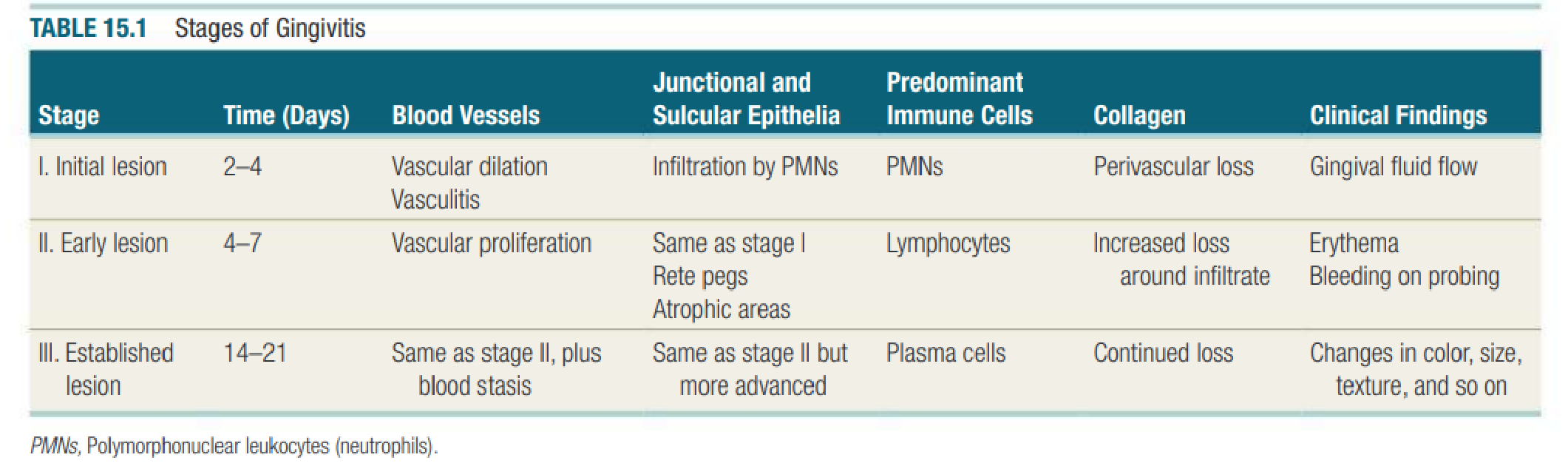
TABLE 15.1 Stages of Gingivitis
| Stage | Time (Days) | Blood Vessels | Junctional and Sulcular Epithelia | Predominant Immune Cells | Collagen | Clinical Findings |
|---|---|---|---|---|---|---|
| I. Initial lesion | 2−4 | Vascular dilation Vasculitis | Infiltration by PMNs | PMNs | Perivascular loss | Gingival fluid flow |
| II. Early lesion | 4−7 | Vascular proliferation | Same as stage I Rete pegs Atrophic areas | Lymphocytes | Increased loss around infiltrate | Erythema Bleeding on probing |
| III. Established lesion | 14−21 | Same as stage II, plus blood stasis | Same as stage II but more advanced | Plasma cells | Continued loss | Changes in color, size, texture, and so on |
PMNs, Polymorphonuclear leukocytes (neutrophils).
 |  |
 |  |
The Advanced Lesion and Periodontitis40
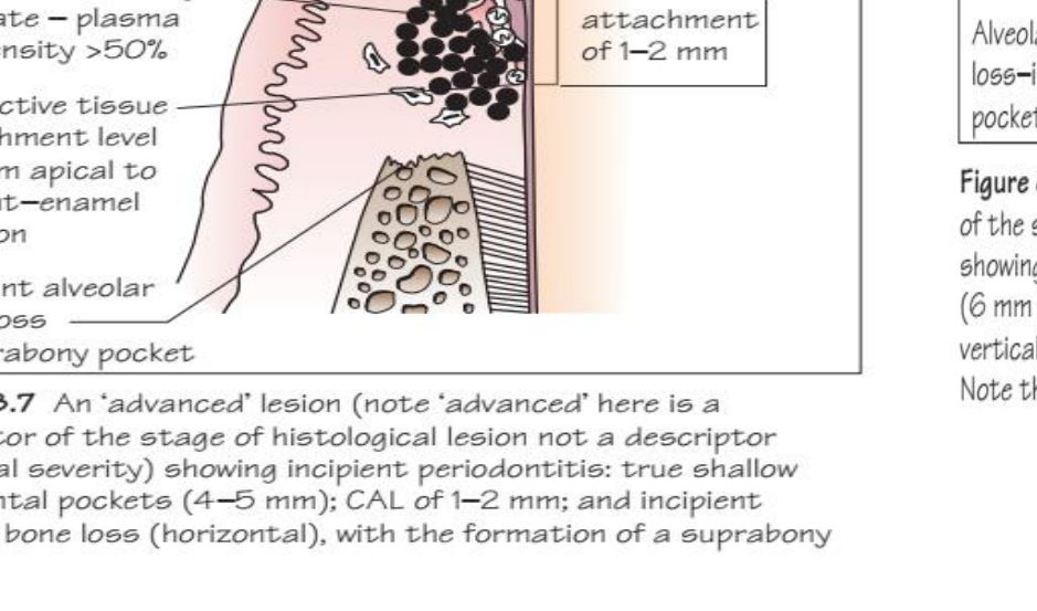
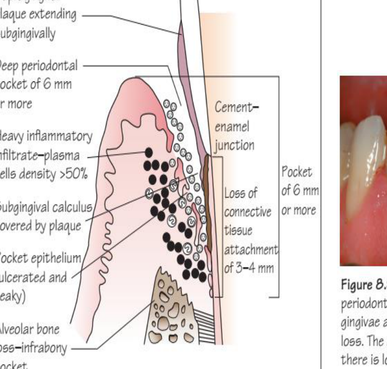
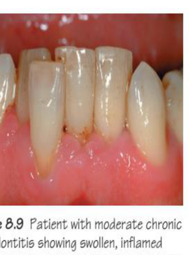
ADVANCED LESION x Periodontitis
Advanced lesion
- Apical plaque growth
- Apical migration of JE from CEJ
- Lateral & apical extension of infiltrate
- Alveolar bone loss starts
- Extensive collagen fiber damage
- Plasma cells predominantly (>50%)
 |  |
 |
Conclusion41
Thank you!!!
THE UNIVERSITY OF WESTERN AUSTRALIA
SEEK WISDOM

Footnotes
-
Original PDF page 1: L5Etiopathogenesis 2, p.1 ↩
-
Original PDF page 2: L5Etiopathogenesis 2, p.2 ↩
-
Original PDF page 3: L5Etiopathogenesis 2, p.3 ↩
-
Original PDF page 4: L5Etiopathogenesis 2, p.4 ↩
-
Original PDF page 5: L5Etiopathogenesis 2, p.5 ↩
-
Original PDF page 6: L5Etiopathogenesis 2, p.6 ↩
-
Original PDF page 7: L5Etiopathogenesis 2, p.7 ↩
-
Original PDF page 8: L5Etiopathogenesis 2, p.8 ↩
-
Original PDF page 9: L5Etiopathogenesis 2, p.9 ↩
-
Original PDF page 10: L5Etiopathogenesis 2, p.10 ↩
-
Original PDF page 11: L5Etiopathogenesis 2, p.11 ↩
-
Original PDF page 12: L5Etiopathogenesis 2, p.12 ↩
-
Original PDF page 13: L5Etiopathogenesis 2, p.13 ↩
-
Original PDF page 14: L5Etiopathogenesis 2, p.14 ↩
-
Original PDF page 15: L5Etiopathogenesis 2, p.15 ↩
-
Original PDF page 16: L5Etiopathogenesis 2, p.16 ↩
-
Original PDF page 17: L5Etiopathogenesis 2, p.17 ↩
-
Original PDF page 18: L5Etiopathogenesis 2, p.18 ↩
-
Original PDF page 19: L5Etiopathogenesis 2, p.19 ↩
-
Original PDF page 20: L5Etiopathogenesis 2, p.20 ↩
-
Original PDF page 21: L5Etiopathogenesis 2, p.21 ↩
-
Original PDF page 22: L5Etiopathogenesis 2, p.22 ↩
-
Original PDF page 23: L5Etiopathogenesis 2, p.23 ↩
-
Original PDF page 24: L5Etiopathogenesis 2, p.24 ↩
-
Original PDF page 25: L5Etiopathogenesis 2, p.25 ↩
-
Original PDF page 26: L5Etiopathogenesis 2, p.26 ↩
-
Original PDF page 27: L5Etiopathogenesis 2, p.27 ↩
-
Original PDF page 28: L5Etiopathogenesis 2, p.28 ↩
-
Original PDF page 29: L5Etiopathogenesis 2, p.29 ↩
-
Original PDF page 30: L5Etiopathogenesis 2, p.30 ↩
-
Original PDF page 31: L5Etiopathogenesis 2, p.31 ↩
-
Original PDF page 32: L5Etiopathogenesis 2, p.32 ↩
-
Original PDF page 33: L5Etiopathogenesis 2, p.33 ↩
-
Original PDF page 34: L5Etiopathogenesis 2, p.34 ↩
-
Original PDF page 35: L5Etiopathogenesis 2, p.35 ↩
-
Original PDF page 36: L5Etiopathogenesis 2, p.36 ↩
-
Original PDF page 37: L5Etiopathogenesis 2, p.37 ↩
-
Original PDF page 38: L5Etiopathogenesis 2, p.38 ↩
-
Original PDF page 39: L5Etiopathogenesis 2, p.39 ↩
-
Original PDF page 40: L5Etiopathogenesis 2, p.40 ↩
-
Original PDF page 41: L5Etiopathogenesis 2, p.41 ↩