Principles of Periodontal Diagnosis and Treatment Planning12
 |  |
Lindhe’s Clinical Periodontology and Implant Dentistry3
- Authors: Tord Berglundh, William V. Giannobile, Mariano Sanz, and Niklaus P. Lang
- Publisher: John Wiley & Sons, Incorporated
- Date: 2021-11-08
 |  |
 |
Journal of Clinical Periodontology (EFP)
- Volume 47, Issue S22
- Clinical Practice Guideline
- Special Issue: Treatment of Stage I-III Periodontitis
- The EFP S3 Level Clinical
- Pages: 1-391
- July 2020
- Issue Edited by: David Herrera Gonzalez, Mariano Sanz, Maurizio Tonetti
BDJ Minimum Intervention Dentistry Themed Issue
- Verifiable CPD Paper – Clinical
- Evidence-based, personalised and minimally invasive treatment for periodontitis patients – the new EFP S3-level clinical treatment guidelines
- Authors: Moritz Kertschull*1,2,3 and Iain Chapple1,2
Periodontology: The Complete Summary
- Edited by Fernando Suárez, DDS, MS
- Study Guide
- Publisher: Quintessence Publishing
Periodontology at a Glance
- Authors: Valeria Cervello, Araceli Tognai and Roberta F. Genco
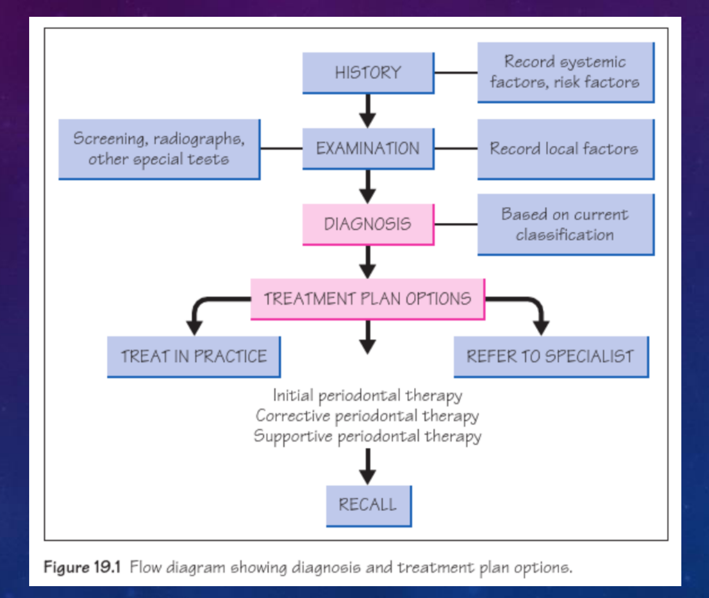
graph TD
    A[HISTORY] --> B(Record systemic factors, risk factors)
    A --> C[EXAMINATION]
    D(Screening, radiographs, other special tests) --- C
    C --> E(Record local factors)
    C --> F[DIAGNOSIS]
    F --> G(Based on current classification)
    F --> H[TREATMENT PLAN OPTIONS]
    H --> I[TREAT_IN_PRACTICE]
    H --> J[REFER TO SPECIALIST]
    H --> K[Initial periodontal therapy<br>Corrective periodontal therapy<br>Supportive periodontal therapy]
    K --> L[RECALL]
Aims and Goals of Periodontal Therapy45
- Design a treatment plan to achieve the best possible biologic, functional, and esthetic outcomes, while preserving original structural characteristics whenever possible.
- Develop a comprehensive treatment plan with:
- Short-term goals: Immediate resolution of acute symptoms.
- Long-term goals: Maintenance of a healthy and functional periodontal environment.
- Treat and correct pathological processes related to periodontal tissues.
- Eliminate gingival inflammation and address conditions derived from it.
- Identify and manage risk factors.
GOALS:
- Preserve, improve and maintain natural dentition, implants and surrounding tissues.
- Obtain healthy periodontium/peri-implant tissues: absence of inflammation and progressive attachment/bone loss.
PROCEDURES TO ESTABLISH AND MAINTAIN HEALTH

Clinical Endpoints of Therapy6
-
- Attainment of sustained high levels of achievement in personal plaque control, reflected as sustained full-mouth bleeding on probing scores around 10% of teeth.
(The absence of bleeding on probing over repeated examinations is the best indicator of periodontal stability currently available.)
-
- Absence of an increase in attachment loss and/or bone loss.
-
- No periodontal pockets more than 4 mm with bleeding on probing or no deep periodontal pockets greater than or equal to 6 mm [≥6 mm]
-
- Tooth hypermobility should be such that it does not impair the patient’s plaque control efforts and allows the patient to function to an acceptable level in comfort.
-
- Appropriate control of risk factors (smoking and diabetes)
Periodontal Examination and Screening7
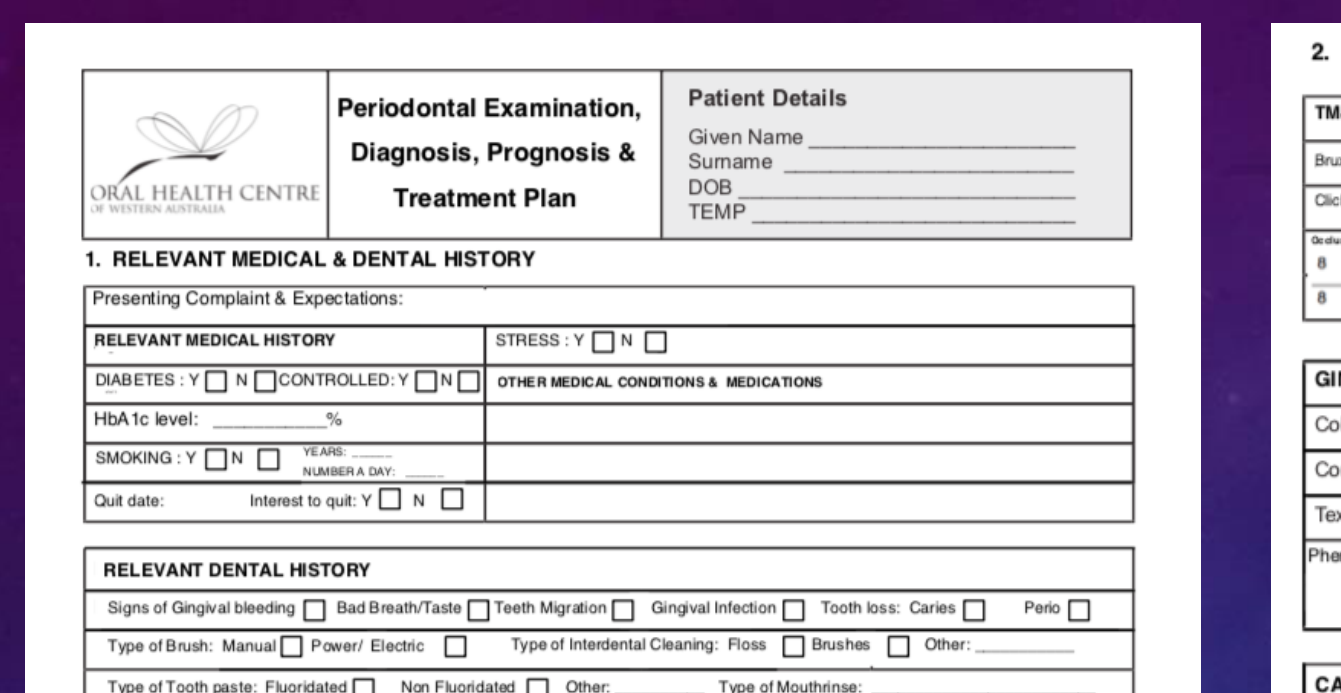
Periodontal management form (e-form)
1. RELEVANT MEDICAL & DENTAL HISTORY
RELEVANT MEDICAL HISTORY
DIABETES: ☐ Y ☐ N ☐ CONTROLLED: ☐ Y ☐ N ☐ OTHER MEDICAL CONDITIONS & MEDICATIONS
HbA1c level: __________%
SMOKING: ☐ Y ☐ N ☐ YEARS __________ NUMBER A DAY: __________
Quit date: __________ Interest to quit: ☐ Y ☐ N ☐ QUARTER __________
RELEVANT DENTAL HISTORY
Signs of Gingival bleeding ☐ Bad Breath/Taste ☐ Teeth Migration ☐ Gingival Infection ☐ Tooth loss: Caries ☐ Perio ☐
Type of Brush: Manual ☐ Power/ Electric ☐ Type of Interdental Cleaning: Floss ☐ Brushes ☐ Other __________
Type of Tooth paste: Fluoridated ☐ Non Fluoridated ☐ Other __________ Type of Mouthrins: __________
CLINICAL PARAMETERS PREVALENCE
Bacterial Biofilm Score % __________
BOP % __________
Periodontal Probing Depth 1-3 mm % __________
Periodontal Probing Depth 4-6 mm % __________
Periodontal Probing Depth 7 or more mm % __________
CPTN / PSR
0 = Healthy 3 = 4 - 5 mm
1 = BOP (no recession) 4 = 6 mm or deeper
2 = Calculus in presence * = Recession/furcation
GINGIVAL RECESSION DEFECTS
| Gingival Site | REC Depth | GT | KTW | CEJ (A / B) | Step (+/-) |
|---|---|---|---|---|---|
| No recession | |||||
| RT1 | |||||
| RT2 | |||||
| RT3 |
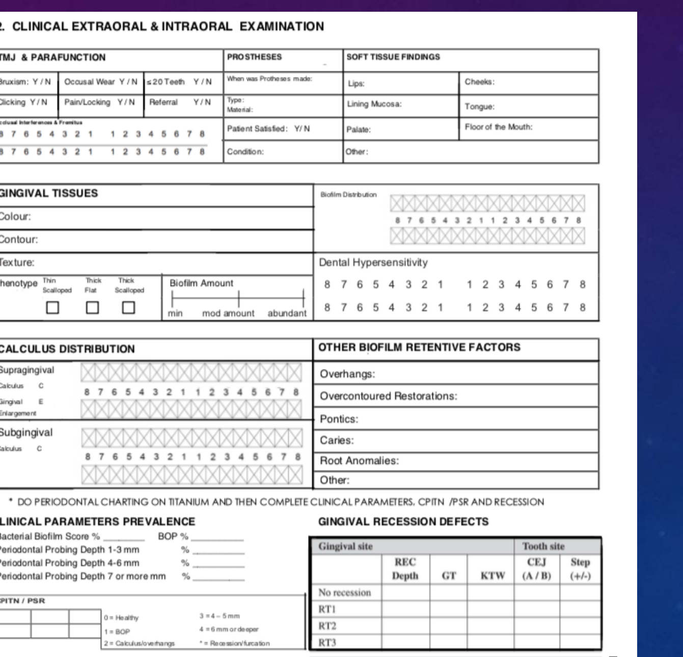
2. CLINICAL EXTRAORAL & INTRAORAL EXAMINATION
TMJ & PARAFUNCTION
Bruxism: ☐ Y ☐ N | Occlusal Wear: ☐ Y ☐ N | ≥20 Teeth: ☐ Y ☐ N
Clicking: ☐ Y ☐ N | Pain/Locking: ☐ Y ☐ N | Referral: ☐ Y ☐ N
Occlusal Interferences & Furcations: [Scale 8-0]
PROSTHESES
When was Prosthesis made: __________ | Lips: __________
Type: __________ | Lining Mucosa: __________
Material: __________ | Tongue: __________
Patient Satisfied: ☐ Y ☐ N | Palate: __________
Condition: __________ | Floor of the Mouth: __________
SOFT TISSUE FINDINGS
Lips: __________
Lining Mucosa: __________
Tongue: __________
Palate: __________
Floor of the Mouth: __________
Condition: __________
Other: __________
GINGIVAL TISSUES
Colour: __________ Contour: __________ Texture: __________ Phenotype: ☐ Thin Scallop ☐ Flat Thick ☐ Thick Scallop Biofilm Amount: __________
Biofilm Distribution: [Chart 8-0]
Dental Hypersensitivity: __________ [Scale 8-0]
CALCULUS DISTRIBUTION
Supragingival Calculus C: __________ Gingiv E: __________ Enlargement: __________
Subgingival Calculus C: __________
[Chart 8-0]
OTHER BIOFILM RETENTIVE FACTORS
Overhangs: __________
Overcontoured Restorations: __________
Pontics: __________
Caries: __________
Root Anomalies: __________
Other: __________
*DO PERIODONTAL CHARTING ON TITANIUM AND THEN COMPLETE CLINICAL PARAMETERS CPTN /PSR AND RECESSION
 |  |
Radiographic Findings
3. RADIOGRAPHIC FINDINGS
| NORMAL BONE LEVEL | HORIZONTAL BONE LOSS | ANGULAR BONE LOSS |
|---|---|---|
| 8 7 6 5 4 3 2 1 | 1 2 3 4 5 6 7 8 8 7 6 5 4 3 2 1 | 1 2 3 4 5 6 7 8 | 8 7 6 5 4 3 2 1 | 1 2 3 4 5 6 7 8 8 7 6 5 4 3 2 1 | 1 2 3 4 5 6 7 8 | 8 7 6 5 4 3 2 1 | 1 2 3 4 5 6 7 8 8 7 6 5 4 3 2 1 | 1 2 3 4 5 6 7 8 |
| MILD BONE LOSS | MODERATE BONE LOSS | SEVERE / ADVANCED BONE LOSS |
| 8 7 6 5 4 3 2 1 | 1 2 3 4 5 6 7 8 8 7 6 5 4 3 2 1 | 1 2 3 4 5 6 7 8 | 8 7 6 5 4 3 2 1 | 1 2 3 4 5 6 7 8 8 7 6 5 4 3 2 1 | 1 2 3 4 5 6 7 8 | 8 7 6 5 4 3 2 1 | 1 2 3 4 5 6 7 8 8 7 6 5 4 3 2 1 | 1 2 3 4 5 6 7 8 |
LOCALISED BONE LOSS ☐ (30% AND LESS TEETH) GENERALISED BONE LOSS ☐ (MORE THAN 30% TEETH)
DENTAL CARIES: ______________________________________ ENDODONTIC: _______________________________________ OVERHANGS: ________________________________________ OPEN CONTACTS: _____________________________________ DEFICIENT RESTORATIONS: ______________________________________ OTHER FINDINGS: ____________________________________
4. PERIODONTAL DIAGNOSIS & PROGNOSIS
☐ CLINICAL GINGIVAL HEALTH (BOP < 10% PROBING ≤ 3mm) ☐ CLINICAL GINGIVAL HEALTH ON A REDUCED PERIODONTIUM (BOP < 10% ON NON-PERIODONTITIS PATIENT RECESSION PROBING ≤ 3mm)
☐ GINGIVITIS: (BOP MORE 10% NO POCKETS) ☐ DENTAL BIOFILM ASSOCIATED GINGIVITIS (LOCALISED ☐ 30% AND LESS BOP / GENERALISED ☐ MORE THAN 30% BOP) ☐ OTHER FORMS OF GINGIVITIS: _____________________________
☐ PERIODONTITIS:
| INTERDENTAL CAL | RADIOGRAPHIC BONE LOSS | TOOTH LOSS | COMPLEXITY | |
|---|---|---|---|---|
| ☐ STAGE I | 1-2 mm ☐ | < 15% HORIZONTAL BONE LOSS | ☐ NO TOOTH LOSS | ☐ MAX PROBING DEPTH ≤ 4mm |
| ☐ STAGE II | 3-4 mm ☐ | 15-33% HORIZONTAL BONE LOSS | ☐ NO TOOTH LOSS | ☐ MAX PROBING DEPTH ≤ 5mm |
| ☐ STAGE III | ≥ 5 mm ☐ | MIDDLE ROOT THIRD AND MORE ☐ ANGULAR | ☐ ≤ 4 TEETH | ☐ MAX PROBING DEPTH ≥ 6mm ☐ FURCATION GRADE II or III ☐ MODERATE RIDGE DEFECT ☐ |
| ☐ STAGE IV | ≥ 5 mm ☐ | MIDDLE ROOT THIRD AND BEYOND ☐ ANGULAR | ☐ ≥ 5 TEETH | ☐ MAX PROBING DEPTH ≥ 6 mm ☐ FURCATION GRADE II or III ☐ SEVERE RIDGE DEFECTS ☐ |
LESS THAN 20 TEETH REMAINING ☐ MASTICATORY DYSFUNCTION ☐ 2ry OCCLUSAL TRAUMA ☐ BITE COLLAPSE ☐
DISTRIBUTION: LOCALISED ☐ (30% AND LESS TEETH) GENERALISED ☐ (MORE THAN 30% TEETH) INCISIVE MOLAR ☐
| EVIDENCE OF BONE LOSS | % BONE LOSS / AGE | CASE PHENOTYPE | SMOKING | DIABETES | |
|---|---|---|---|---|---|
| ☐ GRADE A | NO LOSS 5 YEARS ☐ | < 0.25 ☐ | HEAVY FILM DEPOSITS LOW DESTRUCTION ☐ | ☐ NON SMOKING | ☐ NORMO GLYCEMIC |
| ☐ GRADE B | < 2 mm 5 YEARS ☐ | 0.25 TO 1.0 ☐ | FILM DEPOSITS CORRESPOND TO DESTRUCTION ☐ | ☐ < 10 CIGARETTES DAY | ☐ HbA1c < 7.0 % |
| ☐ GRADE C | ≥ 2 mm 5 YEARS ☐ | ≥ 1.0 ☐ | LOW FILM DEPOSITS HIGH DESTRUCTION ☐ | ☐ ≥ 10 CIGARETTES DAY | ☐ HbA1c ≥ 7.0 % |
OTHER MODIFYING FACTORS: _________________________________________________________
☐ CURRENTLY STABLE PERIODONTITIS: (REDUCED PERIODONTIUM BOP < 10% POCKETS 4 mm OR LESS) ☐ CURRENTLY REMISSION PERIODONTITIS: (GINGIVAL INFLAMMATION ON A REDUCED PERIODONTIUM BOP ≥ 10% ON A TREATED PERIODONTITIS CASE POCKETS 4 mm OR LESS) ☐ CURRENTLY UNSTABLE PERIODONTITIS: (BOP > 10% AND POCKETS 4-5 mm OR MORE) ☐ OTHER FORMS OF PERIODONTITIS: _____________________________
☐ PERI-IMPLANT HEALTH ☐ PERI-IMPLANT MUCOSITIS ☐ PERI-IMPLANTITIS ☐ PERI-IMPLANT ANOMALIES
OVERALL PERIODONTAL PROGNOSIS: GOOD ☐ FAIR ☐ POOR ☐ QUESTIONABLE ☐ HOPELESS ☐
REFERRAL REQUIRED describe unit and needs _________________________________________________________
5. PERIODONTAL TREATMENT PLAN
| EMERGENCIES PHASE OF PERIODONTAL TREATMENT | DATE | UPDATED MEDICAL | ATS | ITEM COMPLETED - TUTOR SIGNATURE |
|---|---|---|---|---|
| RISK MANAGEMENT PHASE OF PERIODONTAL TREATMENT | ||||
| FUNDAMENTAL PHASE OF PERIODONTAL TREATMENT | ||||
| RE-EVALUATION PHASE OF PERIODONTAL TREATMENT | ||||
| CORRECTIVE PHASE OF PERIODONTAL TREATMENT | ||||
| MAINTENANCE PHASE: | ||||
PROPOSED MAINTENANCE INTERVAL: 3 Months ☐ 6 months ☐ 12 months ☐ Other: ___________
Basic Periodontal Examination8
Basic Periodontal Examination (BPE)
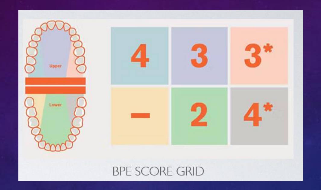
- An example BPE score grid might look like this:
| 4 | 3 | 3* |
|---|---|---|
| - | 2 | 4* |
Both the number and the * should be recorded if a furcation is detected. E.g. the score for a sextant could be 3* (indicating a probing depth 3.5-5.5mm plus a furcation involvement in the sextant).
 |  |
Recording and Scoring the BPE
How to record the BPE
-
The dentition is divided into 6 sextants and the highest score for each sextant is recorded:
- Upper right (17 to 14)
- Lower right (47 to 44)
- Upper anterior (13 to 23)
- Lower anterior (43 to 33)
- Upper left (24 to 27)
- Lower left (34 to 37)
-
All teeth in each sextant are examined (with the exception of 3rd molars unless 1st and/or 2nd molars are missing).
-
For a sextant to qualify for recording, it must contain at least 2 teeth.
-
A World Health Organisation (WHO) BPE probe is used. This has a
ball end0.5mm in diameter and a black band from 3.5mm to 5.5mm. Light probing force should be used (20-25 grams). -
The probe should be
walked aroundthe teeth in each sextant. All sites should be examined to ensure that the highest score in the sextant is recorded before moving on to the next sextant. If a code 4 is identified in a sextant, continue to examine all sites in the sextant. This will help to gain a fuller understanding of the periodontal condition and will make sure that furcation involvements are not missed.
Scoring Codes
| Code | Description |
|---|---|
| 0 | Pockets <3.5mm No calculus/overhangs, no bleeding on probing (black band entirely visible) |
| 1 | Pockets <3.5mm No calculus/overhangs, bleeding on probing (black band entirely visible) |
| 2 | Pockets <3.5mm Supra or subgingival calculus/overhangs (black band entirely visible) |
| 3 | Probing depth 3.5-5.5mm (Black band partially visible, indicating pocket of 4-5mm) |
| 4 | Probing depth >5.5mm (Black band disappears, indicating a pocket of 6mm or more) |
| * | Furcation involvement |
Clinical Application of BPE Scores9
How to use BPE
The BPE is a measurement of the depth of the pocket around a tooth. The BPE is also used to assess the health of the gum and bone around the tooth.
- All new patients should have the BPE recorded
- For patients with codes 0, 1 or 2, the BPE should be recorded at every routine examination
- For patients with BPE codes of 3 or 4, more detailed periodontal charting is required
- Code 3: Initial therapy including self-care advice (oral hygiene instruction and risk factor control) then, post-initial therapy, record a 6-point pocket chart in that sextant only
- Code 4: If there is a Code 4 in any sextant then record a 6-point pocket chart throughout the entire dentition
- BPE cannot be used to monitor the response to periodontal therapy because it does not provide information about how sites within a sextant change after treatment. To assess the response to treatment, a 6-point pocket chart should be recorded pre and post- treatment
- For patients who have undergone initial therapy for periodontitis, and who are now in the maintenance phase of care, then full probing depths throughout the entire dentition should be recorded at least annually
Additional Recommendations
- BPE should not be used around implants (4 or 6-point pocket charting should be used)
- Radiographs should be available for all Code 3 and Code 4 sextants. The type of radiograph used is a matter of clinical judgement but crestal bone levels should be visible. Many clinicians would regard periapical views as essential for Code 4 sextants to allow assessment of bone loss as a percentage of root length and visualisation of the periapical tissues
- When a 6-point pocket chart is indicated it is only necessary to record sites of 4mm and above (although 6 sites per tooth should be measured)
- Bleeding on probing should always be recorded in conjunction with a 6-point pocket chart
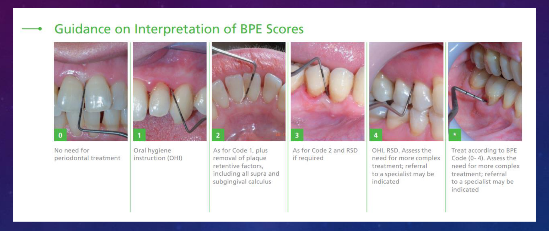
0 No need for periodontal treatment
1 Oral hygiene instruction (OHI)
2 As for Code 1, plus removal of plaque retentive factors, including all supra and subgingival calculus
3 As for Code 2 and RSD if required
4 OHI, RSD. Assess the need for more complex treatment; referral to a specialist may be indicated
* Treat according to BPE Code (0–4). Assess the need for more complex treatment; referral to a specialist may be indicated
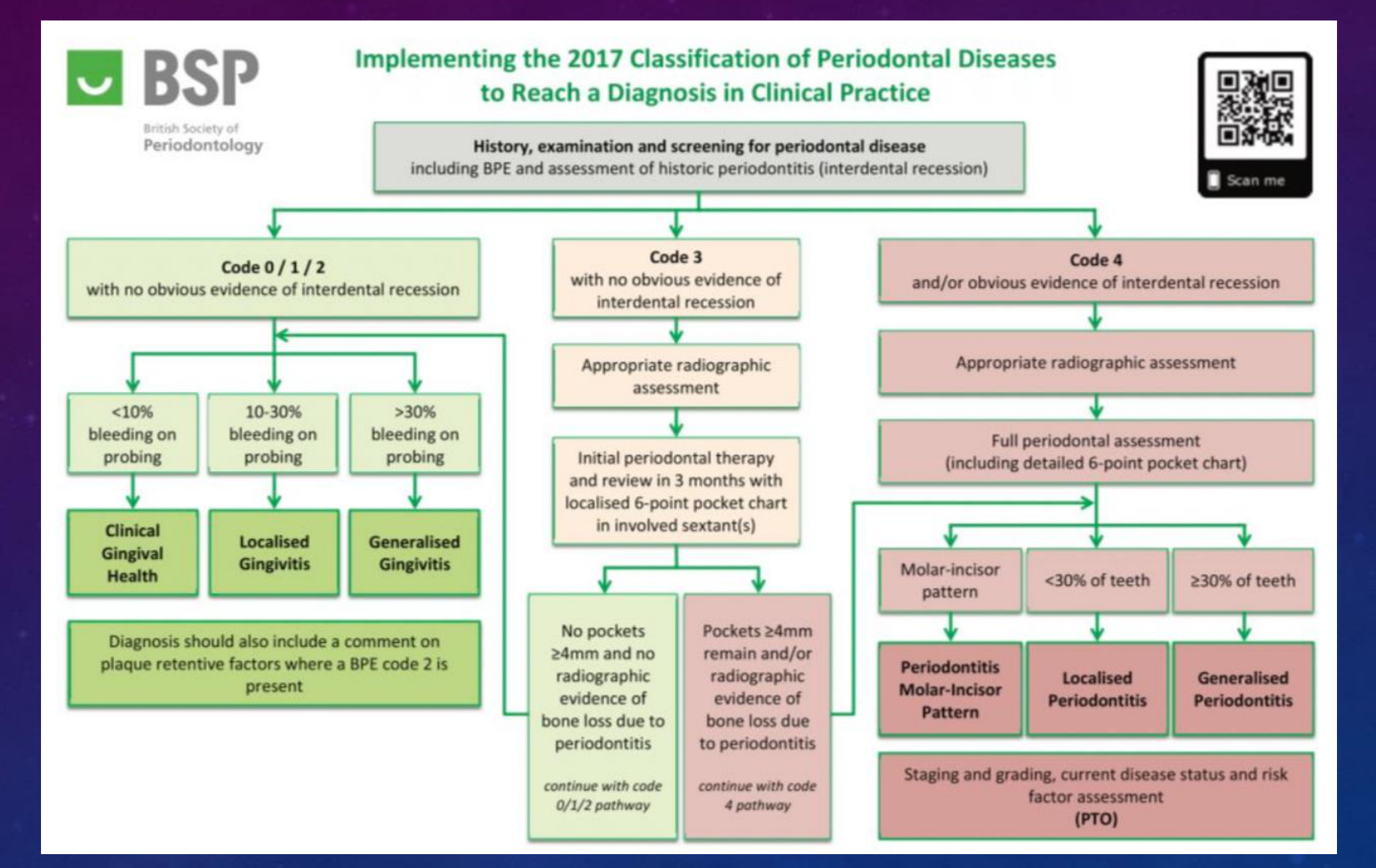
Diagnosis and Treatment Sequencing1011
| History, examination and screening for periodontal disease | Scan me | |||
|---|---|---|---|---|
| including BPE and assessment of historic periodontitis (interdental recession) | ||||
| Code 0 / 1 / 2 with no obvious evidence of interdental recession | Staging and grading, current disease status and risk factor assessment (PTO) | |||
| <10% bleeding on probing | 10-30% bleeding on probing | >30% bleeding on probing | ||
| Clinical Gingival Health | Localised Gingivitis | Generalised Gingivitis | ||
| Diagnosis should also include a comment on plaque retentive factors where a BPE code 2 is present | ||||
| No pockets ≥4mm and no radiographic evidence of bone loss due to periodontitis | Pockets ≥4mm remain and/or radiographic evidence of bone loss due to periodontitis | |||
| Code 3 with no obvious evidence of interdental recession | <30% of teeth Localised Periodontitis | |||
| Appropriate radiographic assessment | ||||
| Initial periodontal therapy and review in 3 months with localised 6-point pocket chart in involved sextant(s) | ||||
| No pockets ≥4mm and no radiographic evidence of bone loss due to periodontitis | Pockets ≥4mm remain and/or radiographic evidence of bone loss due to periodontitis | |||
| continue with code 0/1/2 pathway | ||||
| Code 4 and/or obvious evidence of interdental recession | Molar-incisor pattern Periodontitis Molar-Incisor Pattern | continue with code 4 pathway | ||
| Appropriate radiographic assessment | ||||
| Full periodontal assessment (including detailed 6-point pocket chart) | ≥30% of teeth Generalised Periodontitis | <30% of teeth Localised Periodontitis | ≥30% of teeth Generalised Periodontitis | |
| Molar-incisor pattern Periodontitis Molar-Incisor Pattern | <30% of teeth Localised Periodontitis | ≥30% of teeth Generalised Periodontitis |
Emergency care phase Fundamental phase (Step 1 and 2 EFP Guideline) Re-assessment/re-evaluation phase Corrective phase (Step 3 EFP Guideline) Supportive care phase of periodontal therapy
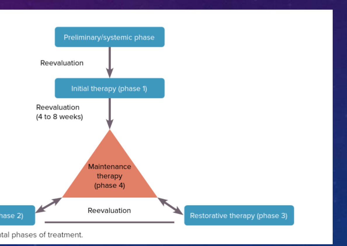
Preliminary/systemic phase Reevaluation Initial therapy (phase 1) Reevaluation (4 to 8 weeks) Maintenance therapy (phase 4) Surgical therapy (phase 2) Reevaluation Restorative therapy (phase 3)
Fig 8-2 Sequence of periodontal phases of treatment.
 |  |
EFP Clinical Practice Guidelines for Stage I-III Periodontitis121314
Treatment of stage I–III periodontitis—The EFP S3 level clinical practice guideline
Mariano Sanz, David Herrera, Moritz Kebschull, Iain Chapple, Søren Jepsen, Tord Beglundh, Anton Sculean, Maurizio S. Tonetti, on behalf of the EFP Workshop Participants and Methodological Consultants
- The 2017 World Workshop on Periodontitis Classification introduced a system linking stages and grades of the disease to tailored prevention and treatment strategies, addressing severity, complexity, and individual risk factors.
- Developed by the European Federation of Periodontology (EFP) using evidence from 15 systematic reviews and the GRADE system, the S3-Level Clinical Practice Guidelines outline a stepwise approach for managing Stage I–III periodontitis.
- Recommendations focus on behavioral changes, biofilm control, surgical interventions, and supportive care to ensure long-term periodontal health.
- These guidelines provide an evidence-based framework for effective treatment, supporting lifelong oral health and informing clinical practice and policy.
Evidence-based, personalised and minimally invasive treatment for periodontitis patients – the new EFP S3-level clinical treatment guidelines
Moritz Kebschull and Iain Chapple
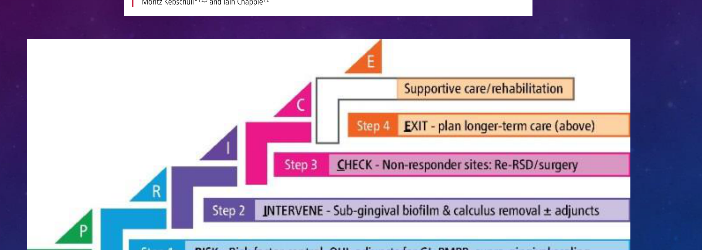
Step 0 – PREREQUISITE TO THERAPY: Educate, classification, diagnosis, risk assess, care plan
Step 1 – RISK: Risk factor control, OHI, adjuncts for GI, PMPR, supra-gingival scaling
Step 2 – INTERVENE: Sub-gingival biofilm & calculus removal ± adjuncts
Step 3 – CHECK: Non-responder sites: Re-RSD/surgery
Step 4 – EXIT: Plan longer-term care (above)
Supportive care/rehabilitation
Preliminary/Emergency Phase: Emergency Dental Treatments
- Pain and acute infections should be managed promptly, including:
- Endodontic treatment for infected or abscessed teeth.
- Treatment of periodontal abscesses.
- Management of necrotizing periodontal diseases (painful and acute).
- Extraction of hopeless teeth (can be postponed if necessary).
 |  |
 |
Step 1: Risk Factor Control and Supragingival Biofilm Removal1516
1st Step
The first step in therapy is aimed at guiding behaviour change by motivating the patient to undertake successful removal of supragingival dental biofilm and risk factor control and may include the following interventions:
- Supragingival dental biofilm control
- Interventions to improve the effectiveness of oral hygiene [motivation, instructions (oral hygiene instructions, OHI)]
- Adjunctive therapies for gingival inflammation
- Professional mechanical plaque removal (PMPR), which includes the professional interventions aimed at removing supragingival plaque and calculus, as well as possible plaque-retentive factors that impair oral hygiene practices.
- Risk factor control, which includes all the health behavioural change interventions eliminating/mitigating the recognized risk factors for periodontitis onset and progression (smoking cessation), improved metabolic control of diabetes, and perhaps physical exercise, dietary counselling and weight loss.
Re-evaluation/Re-assessment
This first step of therapy should be implemented in all periodontitis patients, irrespective of the stage of their disease, and should be re-evaluated frequently in order to:
- Continue to build motivation and adherence, or explore other alternatives to overcome the barriers
- Develop skills in dental biofilm removal and modify as required
- Allow for the appropriate response of the subsequent steps of therapy
Step 2: Subgingival Instrumentation and Adjunctive Therapies1718
2nd Step:
The second step of therapy (cause-related therapy) is aimed at controlling (reducing/eliminating) the subgingival biofilm and calculus (subgingival instrumentation). In addition to this, the following interventions may be included:
- Use of adjunctive physical or chemical agents
- Use of adjunctive host-modulating agents (local or systemic)
- Use of adjunctive subgingival locally delivered antimicrobials
- Use of adjunctive systemic antimicrobials
- This second step of therapy should be used for all periodontitis patients, irrespective of their disease stage, only in teeth with loss of periodontal support and/or periodontal pocket formation.
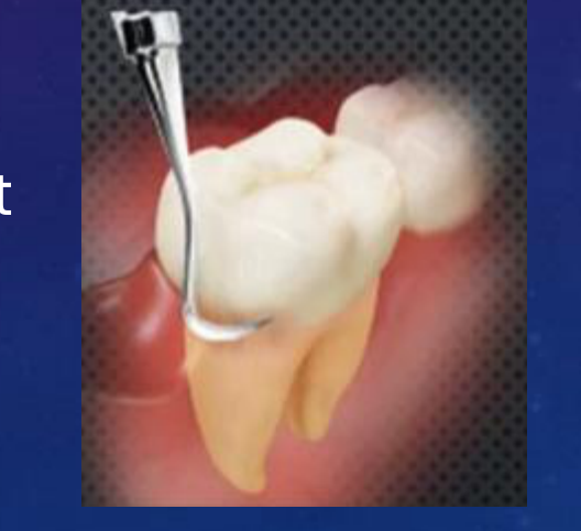
- In specific clinical situations, such as in the presence of deep probing depths, first and second steps of therapy could be delivered simultaneously (such as for preventing periodontal abscess development).
Re-evaluation/Re-assessment
The individual response to the second step of therapy should be assessed once the periodontal tissues have healed (periodontal re-evaluation).
-
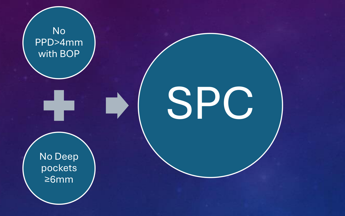
If the endpoints of therapy:
- No periodontal pockets greater than 4 mm with bleeding on probing or
- No deep periodontal pockets greater than or equal to 6 mm [≥6 mm] have not been achieved, the third step of therapy should be considered.
-
If the treatment has been successful in achieving the endpoints of therapy, patients should be placed in a supportive periodontal care (SPC) programme.
Step 3: Corrective Surgical Therapy1920
3rd Step:
The third step of therapy is aimed at treating those areas of the dentition non-responding adequately to the second step of therapy (presence of pockets greater than or equal to 4 mm [≥4 mm] with bleeding on probing or presence of deep periodontal pockets greater than or equal to 6 mm [≥6 mm]), with the purpose of gaining further access to subgingival instrumentation, or aiming at regenerating or resecting those lesions that add complexity in the management of periodontitis (intra-bony and furcation lesions). It may include the following interventions:
- Repeated subgingival instrumentation with or without adjunctive therapies
- Access flap periodontal surgery
- Resective periodontal surgery
- Regenerative periodontal surgery
Additional surgical procedures, such as mucogingival, preprosthetic, preimplant, or implant surgeries, may be performed depending on patient needs. When there is indication for surgical interventions, these should be subject to an additional patient consent and specific evaluation of risk factors or medical contra-indications should be considered. This corrective phase may also entail the orthodontic repositioning of drifted teeth.
Re-evaluation/Re-assessment
| No PPD > 4 mm with BOP | No Deep pockets ≥ 6 mm | → | SPC |
Step 4: Supportive Periodontal Care21
Supportive Periodontal Care (Maintenance Phase)
Objective:
- Maintain periodontal stability in treated periodontitis patients.
- Integrates preventive and therapeutic interventions from earlier therapy steps.
Personalized Care:
- Tailored based on gingival and periodontal status of the patient.
- Regular recall visits scheduled according to individual patient needs.
Monitoring and Re-Treatment:
- Re-treatment may be necessary if recurrent disease is detected.
- Requires proper diagnosis and treatment planning when needed.
- In some cases, extraction of hopeless teeth may be required.
Patient Compliance:
- Adherence to oral hygiene regimens is crucial.
- Encouragement of healthy lifestyle habits to prevent disease recurrence.
Maintenance Phase Procedures22
Supportive Periodontal Care (Maintenance Phase)
- Update medical and smoking history
- Oral hygiene assessment and reinforcement of oral hygiene if necessary
- Full periodontal charting
- Sensibility testing of suspected teeth
- Instrumentation of sites with residual pocket > 4 mm
- Fluoride application on the exposed roots to prevent dental caries
- Regular assessment of prosthetic restorations and radiographic evaluation may also be indicated.
Implementation in Daily Practice23
SPC In Daily Practice:
The recall hour should be planned to meet patient’s individual needs. It consists of four sections:
- EXAMINATION, RE-EVALUATION & DIAGNOSIS (ERD): This section consists of 10–15 mins.
- MOTIVATION, REINSTRUCTION & INSTRUMENTATION: This section consists of 30–40 mins of motivation, reinstruction of oral hygiene, instrumentation concentrated on sites diagnosed with persistent inflammation.
- TREATMENT OF REINFECTED SITES may include small surgical corrections.
- POLISHING OF ENTIRE DENTITION, APPLICATION OF FLUORIDES & DETERMINATION OF FUTURE SPT APPOINTMENT: 5–10 mins should be reserved for this section.
Summary of Treatment Phases and Coding24252627
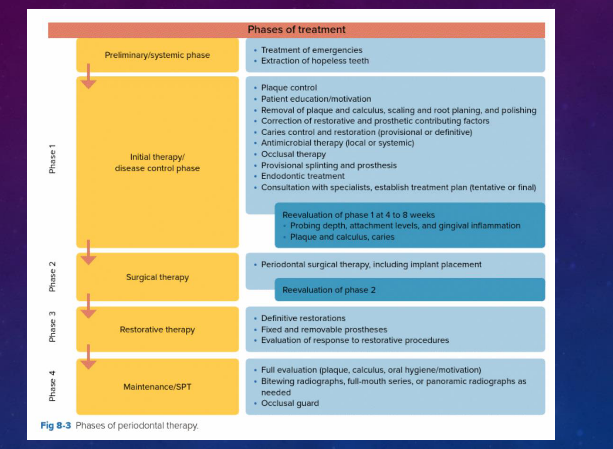
Phases of treatment
- Preliminary/systemic phase → Initial therapy/disease control phase → Surgical therapy → Restorative therapy → Maintenance/SPT
Preliminary/systemic phase:
- Treatment of emergencies
- Extraction of hopeless teeth
Initial therapy/disease control phase:
- Plaque control
- Patient education/motivation
- Removal of plaque and calculus, scaling and root planing, and polishing
- Correction of restorative and prosthetic contributing factors
- Caries control and restoration (provisional or definitive)
- Antimicrobial therapy (local or systemic)
- Occlusal therapy
- Provisional splinting and prosthesis
- Endodontic treatment
- Consultation with specialists, establish treatment plan (tentative or final)
Reevaluation of phase 1 at 4 to 8 weeks:
- Probing depth, attachment levels, and gingival inflammation
- Plaque and calculus, caries
Surgical therapy:
- Periodontal surgical therapy, including implant placement
Reevaluation of phase 2
Restorative therapy:
- Definitive restorations
- Fixed and removable prostheses
- Evaluation of response to restorative procedures
Maintenance/SPT:
- Full evaluation (plaque, calculus, oral hygiene/motivation)
- Bitewing radiographs, full-mouth series, or panoramic radiographs as needed
- Occlusal guard
Fig 8-3 Phases of periodontal therapy.
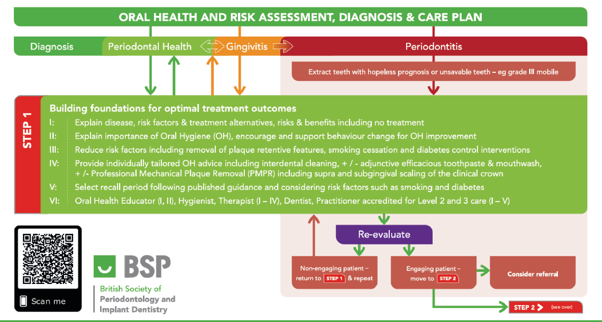
Oral Health and Risk Assessment, Diagnosis & Care Plan
| Diagnosis | Periodontal Health | Gingivitis | Periodontitis |
|---|---|---|---|
| Extract teeth with hopeless prognosis or unsavable teeth – e.g. grade III mobile |
STEP 1 – Building foundations for optimal treatment outcomes
I: Explain disease, risk factors & treatment alternatives, risks & benefits including no treatment
II: Encourage and support behaviour change for OH improvement. Explain importance of Oral Hygiene (OH), encourage and support behaviour change for OH improvement
III: Reduction of risk factors including removal of plaque retentive features, smoking cessation and diabetes control interventions
IV: Provide individually tailored OH advice including interdental cleaning, +/- adjunctive efficacious toothpaste & mouthwash, +/- Professional Mechanical Plaque Removal (PMPR) including supra and subgingival scaling of the clinical crown
V: Select recall period following published guidance and considering risk factors such as smoking and diabetes
VI: Oral Health Educator (I, II), Hygienist, Therapist (I – IV), Dentist, Practitioner accredited for Level 2 and 3 care (I – V)
BSP – British Society of Periodontology and Implant Dentistry
Re-evaluate
- Non-engaging patient – return to STEP 1 & repeat
- Engaging patient – move to STEP 2
- Consider referral
STEP 2 (see over)
© The British Society of Periodontology and Implant Dentistry 2021
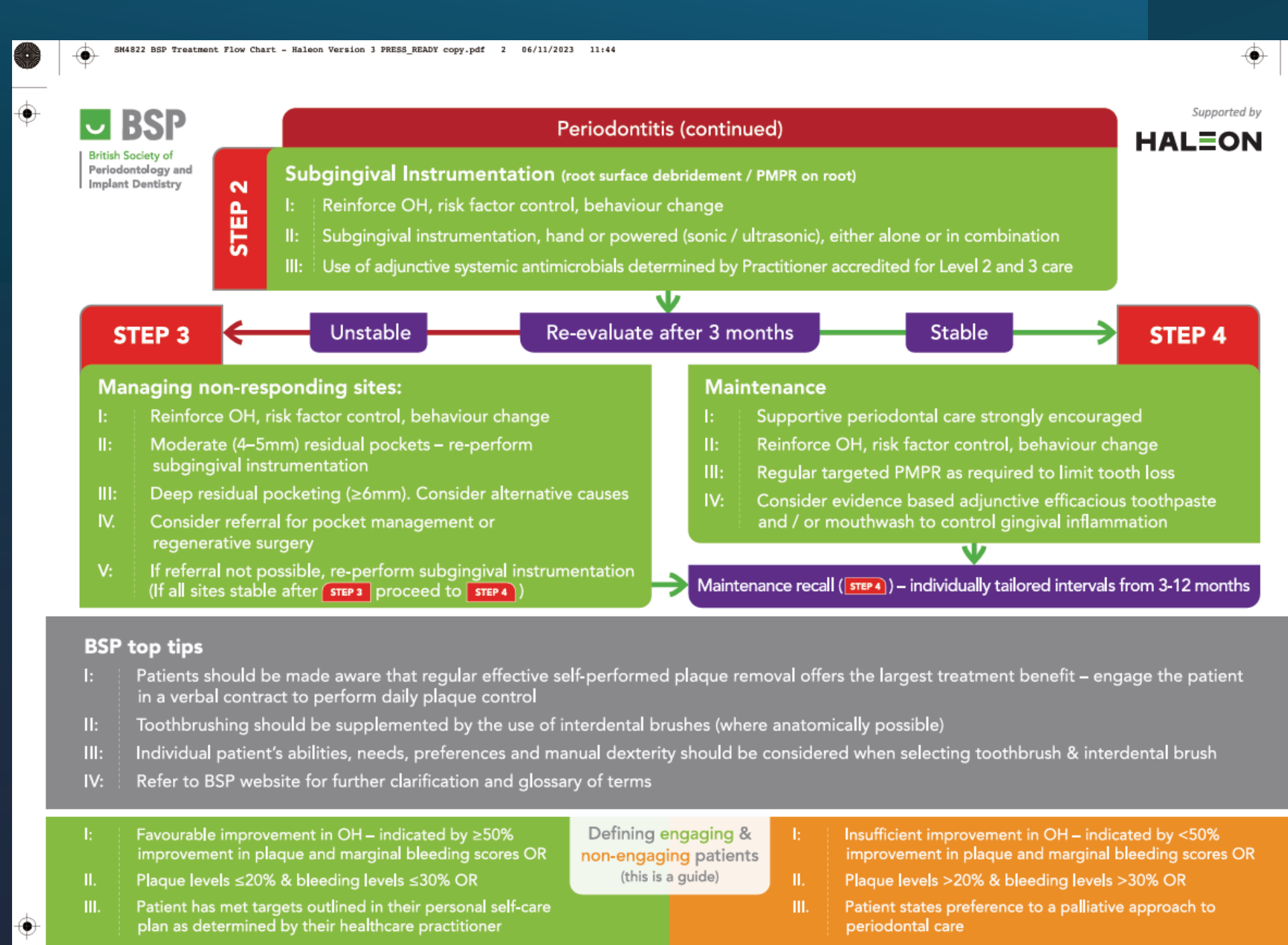
Periodontitis (continued)
STEP 2 – Subgingival Instrumentation (root surface debridement / PMPR on root)
I: Reinforce OH, risk factor control, behaviour change
II: Subgingival instrumentation, hand or powered (sonic / ultrasonic), either alone or in combination
III: Use of adjunctive systemic antimicrobials determined by Practitioner accredited for Level 2 and 3 care
BSP – British Society of Periodontology and Implant Dentistry – Supported by HALEON
STEP 3 – Managing non-responding sites:
I: Reinforce OH, risk factor control, behaviour change
II: Moderate (4–5 mm) residual pockets – re-perform subgingival instrumentation
III: Deep residual pocketing (≥6 mm). Consider alternative causes
IV: Consider referral for pocket management or regenerative surgery
V: If referral not possible, re-perform subgingival instrumentation
(If all sites stable after STEP 3 proceed to STEP 4)
Re-evaluate after 3 months – Unstable → Stable → STEP 4
STEP 4 – Maintenance
I: Supportive periodontal care strongly encouraged
II: Reinforce OH, risk factor control, behaviour change
III: Regular targeted PMPR as required to limit tooth loss
IV: Consider evidence based adjunctive efficacious toothpaste and / or mouthwash to control gingival inflammation
Maintenance recall (STEP 4) – individually tailored intervals from 3–12 months
BSP top tips
I: Patients should be made aware that regular effective self-performed plaque removal offers the largest treatment benefit – engage the patient in a verbal contract to perform daily plaque control
II: Toothbrushing should be supplemented by the use of interdental brushes (where anatomically possible)
III: Individual patient’s abilities, needs, preferences and manual dexterity should be considered when selecting toothbrush & interdental brush
IV: Refer to BSP website for further clarification and glossary of terms
Defining engaging & non-engaging patients (this is a guide)
Engaging patient:
I: Favourable improvement in OH – indicated by ≥50% improvement in plaque and marginal bleeding scores OR
II: Plaque levels ≤20% & bleeding levels ≤30% OR
III: Patient has met targets outlined in their personal self-care plan as determined by their healthcare practitioner
Non-engaging patient:
I: Insufficient improvement in OH – indicated by <50% improvement in plaque and marginal bleeding scores OR
II: Plaque levels >20% & bleeding levels >30% OR
III: Patient states preference to a palliative approach to periodontal care
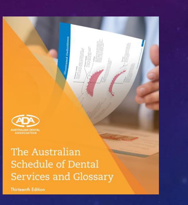
111 Removal of plaque and/or stain – Removal of dental plaque and/or stain from the surfaces of all teeth and/or implants. This item is included in procedures described by items 114, 115, 250 and 251.
113 Recontouring and polishing of pre-existing restoration(s) – per tooth – The reshaping and polishing of pre-existing restorations.
114 Removal of calculus – first appointment – Removal of calculus from the surfaces of teeth and/or implants.
115 Removal of calculus – subsequent appointment – This item describes procedures in item 114 when, because of the extent or degree of calculus, an additional appointment(s) is required to remove deposits from the teeth and/or implants.
250 Active non-surgical periodontal therapy – per quadrant – Non-surgical management of uncontrolled periodontal disease in a quadrant with at least three teeth with clinical attachment loss of at least 3 mm or pocketing of at least 4 mm. This includes procedures described in items 111, 114, 115 and 222 for the quadrant. Items such as 113, 131 and 142 may be itemised separately.
251 Supportive periodontal therapy – per appointment – Maintenance of periodontal health subsequent to active, non-surgical or surgical periodontal therapy. This includes procedures described in items 111, 114, 115 and 222. Items such as 113, 131 and 142 may be itemised separately.
213 Treatment of acute periodontal infection – per appointment – This item describes the treatment of acute periodontal infection(s). It may include establishing drainage and the removal of calculus from the affected tooth (teeth) or implant.
221 Clinical periodontal analysis and recording – This is a special examination performed as part of the diagnosis and management of periodontal disease. The procedure consists of assessing and recording a patient’s periodontal condition.
222 Periodontal debridement – per tooth – The mechanical removal of biofilm and calculus from a periodontally diseased tooth to a level consistent with periodontal health. Where calculus removal is undertaken for other teeth at the same or a subsequent appointment that do not require debridement, it is appropriate to itemise 114/115 for these teeth in conjunction with but separate from the teeth that have undergone periodontal debridement.
223 Non-surgical treatment of peri-implant disease – per implant – The process of debridement using specialised instrumentation to remove the microbial biofilm from the implant and/or abutment surface, which may include localised application of medicaments.
 |  |
 |  |
Case Presentation and Treatment Planning Example28

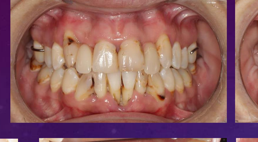
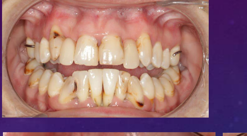
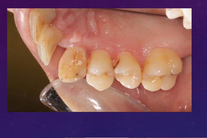
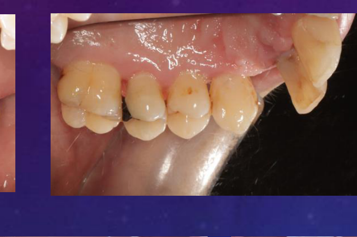
Patient Profile and Initial Examination293031
PATIENT PROFILE
About: Mrs Smith, 37 yrs old, female
Referral from: OHCWA DMD Clinic (Jan 2023)
First Periodontal Consultation: 24-Apr-2023
Patient Concerns:
- Doesn’t want to lose any more teeth and wants to take better care of herself.
- Notices bleeding while brushing.
- Feels that her lower front teeth are moving.
- Reports a bad taste and bad breath.
- Feels that her teeth and gums are sensitive.
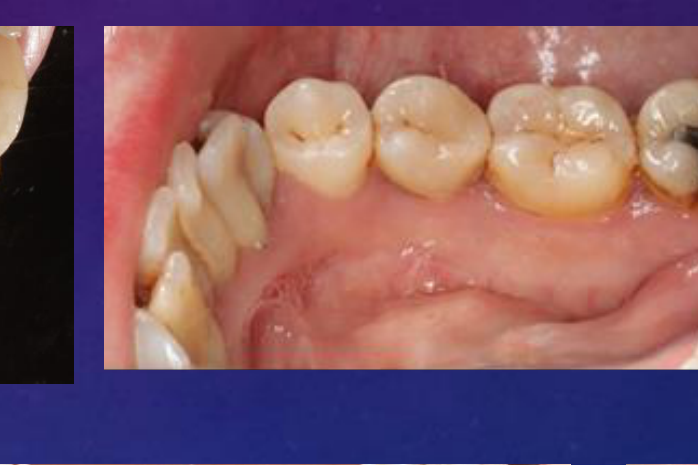
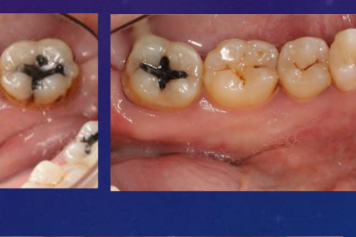
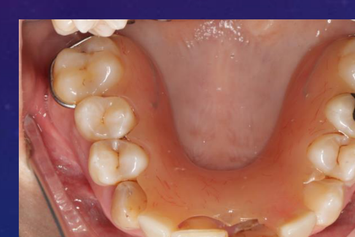
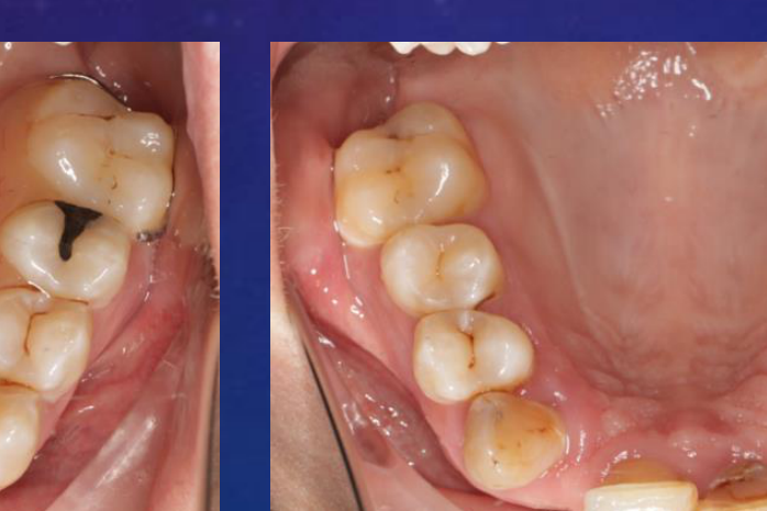
CLINICAL PHOTOGRAPHS – INITIAL EXAMINATION:
 |  |  |
 |  |  |
 |  |  |
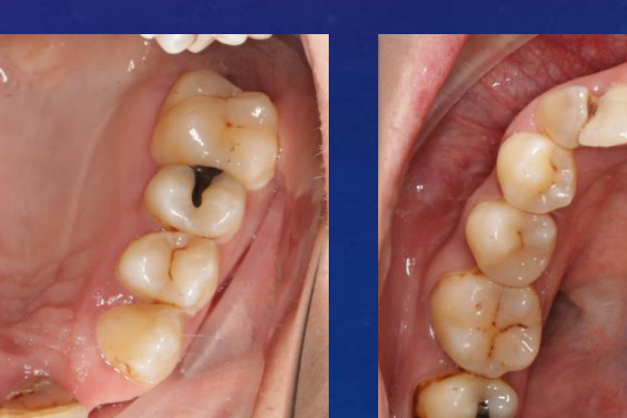 |  |
Social32
- Single
- 4 Children
- Doesn’t work
- Feels stressed
- Highly motivated
- Recently was released from prison
- Smoking: She has been smoking 10-15 cigarettes per day for the past 20 years
- Drug: Used Methamphetamine for 13 years. Quit drugs 3 years ago.
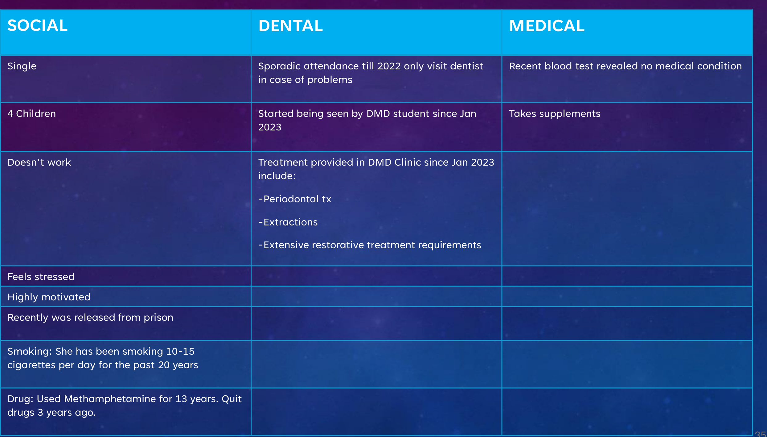
Dental
- Sporadic attendance till 2022 only visit dentist in case of problems
- Started being seen by DMD student since Jan 2023
- Treatment provided in DMD Clinic since Jan 2023 include:
- Periodontal tx
- Extractions
- Extensive restorative treatment requirements
Medical
- Recent blood test revealed no medical condition
- Takes supplements
Clinical Findings and Periodontal Assessment33
PERIODONTAL ASSESSMENT
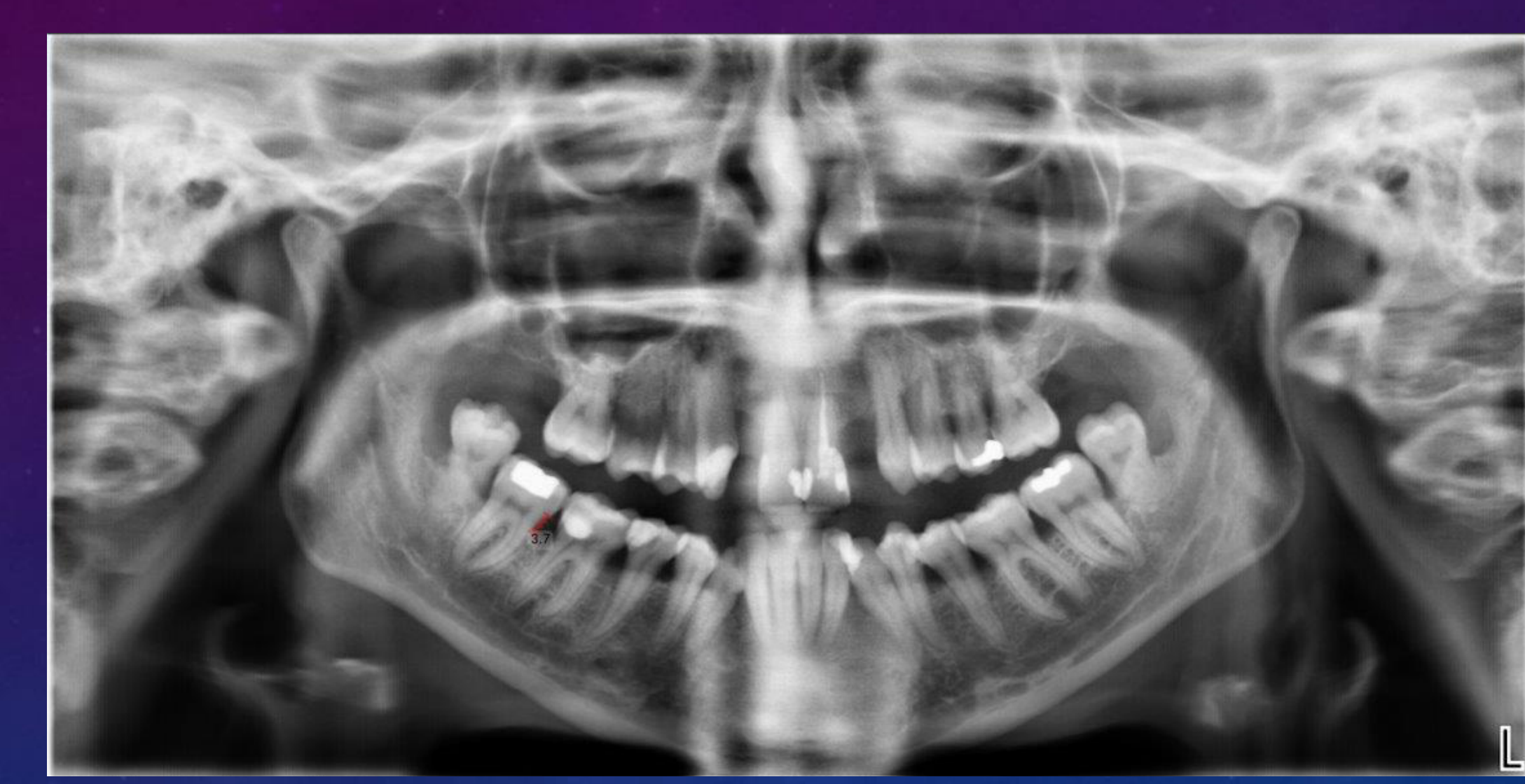
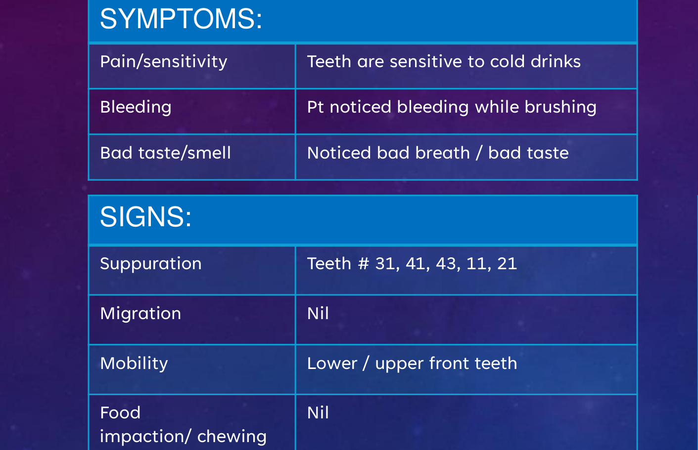
PATIENT SIGNS & SYMPTOMS34
SYMPTOMS:
- Pain/sensitivity: Teeth are sensitive to cold drinks
- Bleeding: Pt noticed bleeding while brushing
- Bad taste/smell: Noticed bad breath / bad taste
SIGNS:
- Suppuration: Teeth # 31, 41, 43, 11, 21
- Migration: Nil
- Mobility: Lower / upper front teeth
- Food impaction/ chewing efficiency: Nil
- Tooth loss: 4 (due to tooth decay)
- Parafunction: She used to clench her teeth when she was on drugs
 | 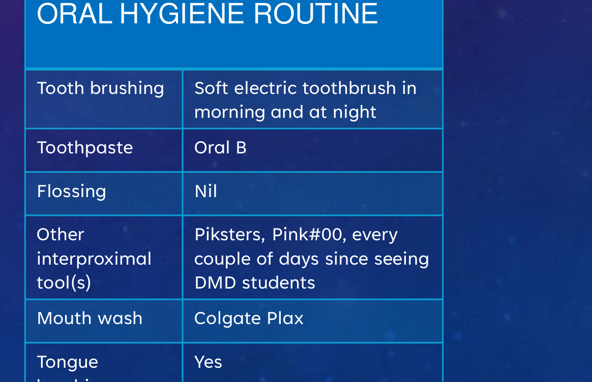 |
ORAL HYGIENE ROUTINE
- Tooth brushing: Soft electric toothbrush in morning and at night
- Toothpaste: Oral B
- Flossing: Nil
- Other interproximal tool(s): Piksters, Pink#00, every couple of days since seeing DMD students
- Mouth wash: Colgate Plax
- Tongue brushing: Yes
INITIAL PERIODONTAL EXAM
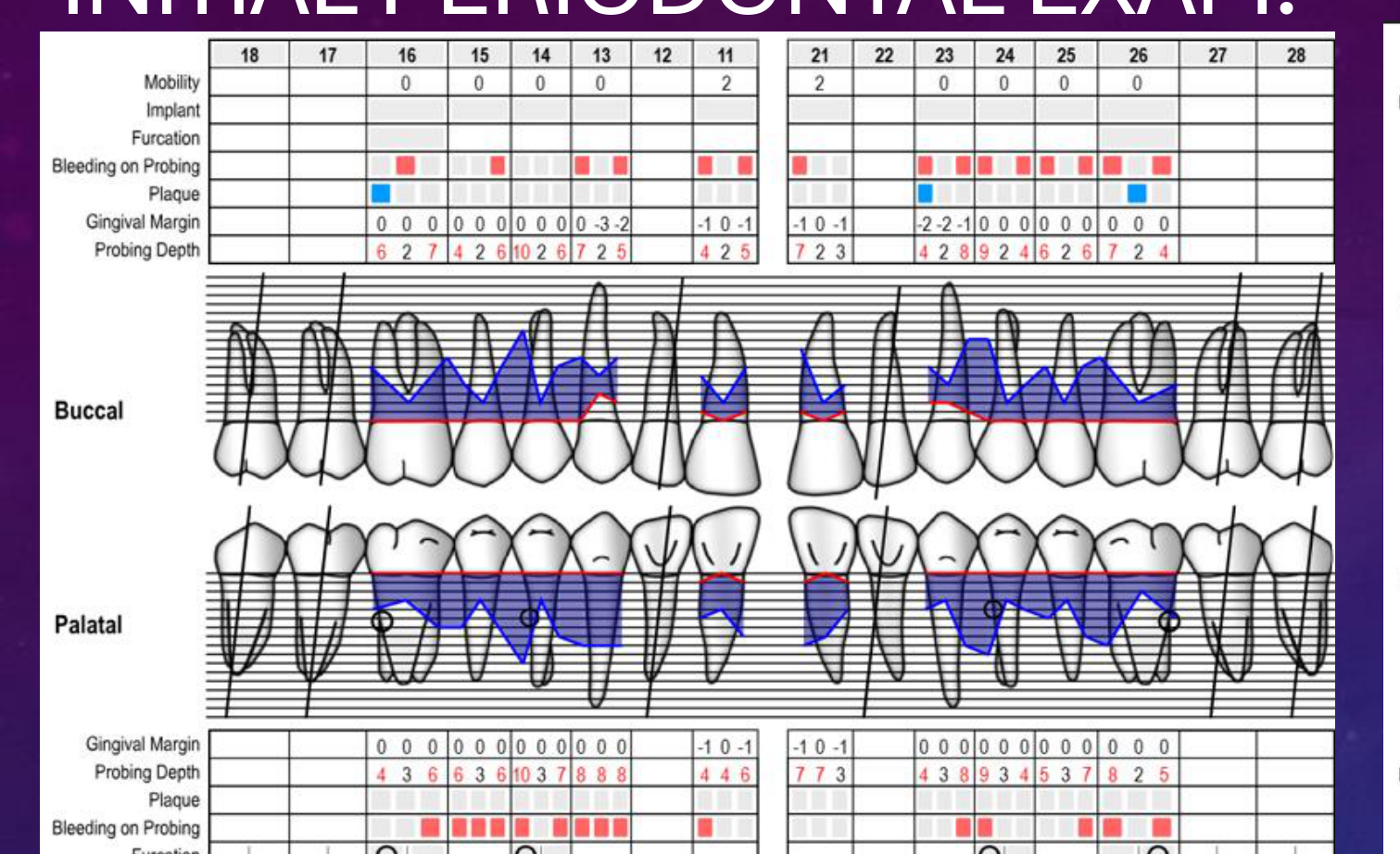
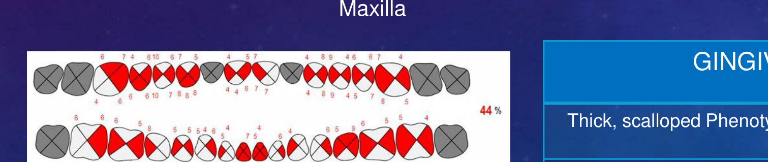
Maxilla35
| 18 | 17 | 16 | 15 | 14 | 13 | 12 | 11 | 21 | 22 | 23 | 24 | 25 | 26 | 27 | 28 | |
|---|---|---|---|---|---|---|---|---|---|---|---|---|---|---|---|---|
| Mobility | 0 | 0 | 0 | 0 | 2 | 2 | 0 | 0 | 0 | 0 | ||||||
| Gingival Margin (Buccal) | 0 0 0 | 0 0 0 | 0 0 0 | 0 0 0 | -3 -2 | -1 0 -1 | -1 0 -1 | -2 -2 -1 | 0 0 0 | 0 0 0 | 0 0 0 | 0 0 0 | ||||
| Probing Depth (Buccal) | 6 2 7 | 4 2 6 | 10 2 6 | 7 2 5 | 4 2 5 | 7 2 3 | 4 2 8 | 9 2 4 | 6 2 6 | 7 2 4 | ||||||
| Gingival Margin (Palatal) | 0 0 0 | 0 0 0 | 0 0 0 | 0 0 0 | 0 0 0 | -1 0 -1 | -1 0 -1 | 0 0 0 | 0 0 0 | 0 0 0 | 0 0 0 | |||||
| Probing Depth (Palatal) | 4 3 6 | 6 3 6 | 10 3 7 | 8 8 8 | 4 4 6 | 7 7 3 | 4 3 8 | 9 3 4 | 5 3 7 | 8 2 5 | ||||||
| Note | SUP | SUP |
 |  |  |
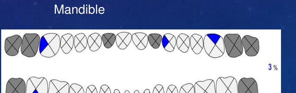 |  |
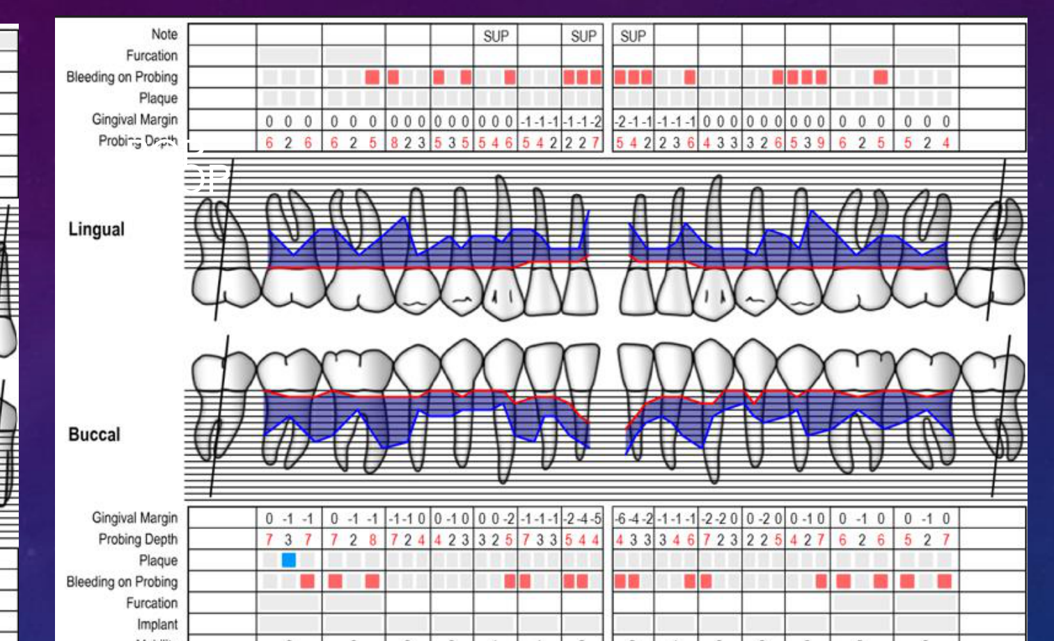
Mandible
| 48 | 47 | 46 | 45 | 44 | 43 | 42 | 41 | 31 | 32 | 33 | 34 | 35 | 36 | 37 | 38 | |
|---|---|---|---|---|---|---|---|---|---|---|---|---|---|---|---|---|
| Gingival Margin (Lingual) | 0 0 0 | 0 0 0 | 0 0 0 | 0 0 0 | 0 0 0 | -1 -1 -1 | -1 -1 -2 | -2 -1 -1 | -1 -1 -1 | -1 -1 0 | 0 0 0 | 0 0 0 | 0 0 0 | 0 0 0 | ||
| Probing Depth (Lingual) | 6 2 6 | 6 2 5 | 8 2 3 | 5 3 5 | 5 4 6 | 5 4 2 | 2 2 7 | 5 4 2 | 2 3 6 | 4 3 3 | 3 2 6 | 5 3 9 | 6 2 5 | 5 2 4 | ||
| Gingival Margin (Buccal) | 0 -1 -1 | 0 -1 -1 | -1 -1 0 | 0 -1 0 | 0 0 -2 | -1 -1 -1 | -2 -4 -5 | -6 -4 -2 | -1 -1 -1 | -2 -2 0 | 0 -2 0 | 0 -1 0 | 0 -1 0 | 0 -1 0 | ||
| Probing Depth (Buccal) | 7 3 7 | 7 2 8 | 7 2 4 | 4 2 3 | 3 2 5 | 7 3 3 | 5 4 4 | 4 3 3 | 3 4 6 | 7 2 3 | 2 2 5 | 4 2 7 | 6 2 6 | 5 2 7 | ||
| Mobility | 0 | 0 | 0 | 0 | 1 | 1 | 2 | 2 | 1 | 0 | 0 | 0 | 0 | 0 |
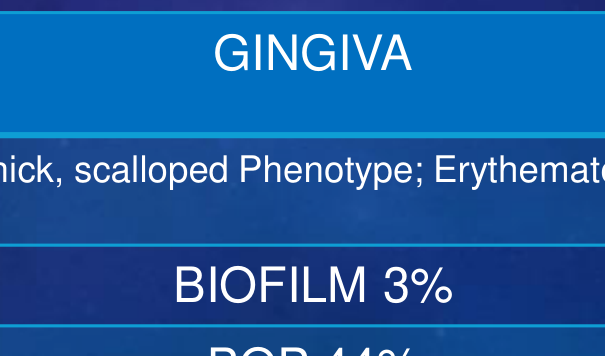
GINGIVA
Thick, scalloped Phenotype; Erythematous
- BIOFILM 3%
- BOP 44%
CLINICAL EXAMINATION (KEY FINDINGS)
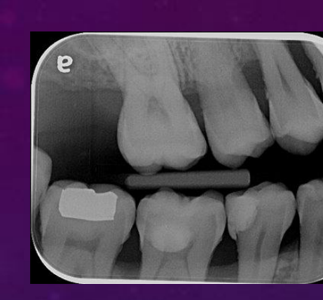
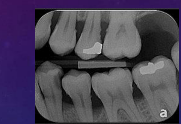
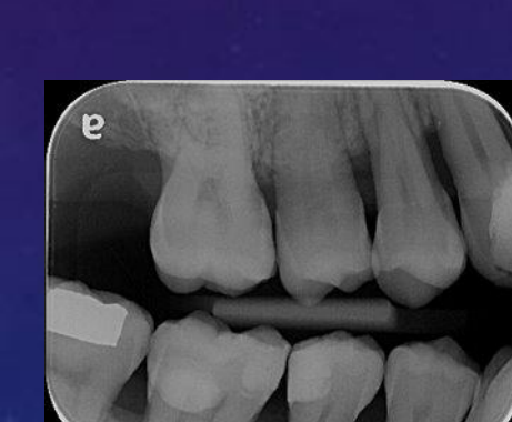
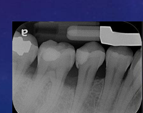
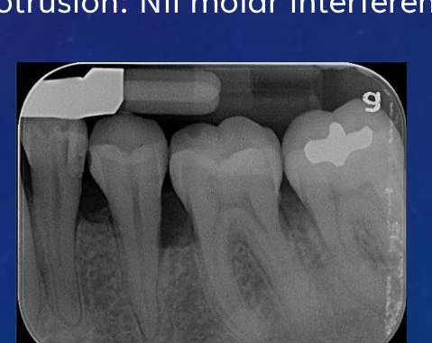
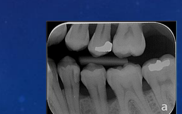
CARIES36
43M, 42D, 21MDBP, 45DO, 46MO, 13MB, 11MBP, 31M, 32D, 34D
 |  |  |
 |  |  |
PROSTHODONTIC
Upper partial acrylic denture demonstrates insufficient adaptation, exhibiting bulkiness and improper fit
ENDODONTIC
21 root-filling, slightly overfilled with no signs and symptoms
OCCLUSAL
Occlusal analysis: Missing 12, 22 Molar Class I RHS, class III LHS Crowded 13–23 Occlusal interferences:
- RHS lateral excursion: canine guidance, Nil interferences
- LHS lateral excursion: Canine guidance, Nil interferences
- Protrusion: Nil molar interference
Diagnosis, Staging, and Grading37
PERIODONTAL DIAGNOSIS
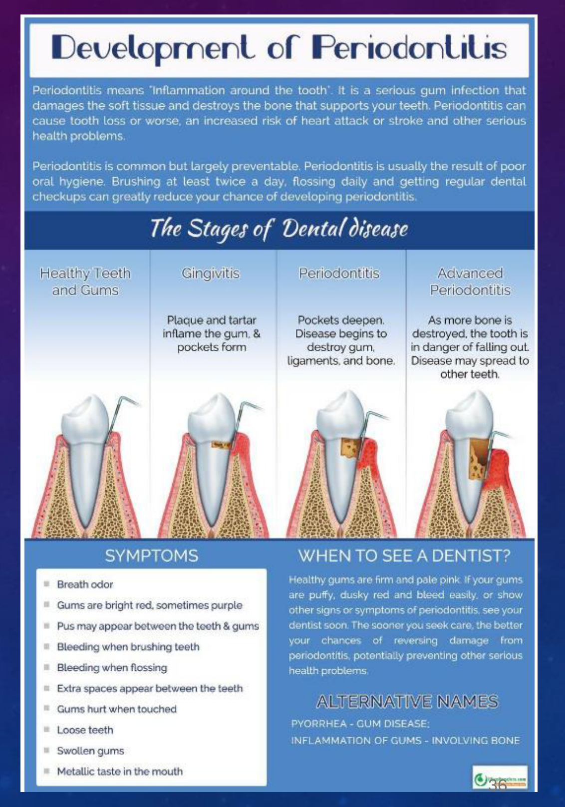
Generalised Stage III, Grade C, Unstable Periodontitis
 |  |
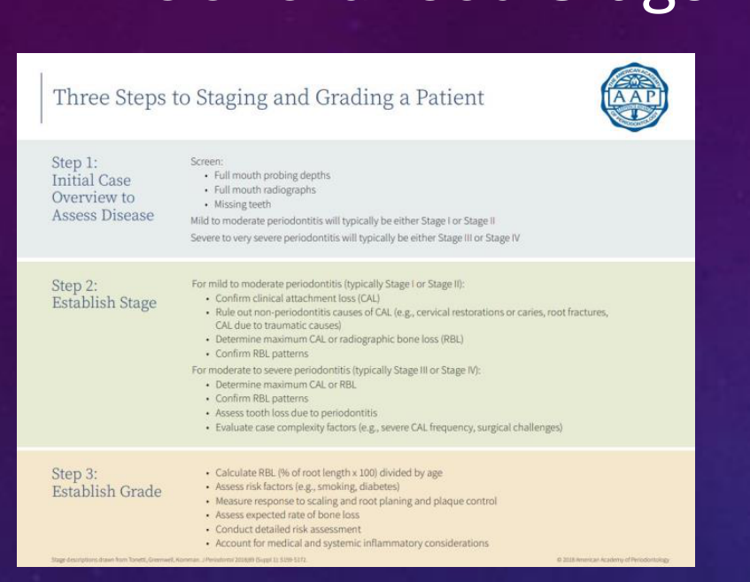
Three Steps to Staging and Grading a Patient38
Step 1: Initial Case Overview to Assess Disease Screen:
- Full mouth probing depths
- Full mouth radiographs
- Missing teeth Mild to moderate periodontitis will typically be either Stage I or Stage II Severe to very severe periodontitis will typically be either Stage III or Stage IV
Step 2: Establish Stage For mild to moderate periodontitis (typically Stage I or Stage II):
- Confirm clinical attachment loss (CAL)
- Rule out non-periodontitis causes of CAL (e.g., cervical restorations or caries, root fractures, CAL due to traumatic causes)
- Determine maximum CAL or radiographic bone loss (RBL)
- Confirm RBL patterns For moderate to severe periodontitis (typically Stage III or Stage IV):
- Determine maximum CAL or RBL
- Confirm RBL patterns
- Assess tooth loss due to periodontitis
- Evaluate case complexity factors (e.g., severe CAL frequency, surgical challenges)
Step 3: Establish Grade
- Calculate RBL (% of root length x 100) divided by age
- Assess risk factors (e.g., smoking, diabetes)
- Measure response to scaling and root planing and plaque control
- Assess expected rate of bone loss
- Conduct detailed risk assessment
- Account for medical and systemic inflammatory considerations
 |  |
 |
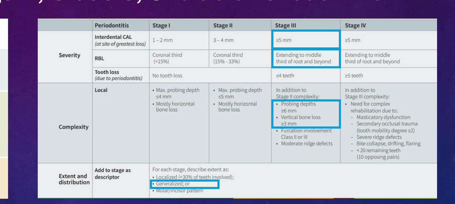
Staging Table
| Periodontitis | Stage I | Stage II | Stage III | Stage IV | |
|---|---|---|---|---|---|
| Severity | Interdental CAL (at site of greatest loss) | 1-2 mm | 3-4 mm | ≥5 mm | ≥5 mm |
| RBL | Coronal third (<15%) | Coronal third (15% - 33%) | Extending to middle third of root and beyond | Extending to middle third of root and beyond | |
| Tooth loss (due to periodontitis) | No tooth loss | ≤4 teeth | ≥5 teeth | ||
| Complexity | Local | Max. probing depth ≤4 mm; Mostly horizontal bone loss | Max. probing depth ≤5 mm; Mostly horizontal bone loss | In addition to Stage II complexity: Probing depths ≥6 mm; Vertical bone loss ≥3 mm; Furcation involvement Class II or III; Moderate ridge defects | In addition to Stage III complexity: Need for complex rehabilitation due to: Masticatory dysfunction; Secondary occlusal trauma (tooth mobility degree ≥2); Severe ridge defects; Bite collapse, drifting, flaring; < 20 remaining teeth (10 opposing pairs) |
| Extent and distribution | Add to stage as descriptor | For each stage, describe extent as: Localized (<30% of teeth involved); Generalized; or Molar/incisor pattern |
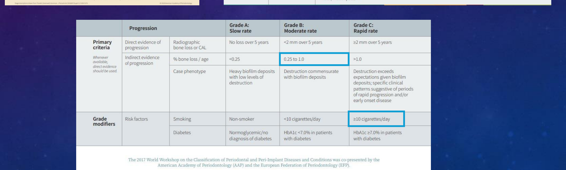
Grading Table
| Progression | Grade A: Slow rate | Grade B: Moderate rate | Grade C: Rapid rate | ||
|---|---|---|---|---|---|
| Primary criteria (Whenever available, direct evidence should be used.) | Direct evidence of progression | Radiographic bone loss or CAL | No loss over 5 years | <2 mm over 5 years | ≥2 mm over 5 years |
| Indirect evidence of progression | % bone loss / age | <0.25 | 0.25 to 1.0 | >1.0 | |
| Case phenotype | Heavy biofilm deposits with low levels of destruction | Destruction commensurate with biofilm deposits | Destruction exceeds expectations given biofilm deposits; specific clinical patterns suggestive of periods of rapid progression and/or early onset disease | ||
| Grade modifiers | Risk factors | Smoking | Non-smoker | <10 cigarettes/day | ≥10 cigarettes/day |
| Diabetes | Normoglycemic/no diagnosis of diabetes | HbA1c <7.0% in patients with diabetes | HbA1c ≥7.0% in patients with diabetes |
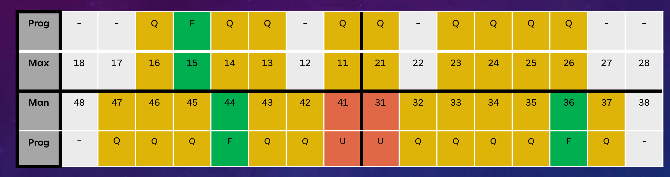
PERIODONTAL PROGNOSIS
| Prog | - | - | Q | F | Q | Q | - | Q | Q | - | Q | Q | Q | Q | - | - |
|---|---|---|---|---|---|---|---|---|---|---|---|---|---|---|---|---|
| Max | 18 | 17 | 16 | 15 | 14 | 13 | 12 | 11 | 21 | 22 | 23 | 24 | 25 | 26 | 27 | 28 |
| Man | 48 | 47 | 46 | 45 | 44 | 43 | 42 | 41 | 31 | 32 | 33 | 34 | 35 | 36 | 37 | 38 |
| Prog | - | Q | Q | Q | F | Q | Q | U | U | Q | Q | Q | Q | F | Q | - |
- F = Favourable
- Q = Questionable
- U = Unfavourable
- H = Hopeless
Treatment Execution and Reassessment
Step 1 and Initial Reassessment
Step 1 EFP Guidelines / Risk Management Phase39
Risk factor control:
- Encourage & discuss relationship between smoking and periodontal disease
- After our discussion about smoking, she demonstrated encouragement and determination.
- She sought additional support from her general practitioner (GP) during her smoking cessation journey and successfully quit smoking following her initial consultation with her GP.
- Began her journey to quit smoking in May 2023.
- Instructions on self-performed biofilm control
- Stress management
- Remove Plaque-retentive factors that impair oral hygiene practices
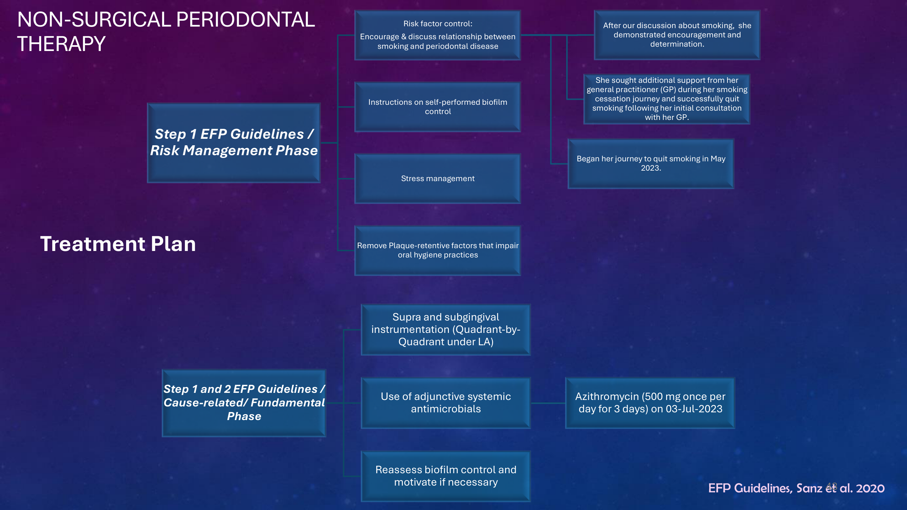
Step 1 and 2 EFP Guidelines / Cause-related/ Fundamental Phase
- Supra and subgingival instrumentation (Quadrant-by-Quadrant under LA)
- Use of adjunctive systemic antimicrobials: Azithromycin (500 mg once per day for 3 days) on 03-Jul-2023
- Reassess biofilm control and motivate if necessary
EFP Guidelines, Sanz et al. 2020
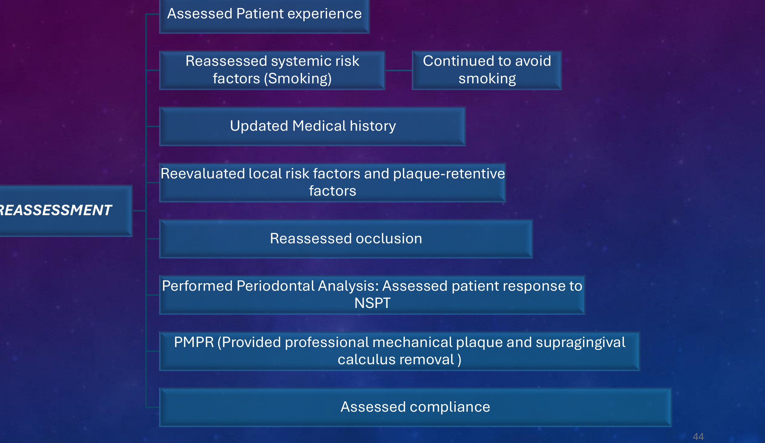
1ST REASSESSMENT40
- Assessed Patient experience
- Reassessed systemic risk factors (Smoking): Continued to avoid smoking
- Updated Medical history
- Reevaluated local risk factors and plaque-retentive factors
- Reassessed occlusion
- Performed Periodontal Analysis: Assessed patient response to NSPT
- PMPR (Provided professional mechanical plaque and supragingival calculus removal)
- Assessed compliance
Step 3: Surgical Periodontal Therapy
SURGICAL PERIODONTAL THERAPY41
Step 3 EFP Guidelines / Corrective Phase
- Repeating non-surgical debridement
- Access surgery
- Resective surgery
- Regenerative surgery
- Pockets ≥6 mm with associated Intrabony defects ≥3 mm
- Furcation defects class II
- Endpoint of therapy not reached: Consider modality of further treatment for residual PPD ≥4mm with BOP or ≥6mm.
EFP Guidelines, Sanz et al. 2020
 |  |
Step 4: Supportive Periodontal Care Reassessment42
REASSESSMENT43
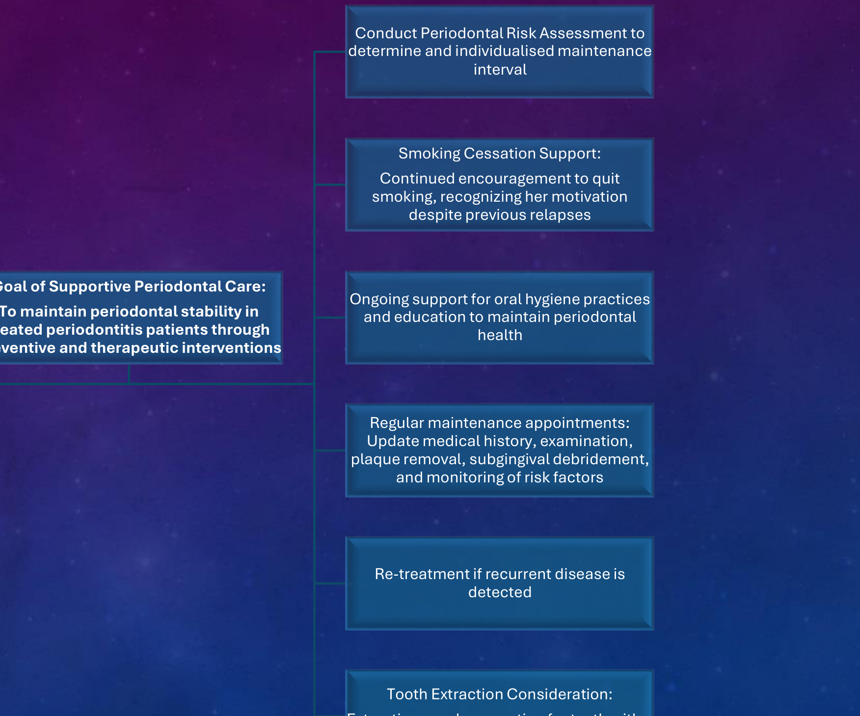
Step 4 EFP Guidelines / SPC
- Conduct Periodontal Risk Assessment to determine and individualised maintenance interval
- Smoking Cessation Support: Continued encouragement to quit smoking, recognizing her motivation despite previous relapses
- Goal of Supportive Periodontal Care: To maintain periodontal stability in treated periodontitis patients through preventive and therapeutic interventions
- Ongoing support for oral hygiene practices and education to maintain periodontal health
- Regular maintenance appointments: Update medical history, examination, plaque removal, subgingival debridement, and monitoring of risk factors
- Re-treatment if recurrent disease is detected
- Tooth Extraction Consideration: Extraction may be an option for teeth with a hopeless prognosis
EFP Guidelines, Sanz et al. 2020
Thank You
☺
Footnotes
-
Original PDF page 1: Treatment Planning-1, p.1 ↩
-
Original PDF page 3: Treatment Planning-1, p.3 ↩
-
Original PDF page 2: Treatment Planning-1, p.2 ↩
-
Original PDF page 4: Treatment Planning-1, p.4 ↩
-
Original PDF page 5: Treatment Planning-1, p.5 ↩
-
Original PDF page 6: Treatment Planning-1, p.6 ↩
-
Original PDF page 7: Treatment Planning-1, p.7 ↩
-
Original PDF page 9: Treatment Planning-1, p.9 ↩
-
Original PDF page 13: Treatment Planning-1, p.13 ↩
-
Original PDF page 14: Treatment Planning-1, p.14 ↩
-
Original PDF page 15: Treatment Planning-1, p.15 ↩
-
Original PDF page 16: Treatment Planning-1, p.16 ↩
-
Original PDF page 17: Treatment Planning-1, p.17 ↩
-
Original PDF page 18: Treatment Planning-1, p.18 ↩
-
Original PDF page 19: Treatment Planning-1, p.19 ↩
-
Original PDF page 20: Treatment Planning-1, p.20 ↩
-
Original PDF page 21: Treatment Planning-1, p.21 ↩
-
Original PDF page 22: Treatment Planning-1, p.22 ↩
-
Original PDF page 23: Treatment Planning-1, p.23 ↩
-
Original PDF page 24: Treatment Planning-1, p.24 ↩
-
Original PDF page 25: Treatment Planning-1, p.25 ↩
-
Original PDF page 26: Treatment Planning-1, p.26 ↩
-
Original PDF page 27: Treatment Planning-1, p.27 ↩
-
Original PDF page 28: Treatment Planning-1, p.28 ↩
-
Original PDF page 29: Treatment Planning-1, p.29 ↩
-
Original PDF page 30: Treatment Planning-1, p.30 ↩
-
Original PDF page 31: Treatment Planning-1, p.31 ↩
-
Original PDF page 32: Treatment Planning-1, p.32 ↩
-
Original PDF page 33: Treatment Planning-1, p.33 ↩
-
Original PDF page 34: Treatment Planning-1, p.34 ↩
-
Original PDF page 36: Treatment Planning-1, p.36 ↩
-
Original PDF page 35: Treatment Planning-1, p.35 ↩
-
Original PDF page 39: Treatment Planning-1, p.39 ↩
-
Original PDF page 37: Treatment Planning-1, p.37 ↩
-
Original PDF page 38: Treatment Planning-1, p.38 ↩
-
Original PDF page 40: Treatment Planning-1, p.40 ↩
-
Original PDF page 42: Treatment Planning-1, p.42 ↩
-
Original PDF page 41: Treatment Planning-1, p.41 ↩
-
Original PDF page 43: Treatment Planning-1, p.43 ↩
-
Original PDF page 44: Treatment Planning-1, p.44 ↩
-
Original PDF page 45: Treatment Planning-1, p.45 ↩
-
Original PDF page 47: Treatment Planning-1, p.47 ↩
-
Original PDF page 46: Treatment Planning-1, p.46 ↩