Mri Ultrasound And Nuclear Medicine Imaging1
Dr Dayea Oh OMF Radiologist
Learning Objectives2
- To understand the basic theory of:
- Magnetic Resonance Imaging (MRI)
- Ultrasound Imaging / Ultrasonography (US)
- Nuclear Medicine Imaging (NMI)
- Scintigraphy
- Positron Emission Tomography (PET)
- Single-Photon Emission Computed Tomography (SPECT)
- To know applications / indications for each of the above advanced imaging modalities
Magnetic Resonance Imaging3
I. Magnetic Resonance Imaging (MRI)
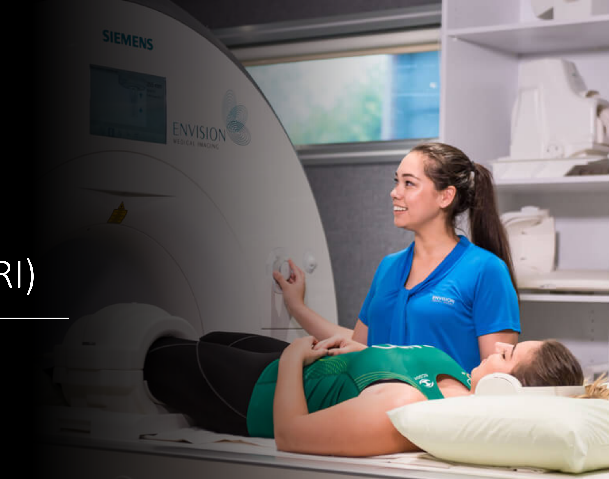
Siemens Envision Medical Imaging
Introduction To Mri Principles4
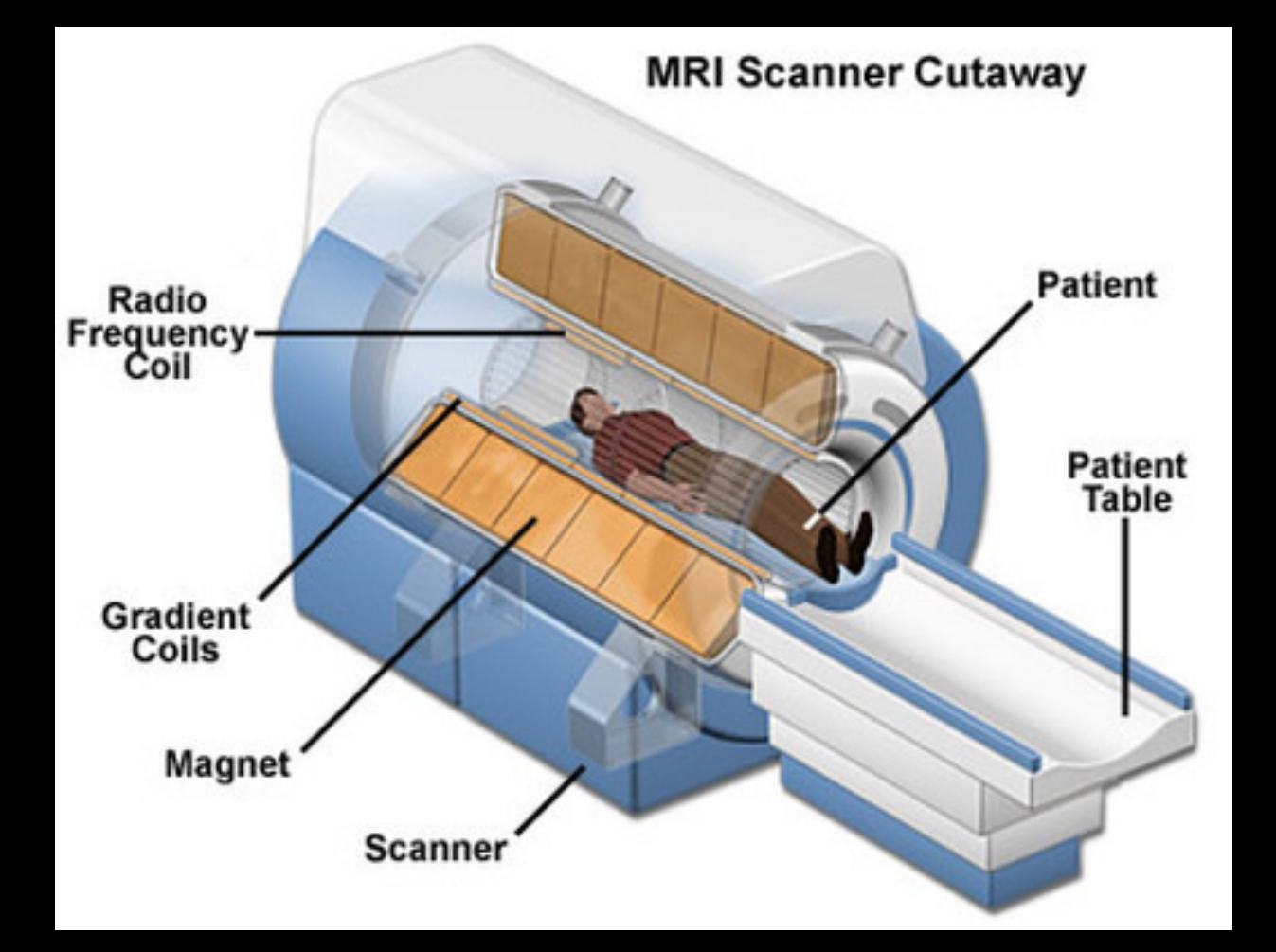
Magnetic Resonance Imaging (MRI)
- Magnetic field
- = “Magnetic”
- Radiofrequency waves
- = “Resonance”
- NO ionising radiation
- Soft tissue imaging
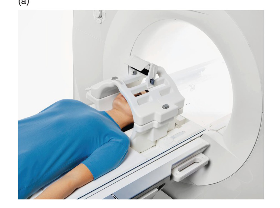
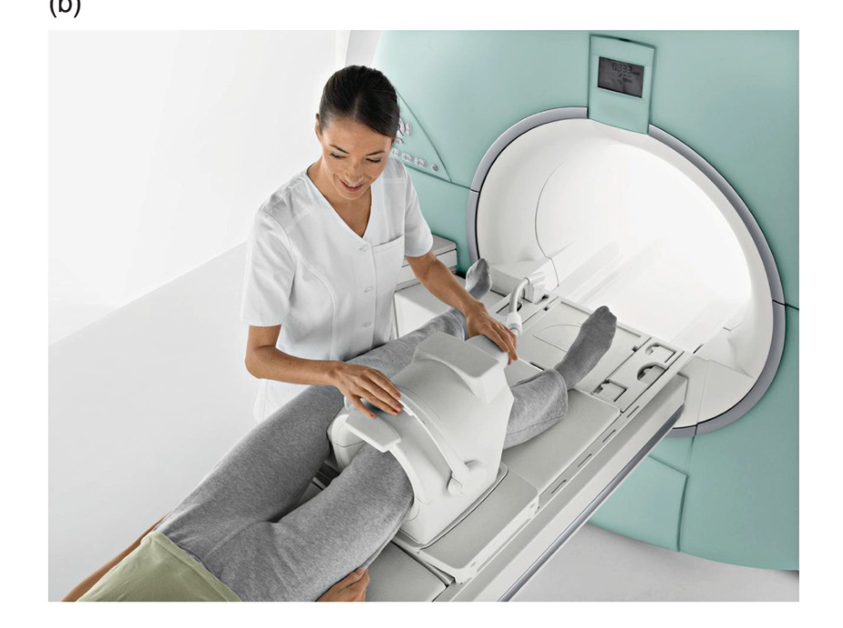
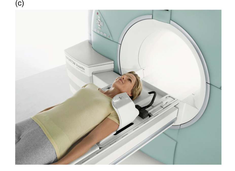
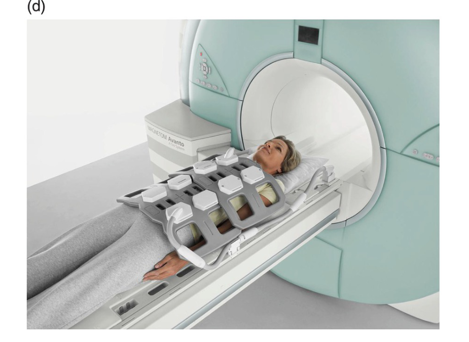
- Bore
Patient Safety And Contraindications5
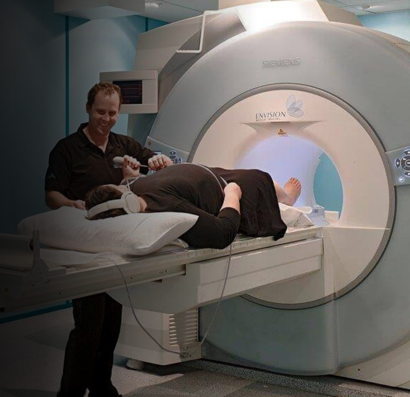
MRI Scan
- Long scanning time
- ✅ Claustrophobia
- Noisy
- Heat in the area being examined
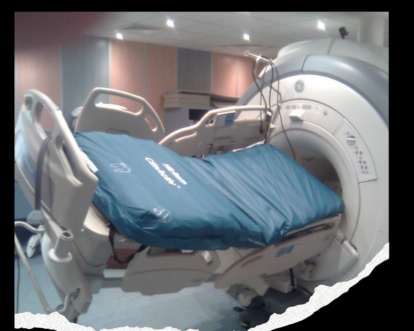
- ✅ Ferromagnetic metals
Pre-MRI Check6
- Pacemaker / Defibrillator (ICD)
- Neurostimulator
- Cochlear Implant
- Stapedectomy (middle ear prosthesis)
- Aneurysm Clip
- Morphine / Insulin Pump
- Glucose Monitor
- Programmable VP Shunt
- Shrapnel
- Retained Metal fragment in eye (eg. from welding / grinding)
Mri Physics And Theory
Step 1. Magnetisation7
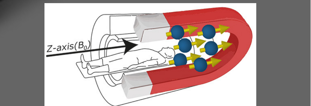
Step One Magnetisation
- STRONG external magnetic field of 1.5T or higher (MRI machine)
- 1 T can lift a car!
- Hydrogen atoms (in water “H2O” and fat)
- Single proton (& 1 electron & zero neutron)
- Naturally spinning and wobbling particles with +ve charge
- Charge with motion creates a magnetic field
Magnetisation & z-axis
Under external magnetic field (MRI scanner)
Step Two Resonance And Excitation8
Step 2. Resonance; precession, excitation & relaxation
- Hydrogen spin & wobble motion = precession
- Larmor’s equation
- is the frequency in MHz,
- is the gyromagnetic ratio in MHz/Tesla,
- is the strength of the magnetic field in Tesla
- H protons precess at the rate of 63.87MHz at 1.5T
- Matching radiofrequency (RF) waves create RESONANCE
- Resonance results in ‘Excitation’
- ‘Relaxation’ releases MR Signals captured by receiver coil
- Different body tissues ‘relax’ at different rates
- The faster the tissue relaxes, the more the MR signals released in a given time
Secondary Magnetic Field (Gradient)
- Distorting / tilting the main magnetic field to acquire slices from different sections of the body
- Different resonance frequencies along the gradient
- Localisation of different anatomical structures
 |  |
 |  |
Step Three Signal Acquisition And Processing
Step 3. Signal Acquisition and Processing
- MR Signal received by receiver coil is converted into a digital form and transmitted to computer
- The computer processes the data and an image is created
Understanding Mri Signal Sequences
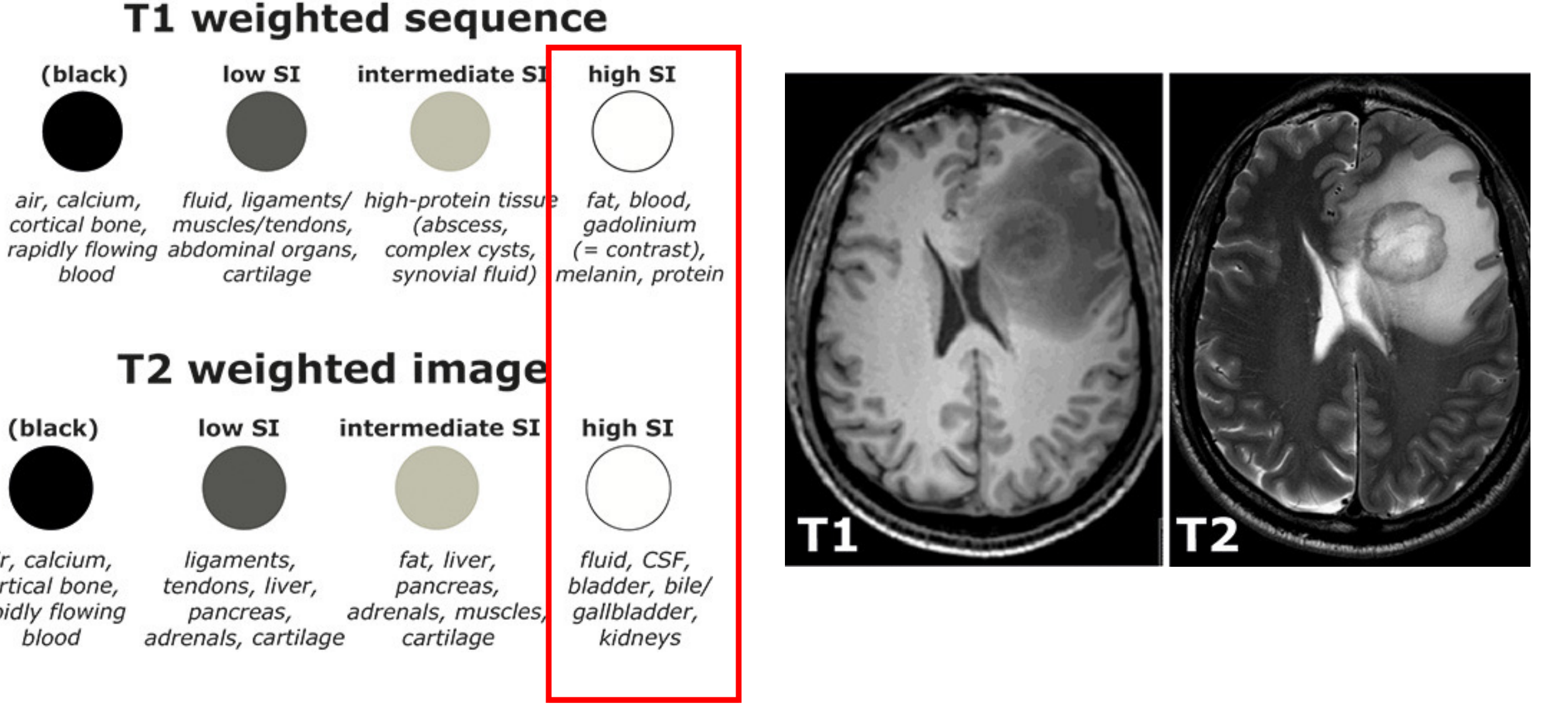
T1 Weighted Sequence9
- (black): air, calcium, cortical bone, rapidly flowing blood
- low SI: fluid, ligaments/muscles/tendons, abdominal organs, cartilage
- intermediate SI: high-protein tissue (abscess, complex cysts, synovial fluid)
- high SI: fat, blood, gadolinium (= contrast), melanin, protein
T2 Weighted Image
- (black): air, calcium, cortical bone, rapidly flowing blood
- low SI: ligaments, tendons, liver, pancreas, adrenals, cartilage
- intermediate SI: fat, liver, pancreas, adrenals, muscles, cartilage
- high SI: fluid, CSF, bladder, bile/gallbladder, kidneys
Mri Applications In Dental And Maxillofacial Radiology10
Applications11
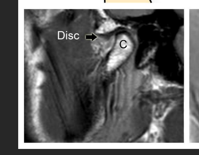
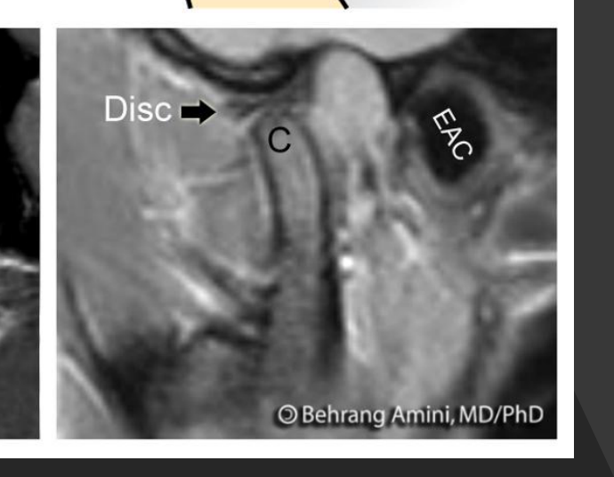
- TMJ disorders
- Tumours and cysts in the head and neck
- Jaw lesions
- Salivary gland lesions
- Paranasal sinus lesions
- Assessment of IDN
- Infection / inflammation eg. osteomyelitis
- Vascular lesions
 |  |
 |
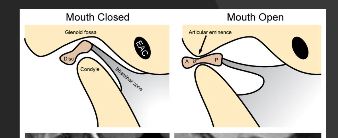
Diagrams
Mouth Closed
- Glenoid fossa
- Disc
- Condyle
- Bilaminar zone
- EAC
Mouth Open
- Articular eminence
- A, P (Anterior, Posterior)
- ILZ (Interalveolar Ligament Zone - inferred)
- Disc
MRI scans showing:
- Disc, C (Condyle), EAC (External Auditory Canal)
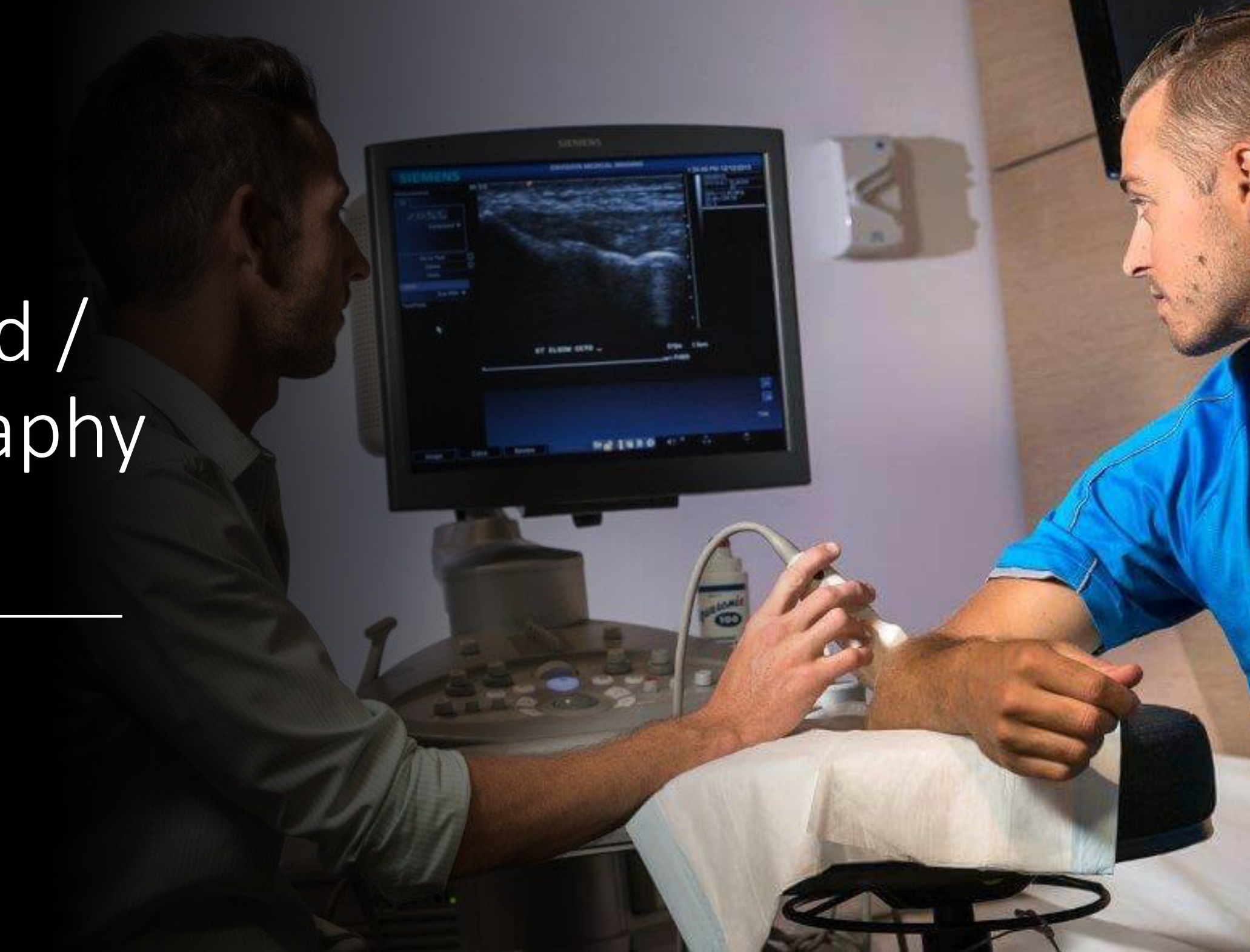
Ultrasound Imaging12
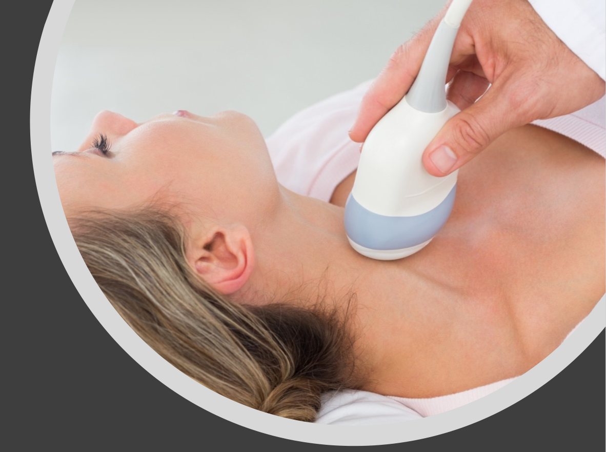
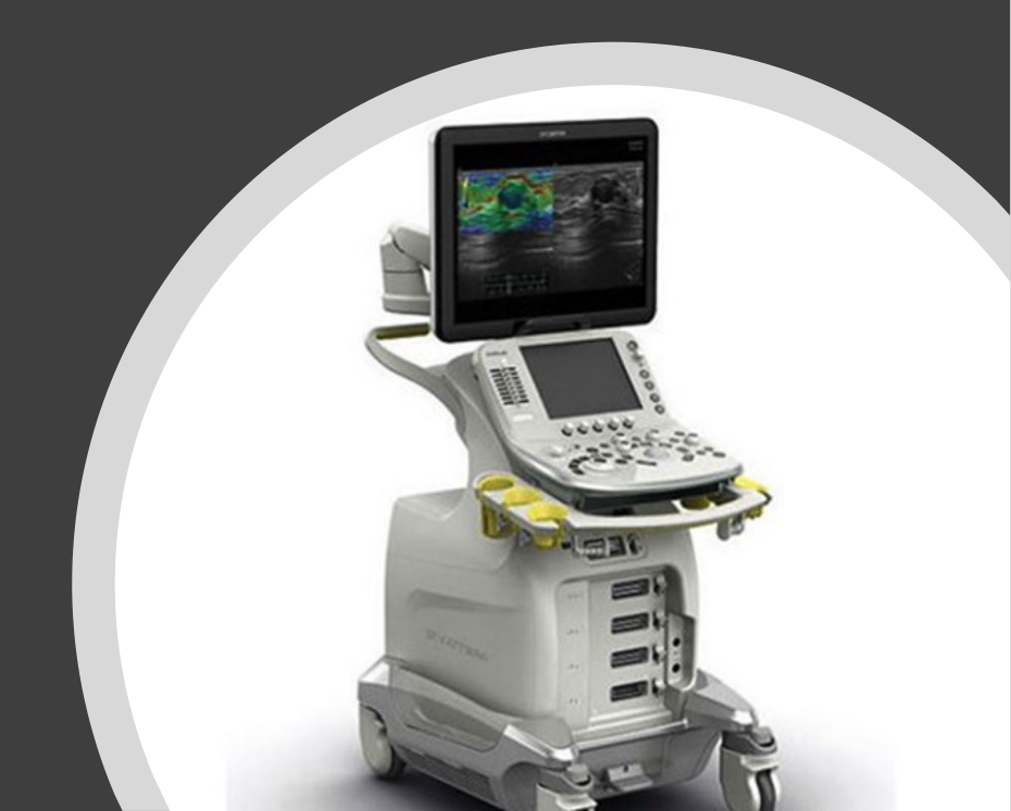
- Non-ionising
- Sound waves
- Soft tissue imaging
- Compact
- Inexpensive
- Real-time imaging
- Technique sensitive
 |  |
Principles Of Ultrasonography13
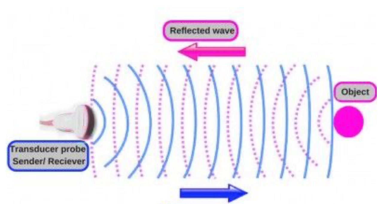
Ultrasound (US) = sound wave with high frequency
- Diagnostic frequency = 2MHz to 30MHz
- Human hearing is 16Hz – 20kHz
Piezoelectric transducer:
- Send US waves into the body
- Receive reflected waves “echoes” from the body
- Convert echoes into electric signal
- Signal sent to computer for image display
US transmission is improved with ultrasound gel “jelly”
Understanding Ultrasound Echogenicity
Understanding US Signals “Echogenecity”14
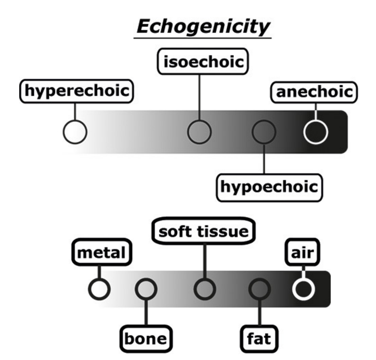
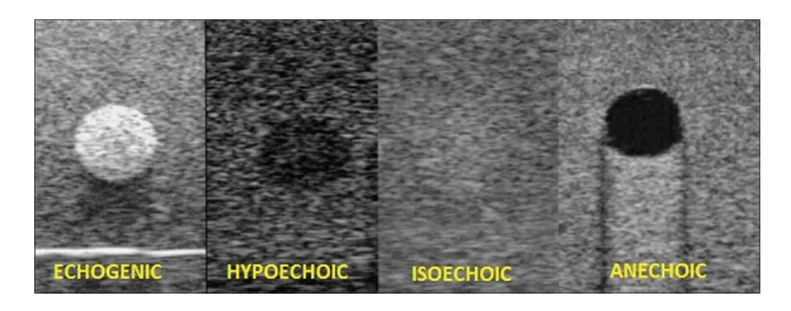
Echogenicity
| Echogenicity Level | Label |
|---|---|
| Leftmost (light) | hyperechoic |
| Light-grey | isoechoic |
| Grey | hypoechoic |
| Dark grey/black | anechoic |
| Tissue/Structure | Classification |
|---|---|
| metal | hyperechoic |
| bone | hyperechoic |
| soft tissue | isoechoic |
| fat | hypoechoic |
| air | anechoic |
 |  |
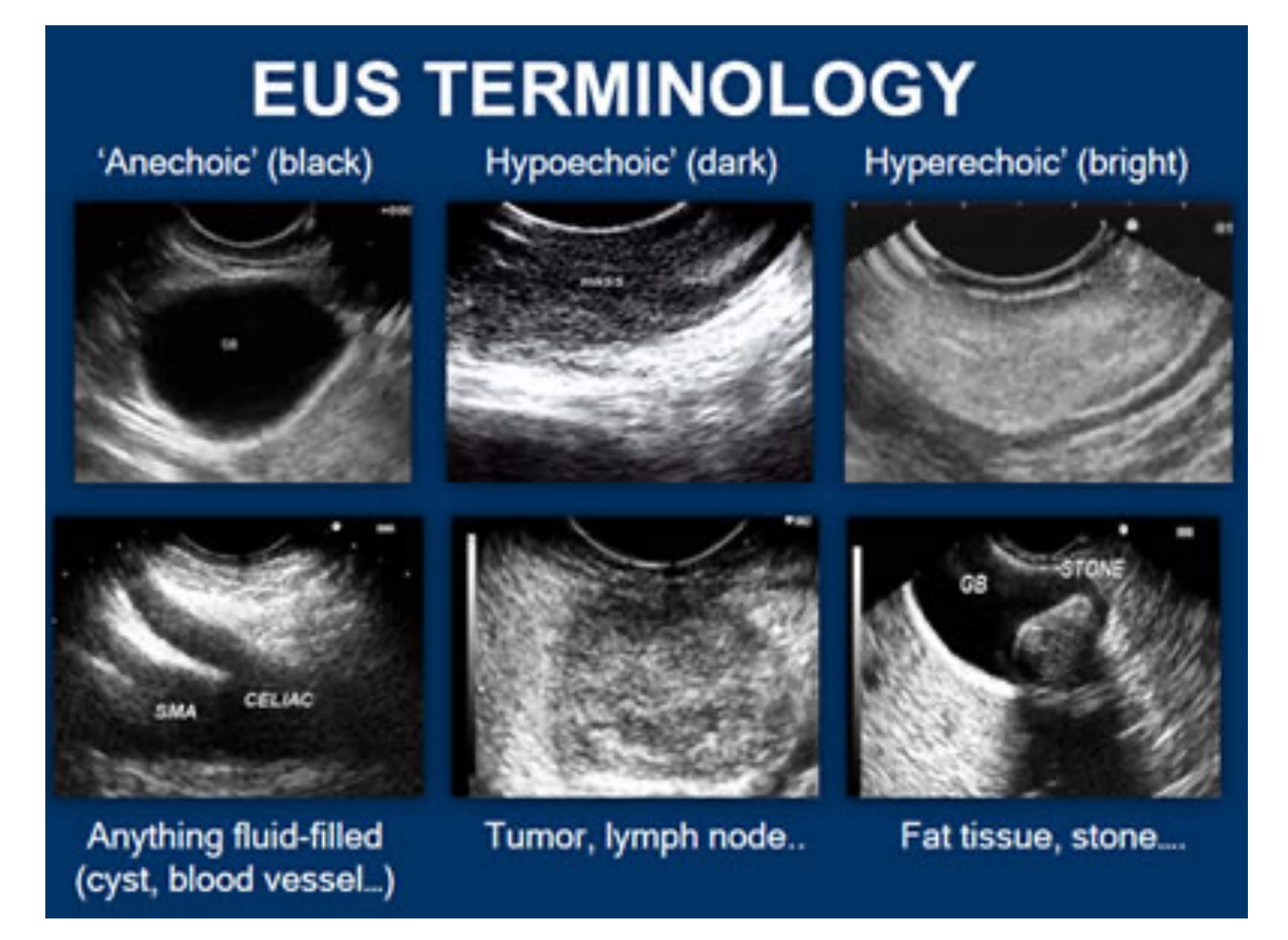
EUS TERMINOLOGY15
| ’Anechoic’ (black) | ‘Hypoechoic’ (dark) | ‘Hyperechoic’ (bright) |
|---|---|---|
| Anything fluid-filled (cyst, blood vessel…) | Tumor, lymph node… | Fat tissue, stone… |
Ultrasound Applications In Dental And Maxillofacial Radiology16
- Salivary gland disease
- Soft tissue swelling of the head and neck
- Tumours, Cysts, Infection, etc.
- Vascular diseases and anomalies of the head and neck
- Carotid artery stenosis (blockage or narrowing)
- Hematoma (collection of blood in tissues from damaged vessels)
- Abnormalities in the blood vessels, lymph node, etc.
- Ultrasound-guided procedures
- Biopsy (Fine needle aspirational / FNA)
- Corticosteroid injection (TM joint)
- TMJ Imaging
- Real-time imaging (disc-condyle relationship on mouth opening)
Nuclear Medicine Imaging17
III. Nuclear Medicine Imaging (NMI)
Principles Of Radiopharmaceuticals
What is Nuclear Medicine Imaging?18
- Radiology “done inside out”
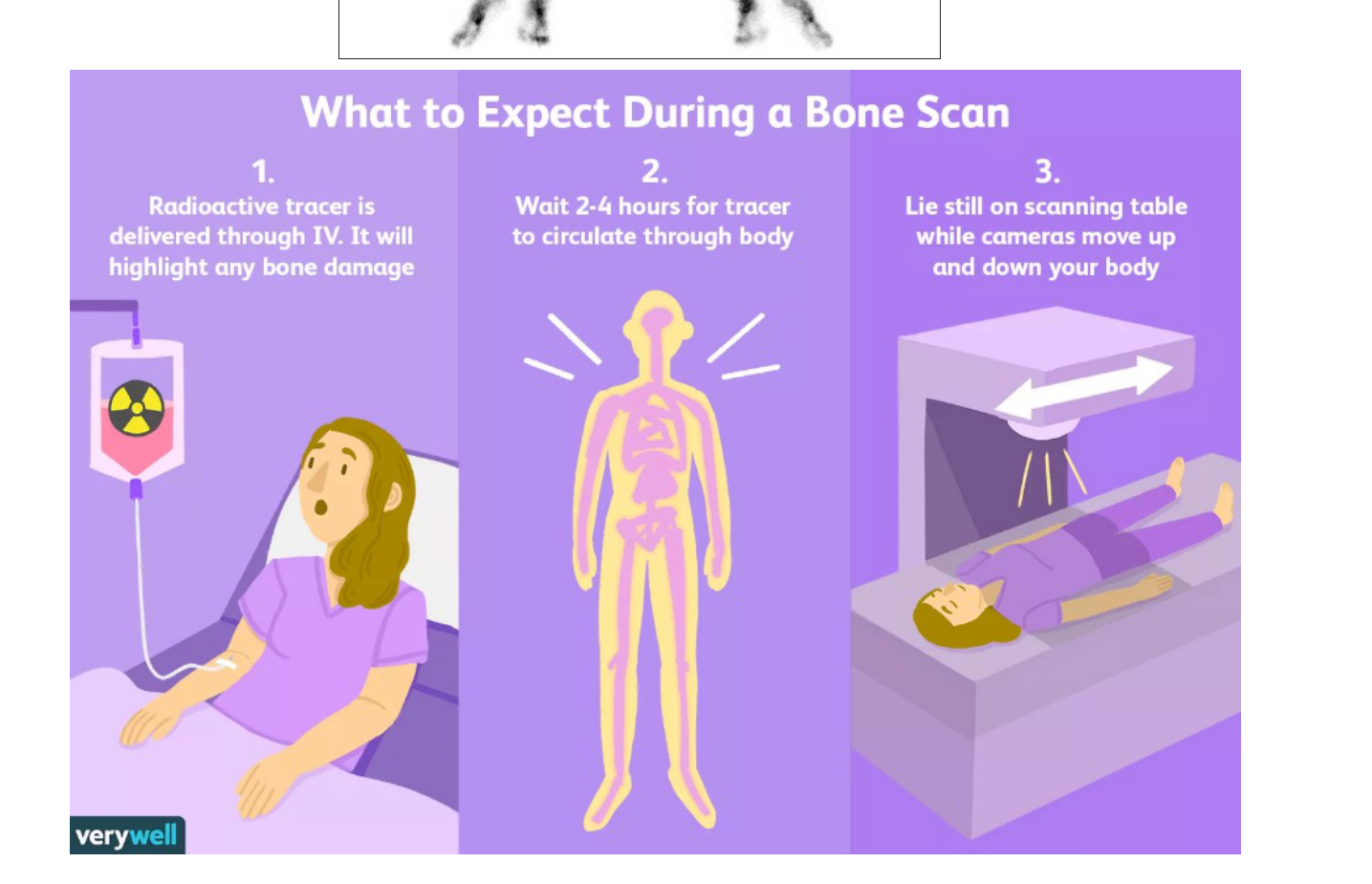
- Radiopharmaceuticals
- 99mTc MDP
- Technetium99m + Methyl diphosphate
- Half life of 6 hrs
- 99mTc MDP
- “Hot spots”
- Three types:
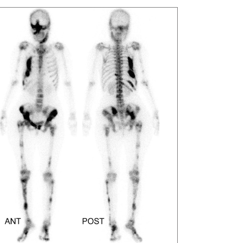
- Scintigraphy (2D imaging eg. Bone scan)
- Single photon emission computed tomography (SPECT)
- Positron emission tomography (PET)
 |  |
Three Dimensional Imaging Modalities1920
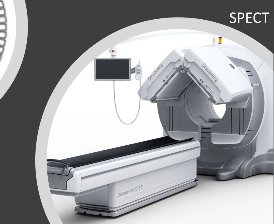
SPECT (single photon emission computed tomography)21
- 3D localisation & estimation of the size & extend of the disease
- Single gamma rays
- Two receptors
 |  |
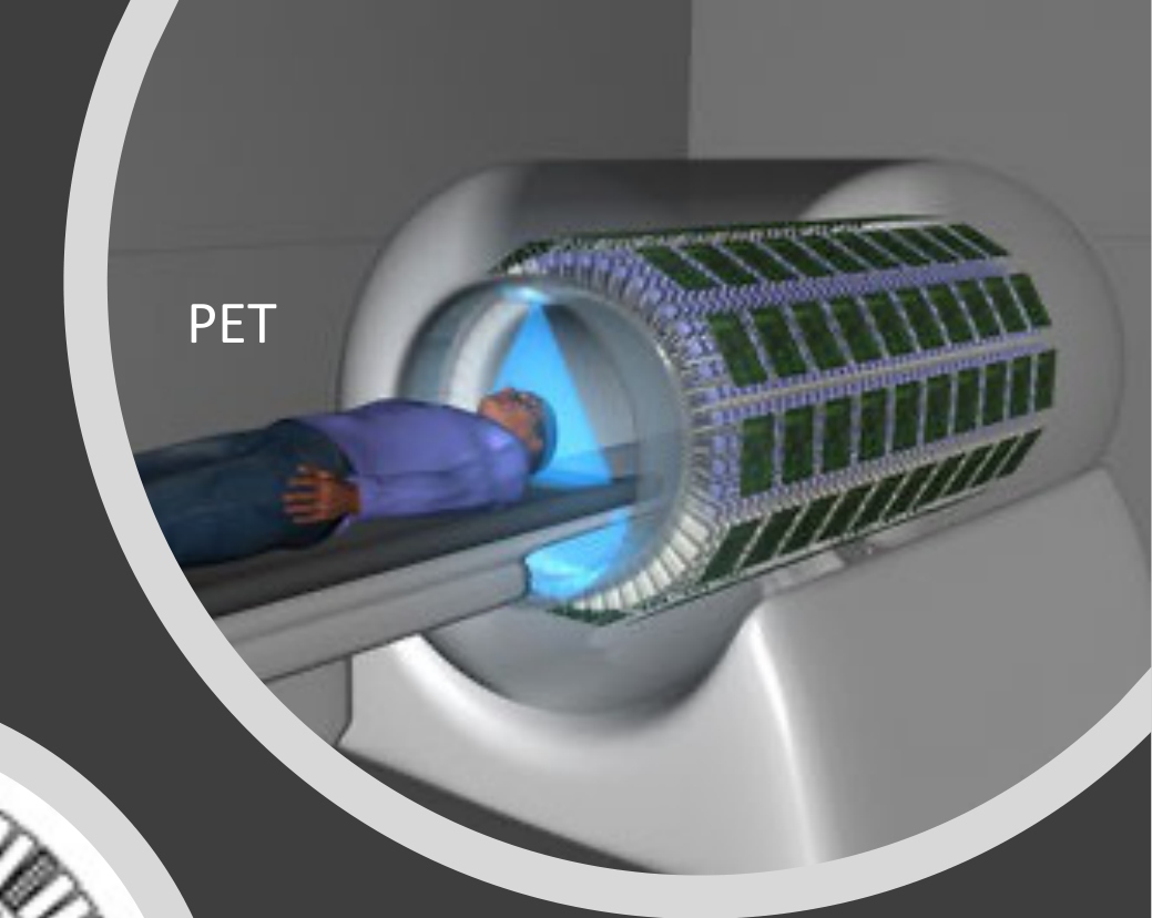 |  |
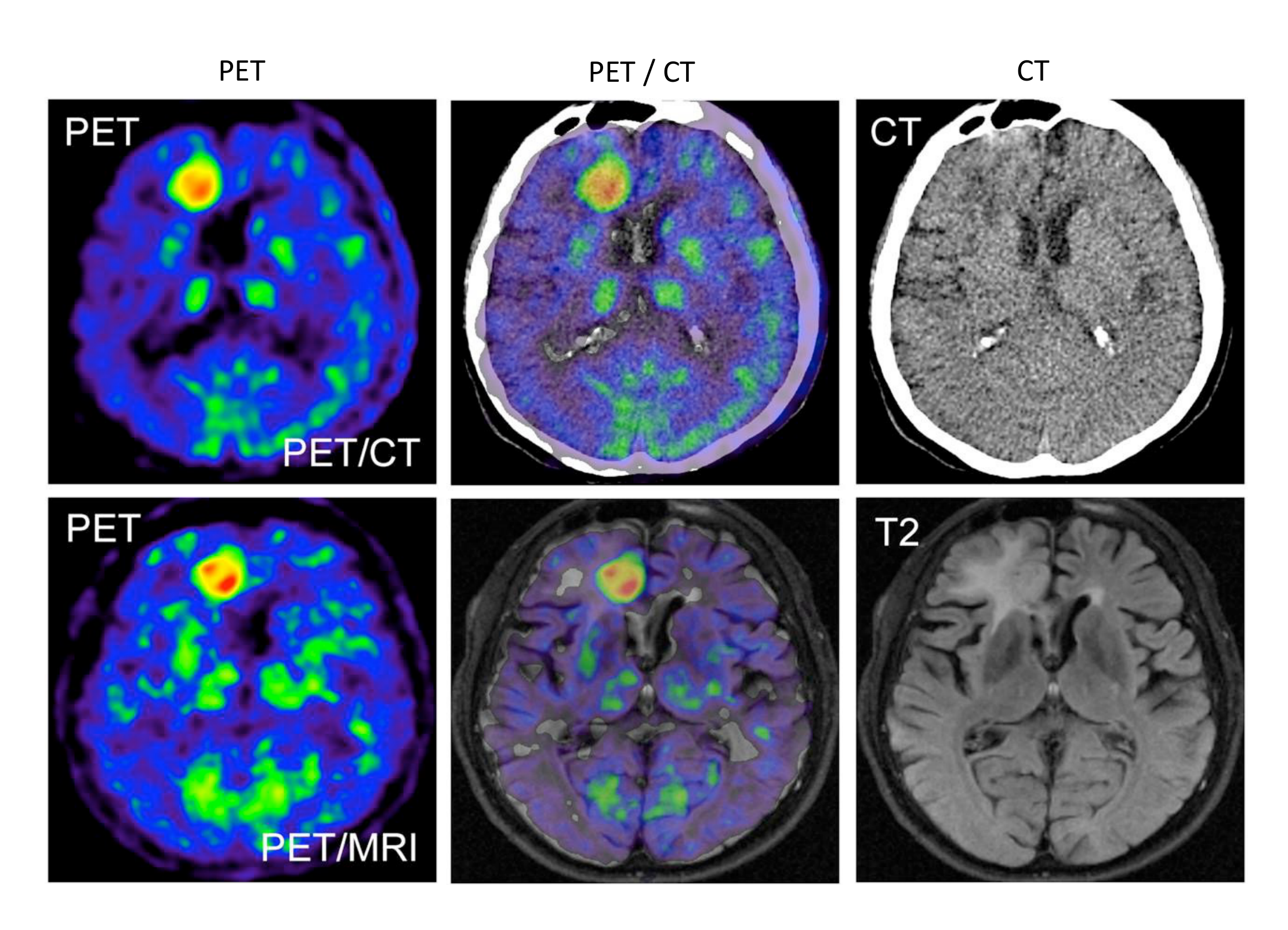
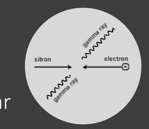
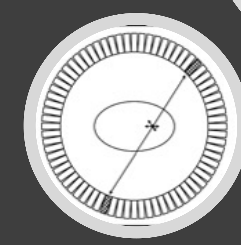
PET (positron emission tomography)
- Like SPECT, but more dimensionally accurate
- Paired gamma rays
- Multiple receptors
Nuclear Medicine Applications In Dental And Maxillofacial Radiology
Nuclear Medicine Imaging in DMFR22
- Progressive or spontaneous growth of the jaws
- Eg. condylar hyperactivity, neoplastic growth
- Osteonecrosis of the Jaws
- Eg. Medication-related osteonecrosis of the jaws (MRONJ), Osteoradionecrosis of the jaws (ORNJ)
- Osteomyelitis
- Eg. infectious (secondary) osteomyelitis, primary osteomyelitis
- Bone fractures
- Bone disorders inc. primary bone tumours
- Head and neck cancer PET
- Eg. oral squamous cell carcinoma (OSCC), lymphoma
Further Reading
 |  |
Footnotes
-
Original PDF page 1: L11 MRI, US and NMI, p.1 ↩
-
Original PDF page 2: L11 MRI, US and NMI, p.2 ↩
-
Original PDF page 3: L11 MRI, US and NMI, p.3 ↩
-
Original PDF page 4: L11 MRI, US and NMI, p.4 ↩
-
Original PDF page 5: L11 MRI, US and NMI, p.5 ↩
-
Original PDF page 6: L11 MRI, US and NMI, p.6 ↩
-
Original PDF page 7: L11 MRI, US and NMI, p.7 ↩
-
Original PDF page 11: L11 MRI, US and NMI, p.11 ↩
-
Original PDF page 13: L11 MRI, US and NMI, p.13 ↩
-
Original PDF page 15: L11 MRI, US and NMI, p.15 ↩
-
Original PDF page 14: L11 MRI, US and NMI, p.14 ↩
-
Original PDF page 16: L11 MRI, US and NMI, p.16 ↩
-
Original PDF page 17: L11 MRI, US and NMI, p.17 ↩
-
Original PDF page 18: L11 MRI, US and NMI, p.18 ↩
-
Original PDF page 19: L11 MRI, US and NMI, p.19 ↩
-
Original PDF page 20: L11 MRI, US and NMI, p.20 ↩
-
Original PDF page 21: L11 MRI, US and NMI, p.21 ↩
-
Original PDF page 22: L11 MRI, US and NMI, p.22 ↩
-
Original PDF page 24: L11 MRI, US and NMI, p.24 ↩
-
Original PDF page 25: L11 MRI, US and NMI, p.25 ↩
-
Original PDF page 23: L11 MRI, US and NMI, p.23 ↩
-
Original PDF page 26: L11 MRI, US and NMI, p.26 ↩