Radiologic Interpretation Part 21
Dr Dayea Oh OMF Radiologist
Learning Objectives2
- Reviewing the systematic approach
- Describing “lesions” considering:
- Location
- Borders
- Morphology
- Internal Architecture / Pattern
- Effects on Adjacent Tissues
- Understanding Surgical Sieve
- Choosing next appropriate diagnostic imaging modality for further evaluation
- Knowing when to urgently refer
Systematic Approach to Interpretation3
-
Any Technical or Positional Error?
-
Address Clinical Concerns
-
Dentoalveolar
- a) Count teeth & identify ectopic and/or impacted teeth
- b) Dental Anomalies (Position, Shape, …)
- c) Periodontal bone loss
- d) Dental pathology (non-carious tooth loss, caries, fracture, periapical pathologies, etc.)
-
Maxilla and Mandible
- a) Abnormal lucencies
- b) Abnormal opacities
- c) Altered trabecular pattern
- d) Fractures
- e) Jaw Asymmetry
-
Maxillary sinuses (esp. mucosal changes)
-
TM Joints (esp. morphological change of condyles)
-
Soft Tissues (swelling, soft tissue calcifications eg. Tonsiloliths, salivary gland stones, etc.)
-
Other sites: Spine, orbits, etc. (structural changes)
Diagnostic Framework for Jaw Lesions
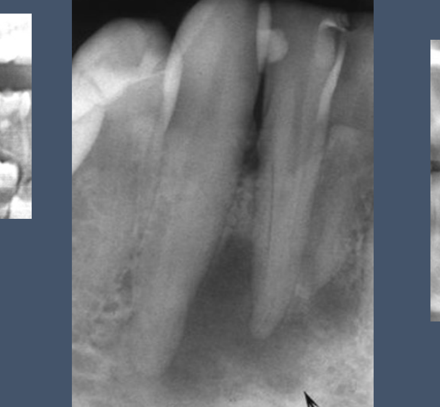
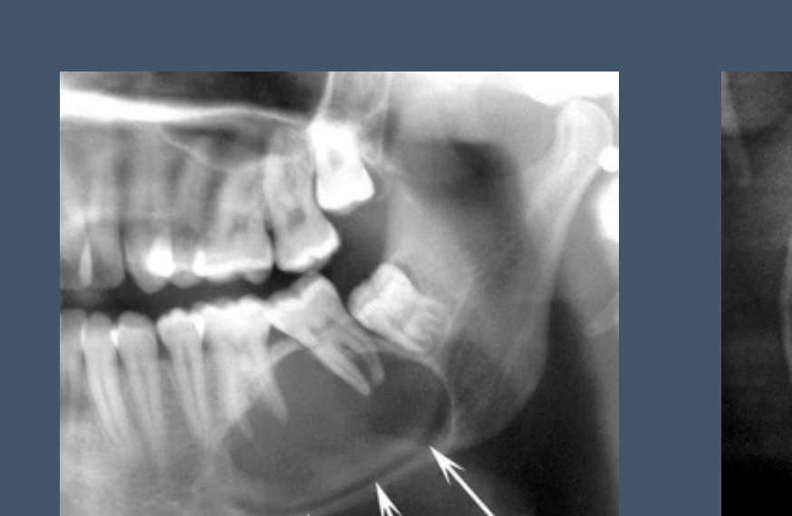
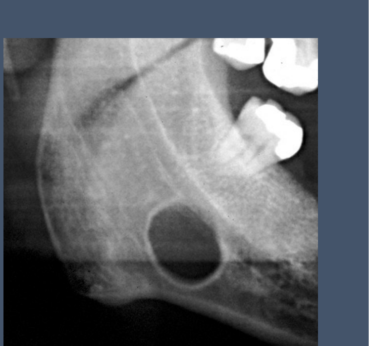
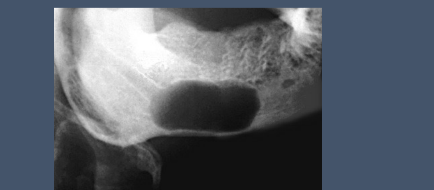
Radiodensity and Localization45
Abnormal Findings (Lesions) in the Jaws
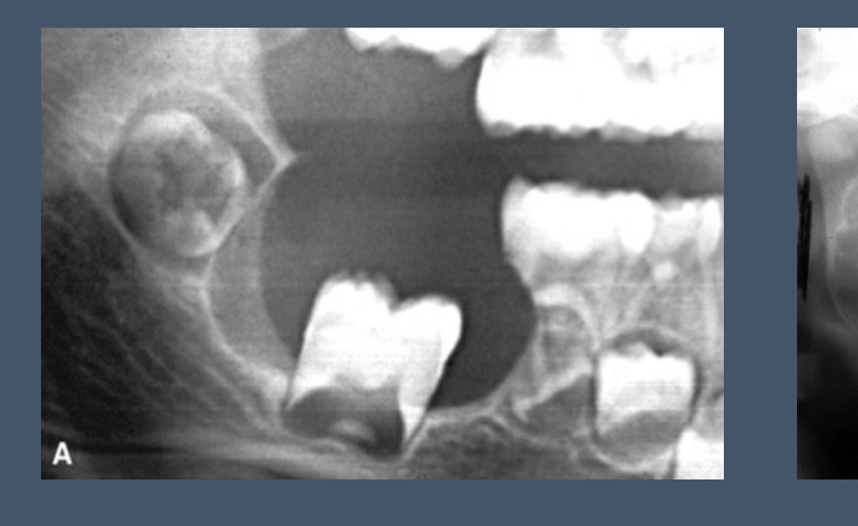
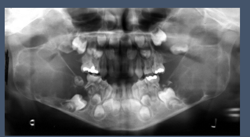
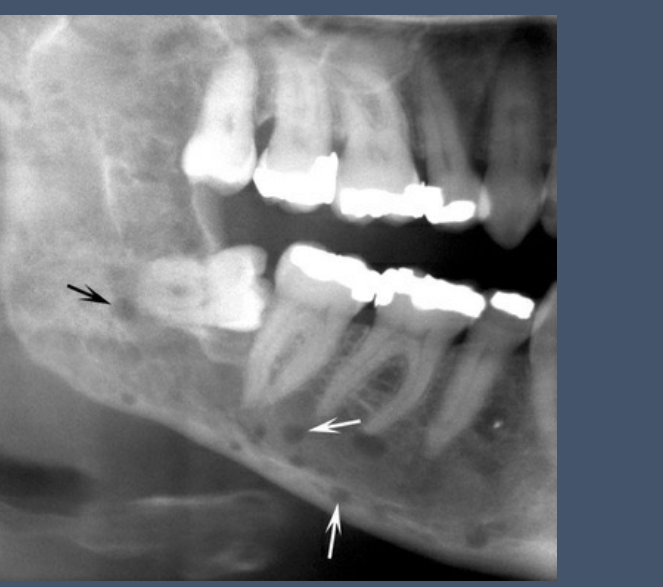
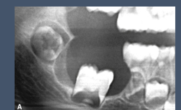
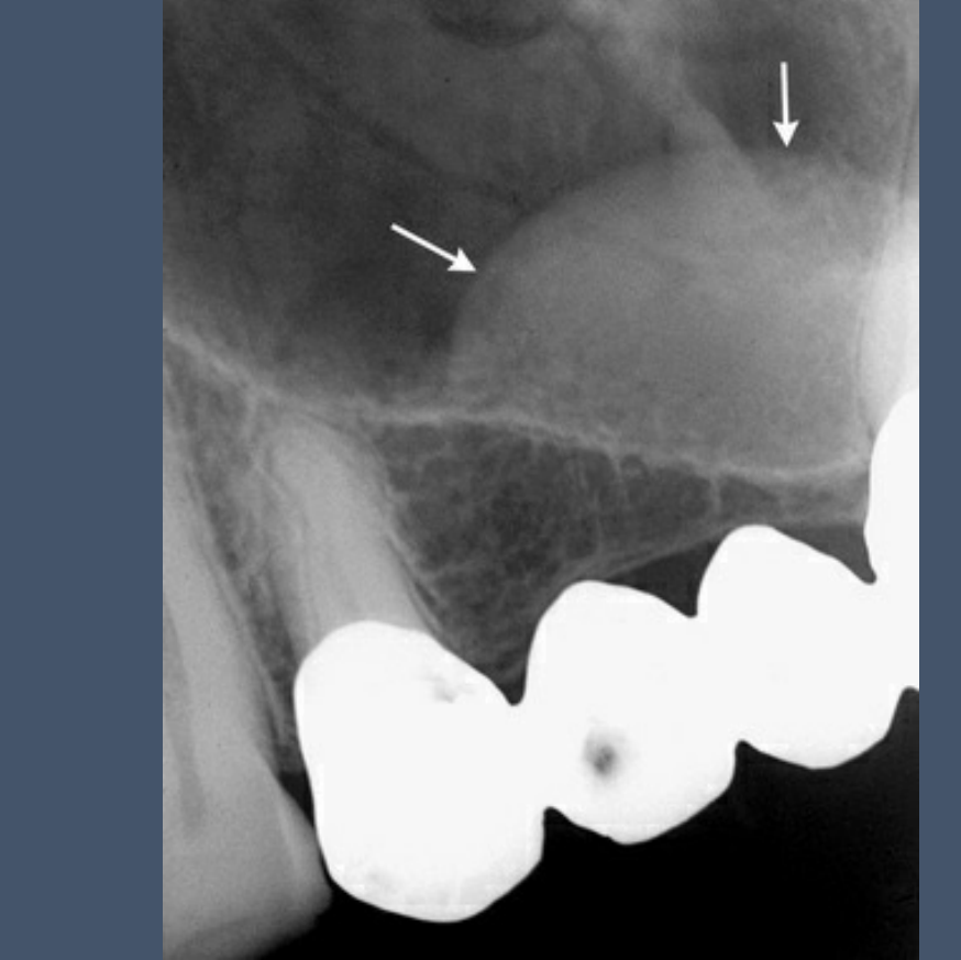
1. Lucent? Opaque? Or Mixed?
2. Localisation
- Unilateral or Bilateral?
- Single or Multiple?
- Epicentre
- Pericoronal / Periapical to Tooth?
- MD Body ➝ Superior / Inferior to Mandibular Canal or Within?
- MX ➝ Inferior or Superior to Maxillary Sinus Floor?
- Other: MD condyle, ramus, angle, etc.
 |  |  |
 |  |  |
 |  |  |
Borders and Morphology
Abnormal Findings (Lesions) in the Jaws6
 |  |
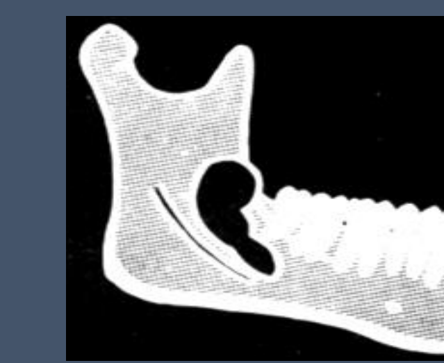 | 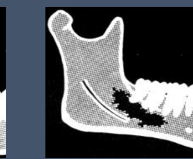 |
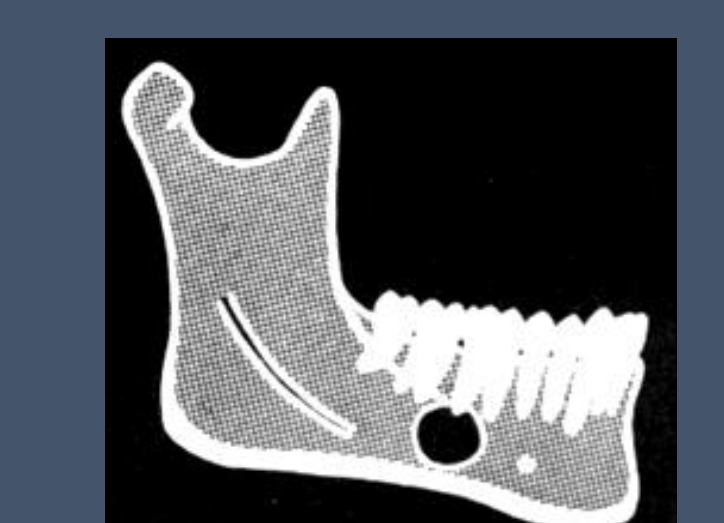
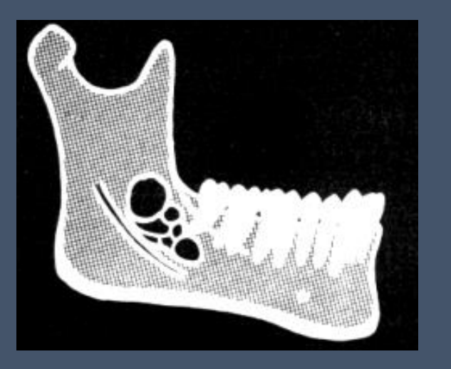
3. Borders
- Well defined / Ill (poorly) defined
- Well defined ⟶ Corticated, Sclerotic, etc.
- Circular, Scalloping, Irregular…
4. Morphology
- Unilocular (circular border), Multilocular, Pseudo-multilocular
- Other: circular, ovoid, triangular, …
Internal Architecture and Patterns
Abnormal Findings (Lesions) in the Jaws7
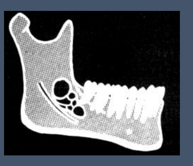 | 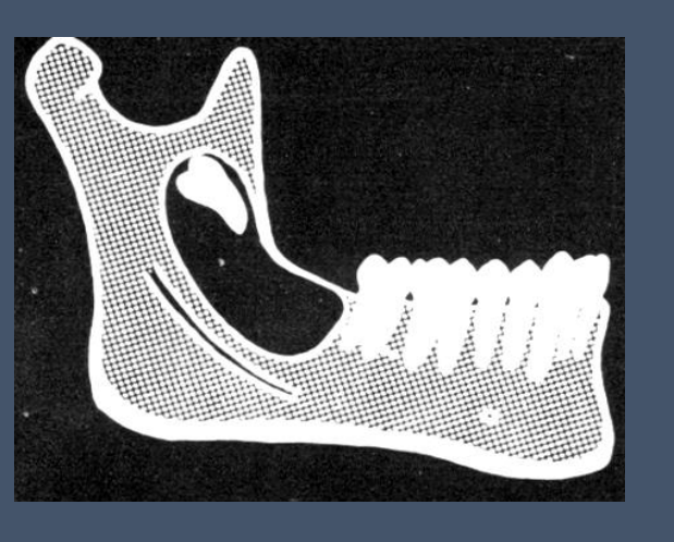 |
5. Internal Architecture / Structure (lucent lesion)
- Completely lucent?
- Septa, calcifications, tooth-like structure, etc.
6. Pattern (opaque lesion)
- Homogenous / Heterogenous
- Altered trabecular bone
- Ground glass, wheel-spoke, popcorn-like, etc.
Effects on Adjacent Structures
Abnormal Findings (Lesions) in the Jaws8
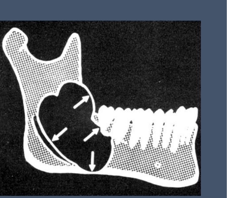
7. Effects on Adjacent Structures
- Teeth: lamina dura, PDL space, root, follicular space
- Mandibular Canal:
- Displacement, compression / flattening, widening, obliteration
- Surrounding Bone:
- Sclerosis
- Cortical plate:
- Expansion, thinning, effacement
- Periosteal reaction / new bone formation
- Lamellar / Onion-skin / Hair-on-end / Sunray / Codman’s triangle, etc.
- Maxillary sinus:
- Floor: lifting, effacement, discontinuity, remodelling, etc.
- Other walls: discontinuity, remodelling, inward bowing, etc.
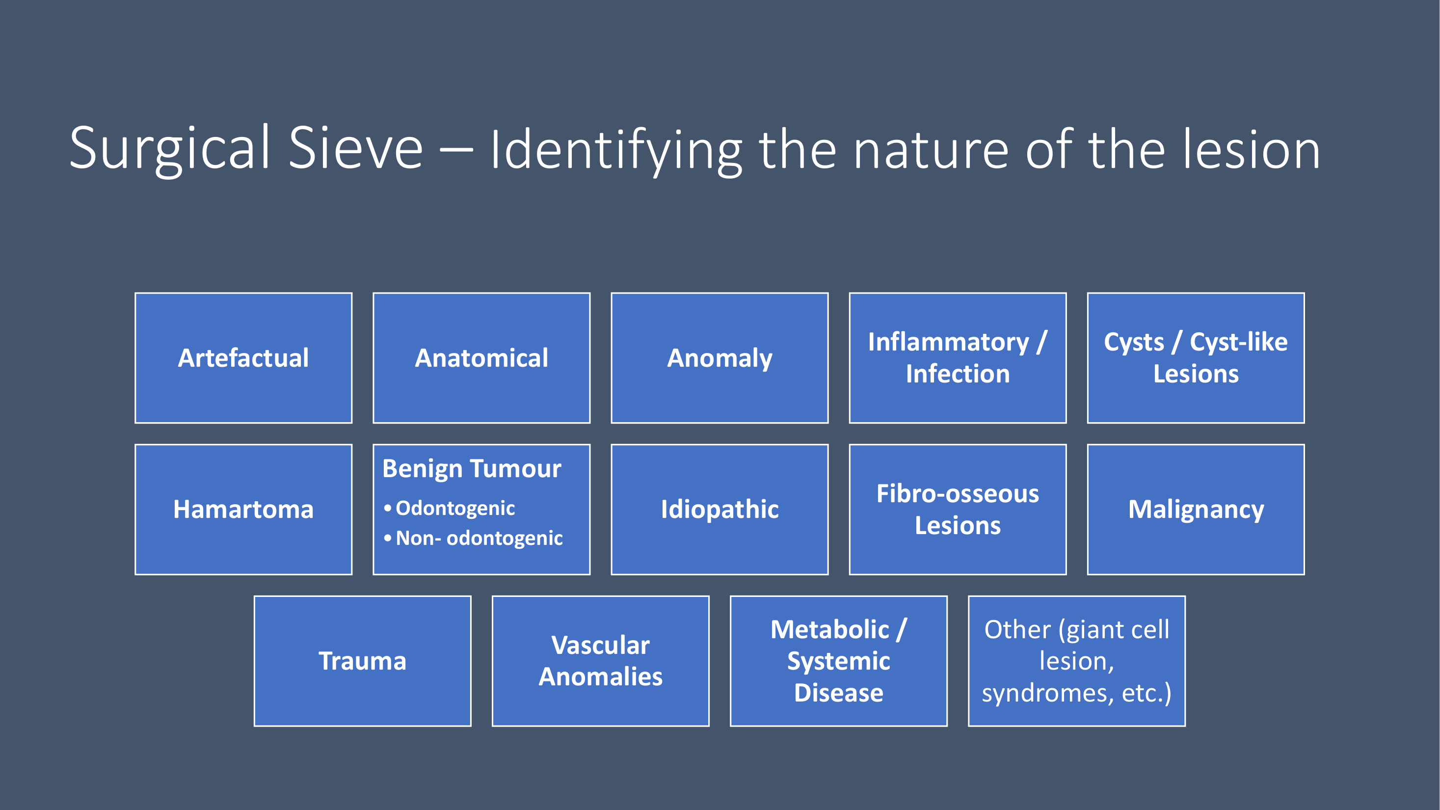
Pathological Categories and Case Examples910
| Artefactual | Anatomical | Anomaly | Inflammatory / Infection | Cysts / Cyst-like Lesions |
|---|---|---|---|---|
| Hamartoma | Benign Tumour • Odontogenic • Non-odontogenic | Idiopathic | Fibro-osseous Lesions | Malignancy |
| Trauma | Vascular Anomalies | Metabolic / Systemic Disease | Other (giant cell lesion, syndromes, etc.) |
 |  |
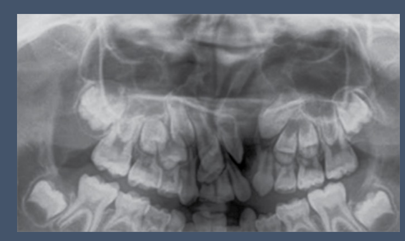
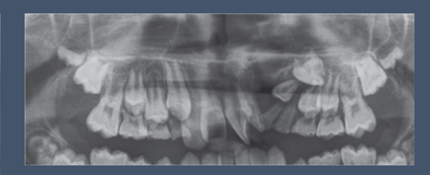
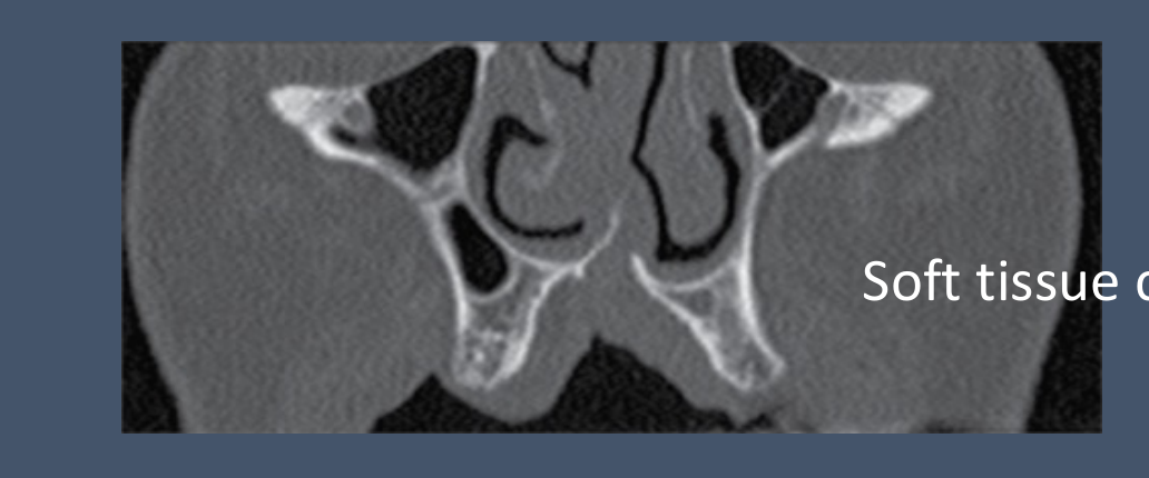
Developmental Anomalies11
- Cleft palate (congenital)
- Always look for other anomalies, especially dental anomalies (e.g., supernumerary teeth, malformed teeth, missing teeth, etc.)
 |  |
 |
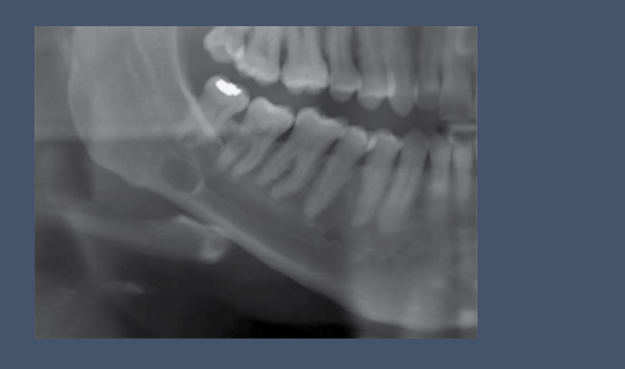
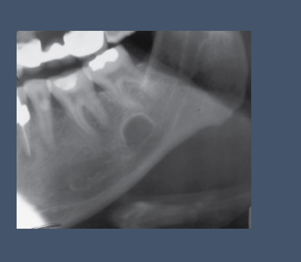
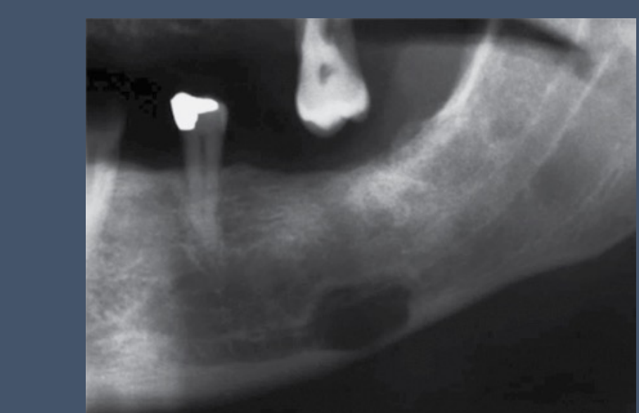
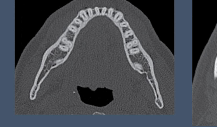
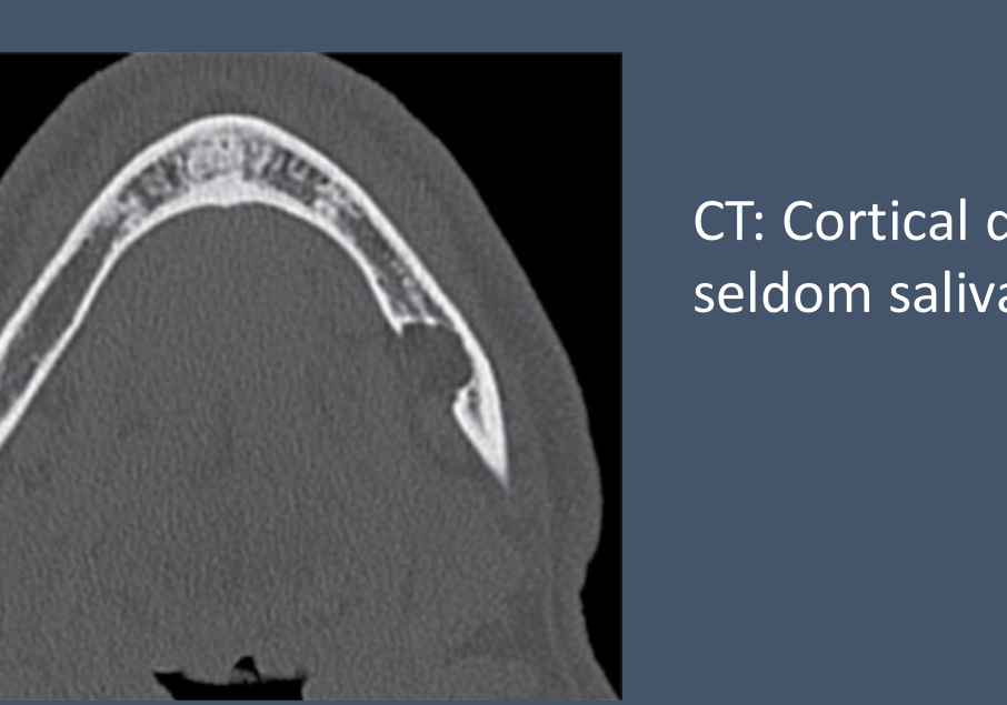
Stafne Defect
OPG12
- Well-defined lucency with heavily/thick corticated border
 |  |  |
 |  |
CT
- Cortical depression (often fat-filled, seldom salivary gland)
- CT review
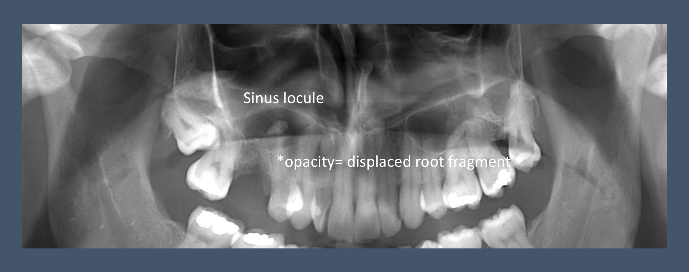
Inflammatory and Infectious Lesions1314
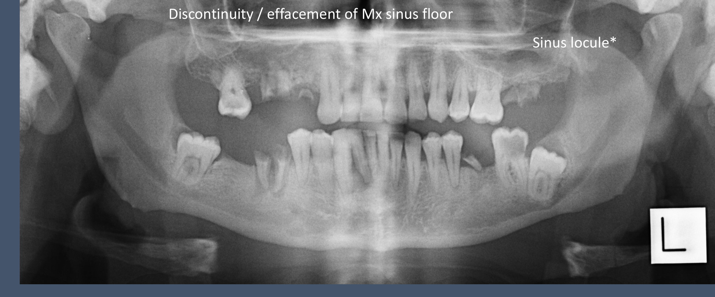
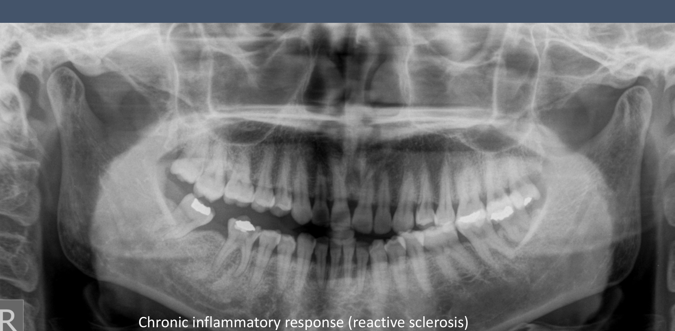
- Lucent → Mixed lucent-sclerotic → Sclerotic
- Discontinuity/effacement of maxillary sinus floor
- Sinus locule*
Inflammatory
 |  |
Cysts and Cyst-like Lesions
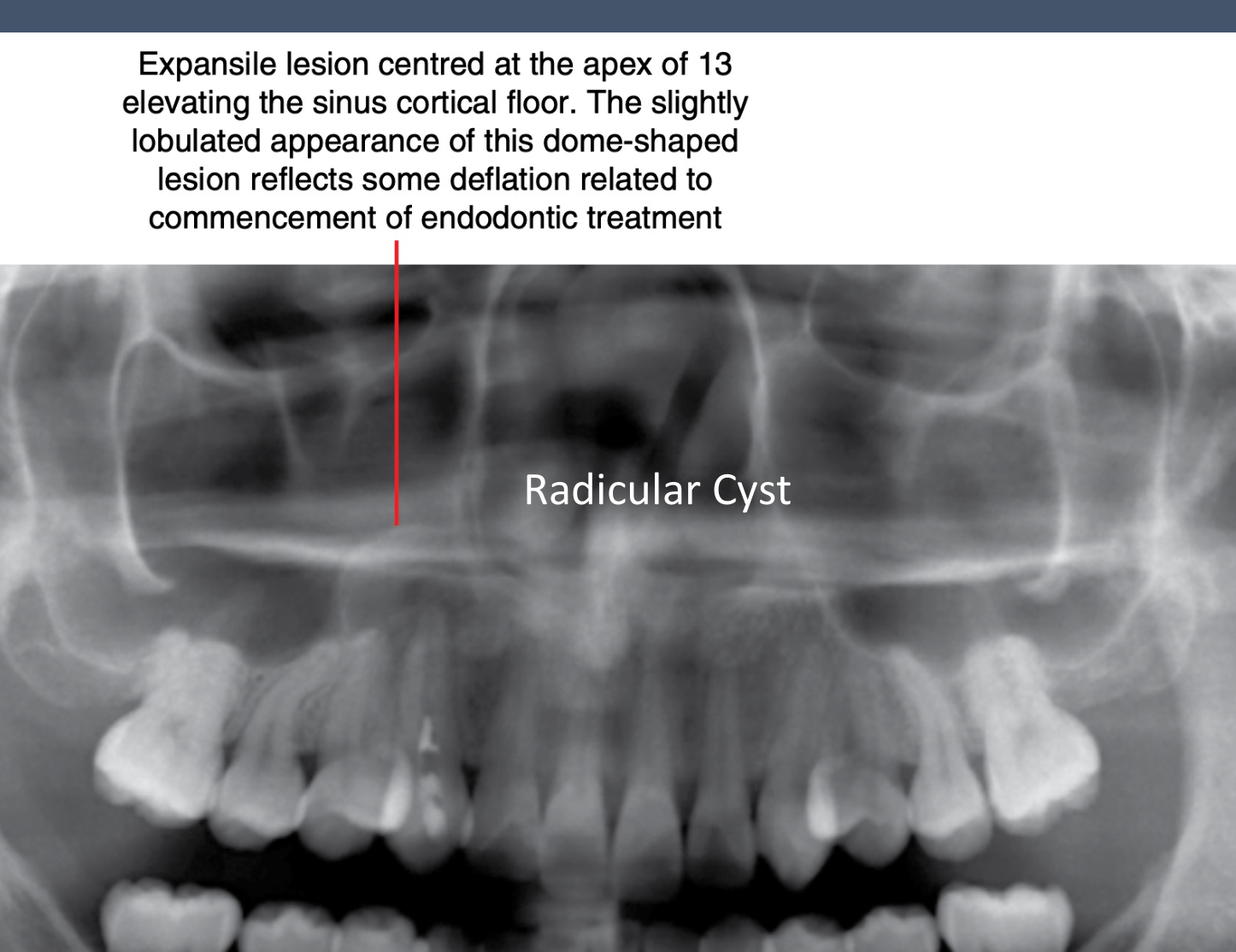
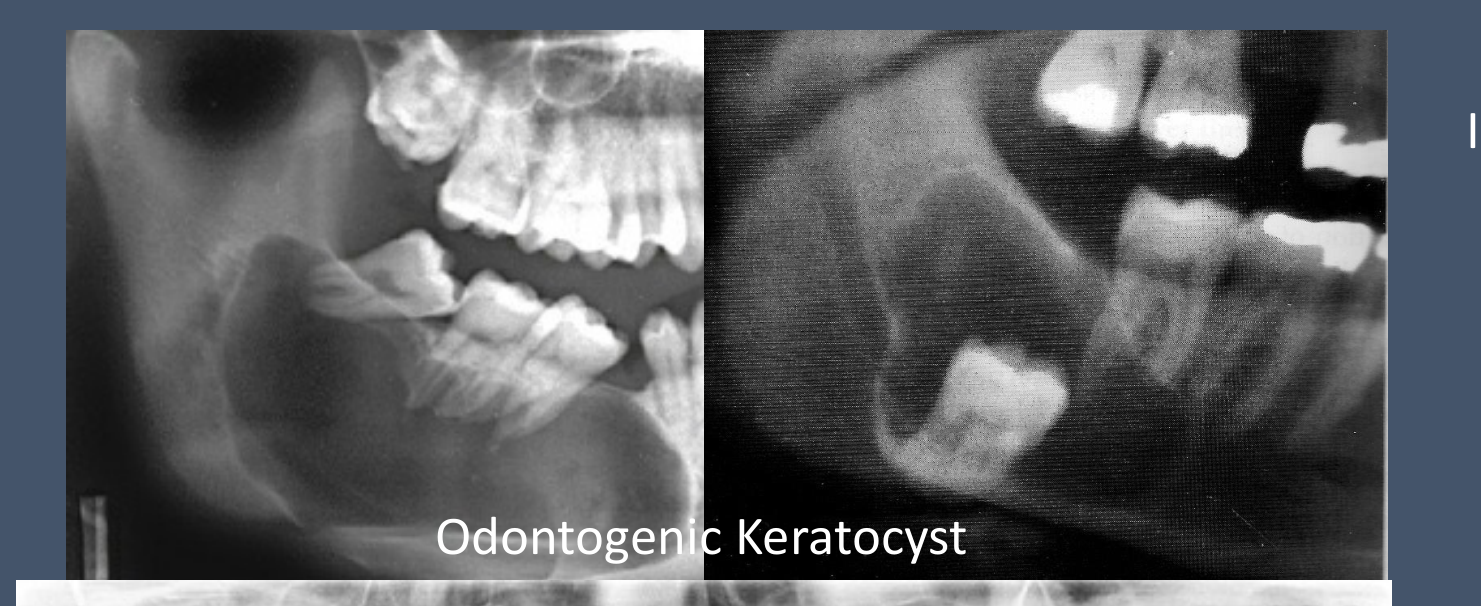
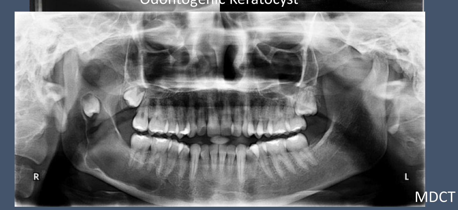
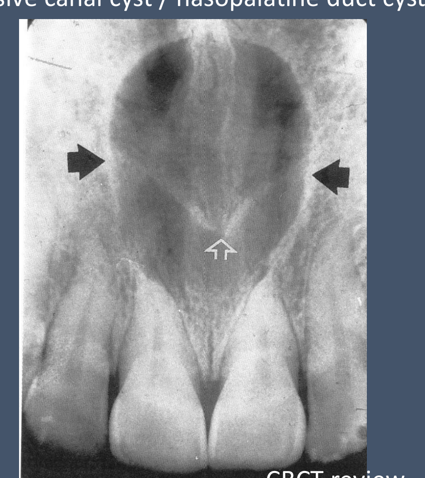
Radicular Cysts15161718
-
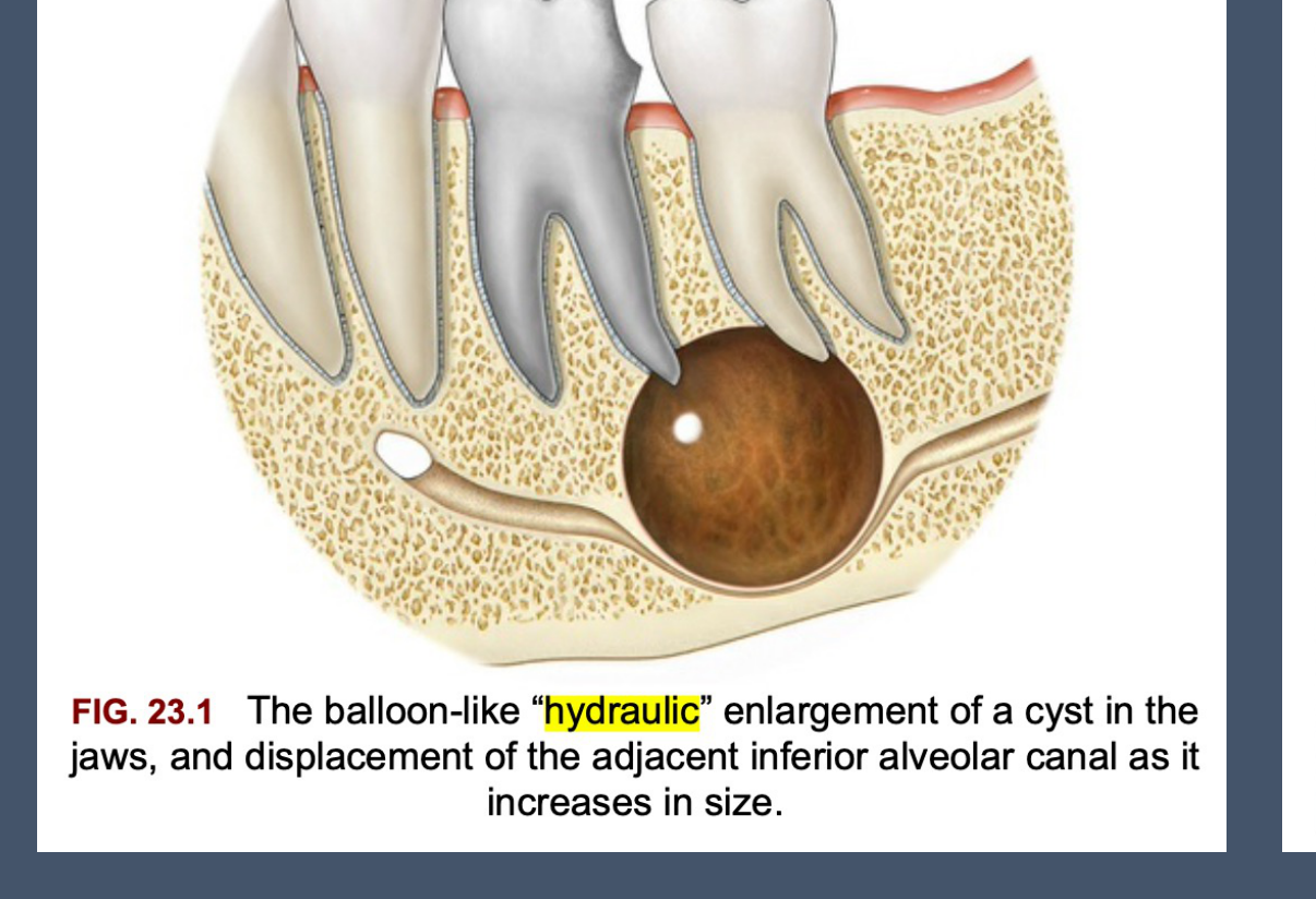
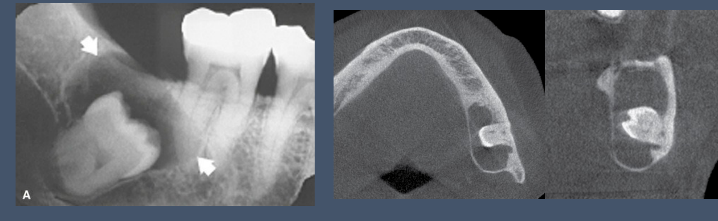
Cysts are expansile and have corticated borders.
-
Corticated borders = slow growing lesion.
-
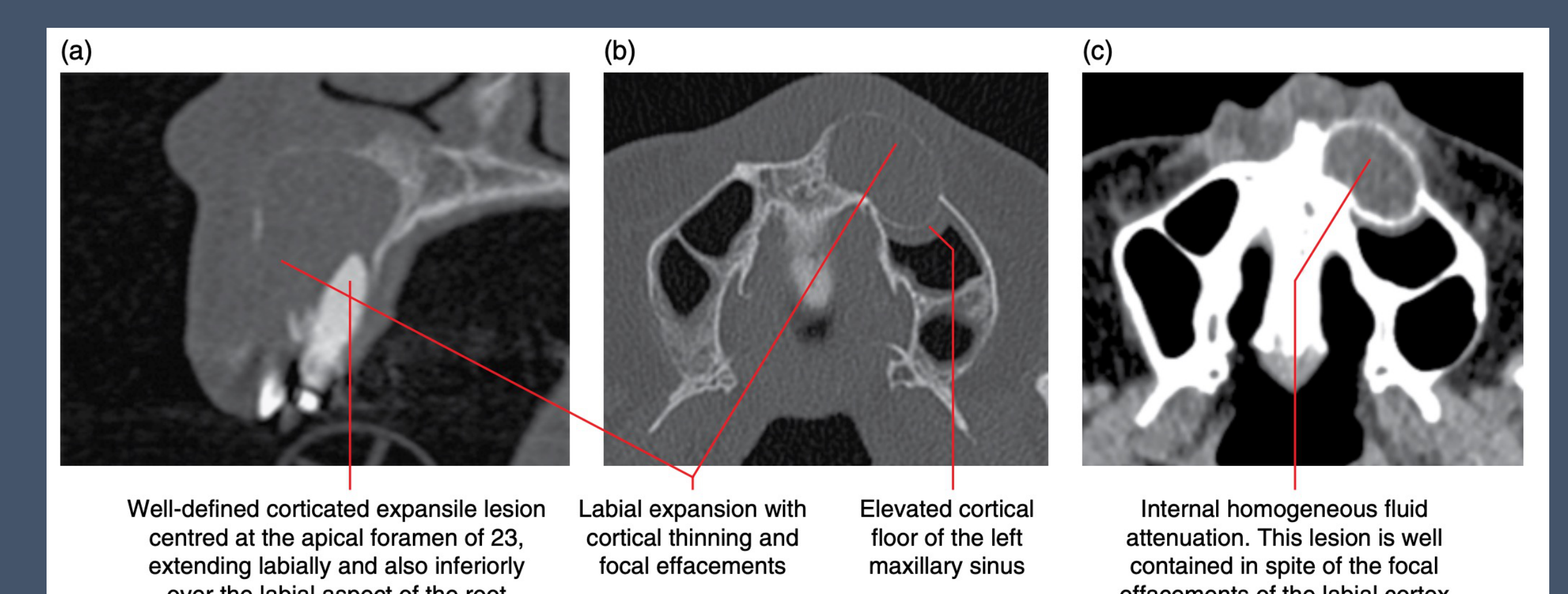
Further investigation with CBCT or MDCT.
-
The balloon-like “hydraulic” enlargement of a cyst in the jaws, and displacement of the adjacent inferior alveolar canal as it increases in size.
-
Well-defined corticated expansile lesion centred at the apical foramen of 23, extending labially and also inferiorly over the labial aspect of the root
-
Labial expansion with cortical thinning and focal effacements
-
Elevated cortical floor of the left maxillary sinus
-
Internal homogeneous fluid attenuation. This lesion is well contained in spite of the focal effacements of the labial cortex
CBCT / MDCT review
CBCT review
 |  |  |
 |  |  |
 |
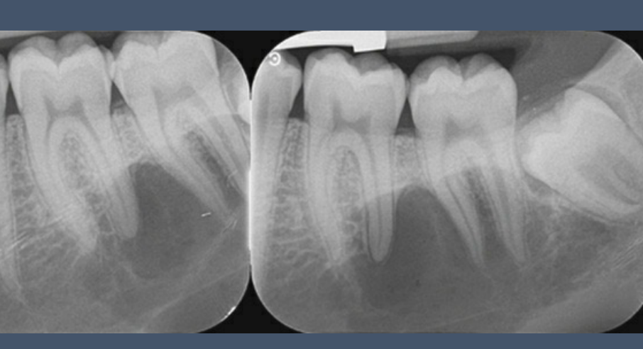
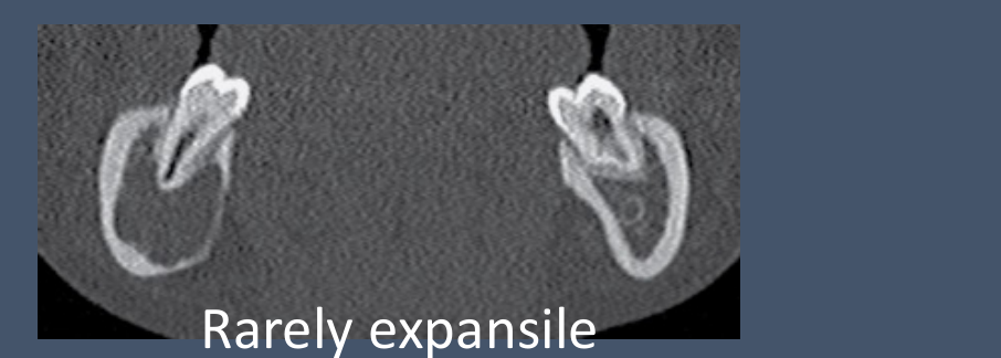
Simple Bone Cysts19
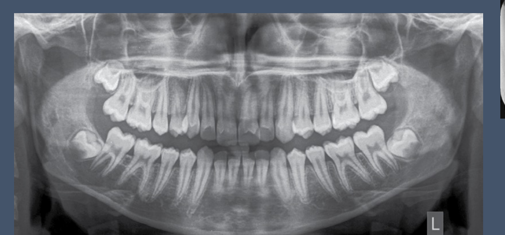
- Simple Bone Cyst: Corticated lucency. Can be mildly expansile.
- MDCT/MRI Review
 |  |
 |
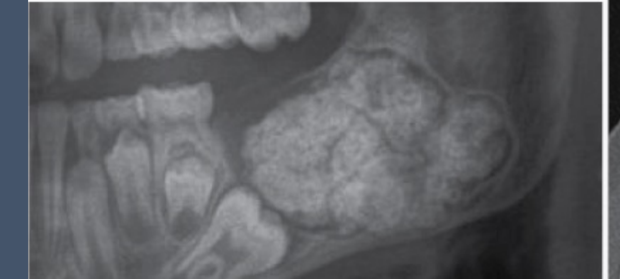
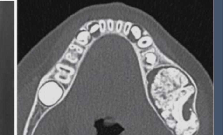
Hamartomas and Odontomas
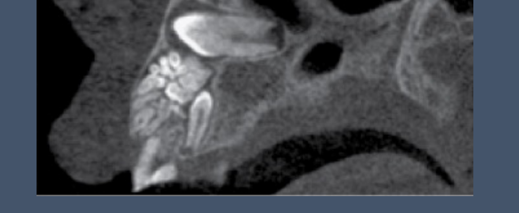
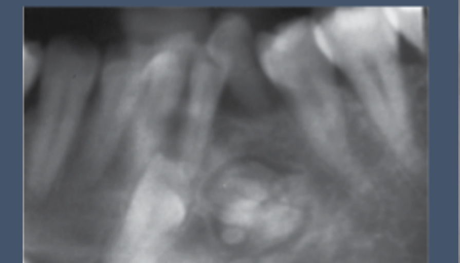
Hamartoma (Tumour-like Lesions)20
- Odontoma (Compound & Complex type): amorphous mix of calcified dental tissues
- CT (CBCT or MSCT/MDCT) Review
- Refer to Surgeon
 |  |
 |  |
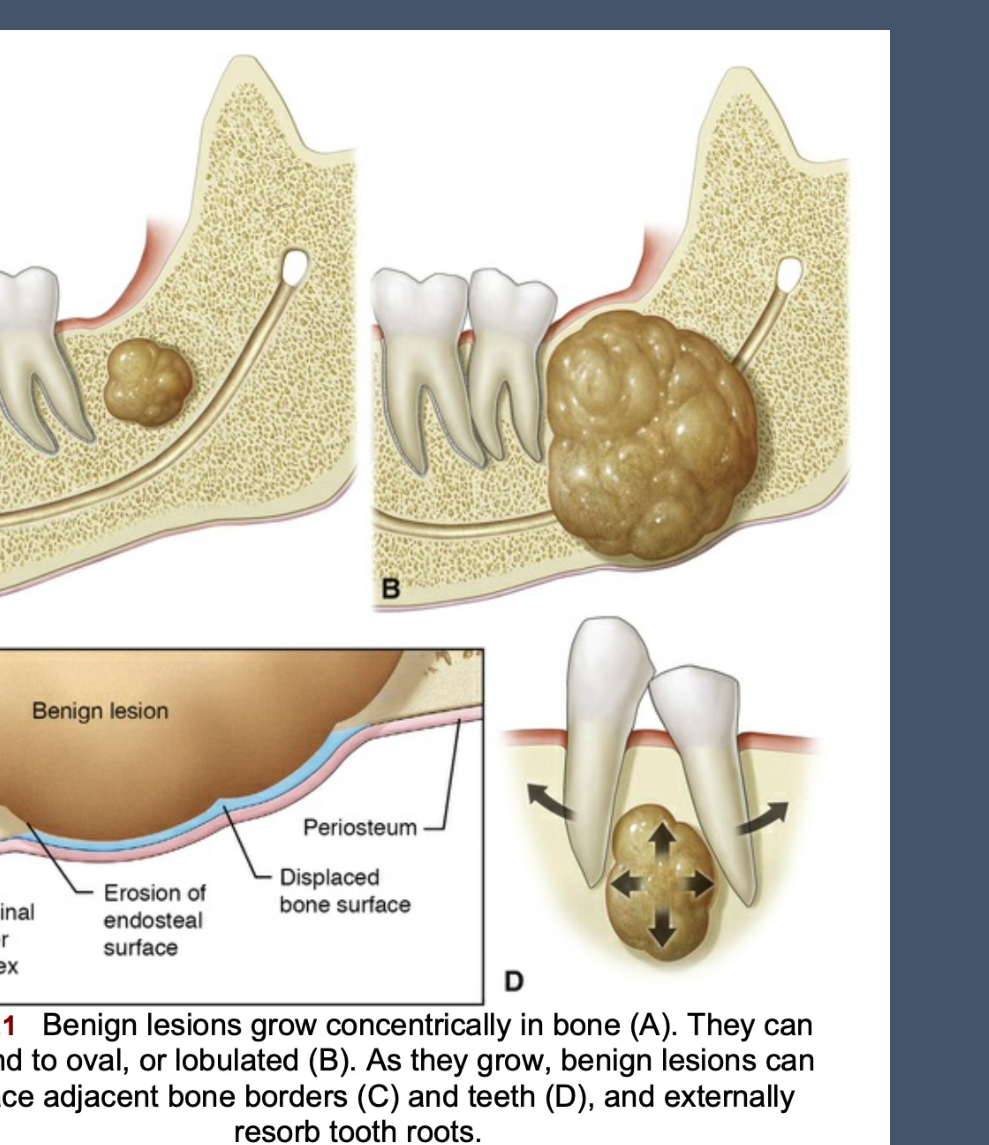
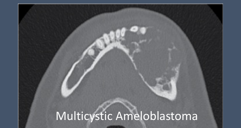
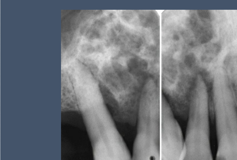
Benign Odontogenic Tumours2122
-
Mass effect
-
Expansile features + resorption
-
Original outer cortex
-
Benign lesion
-
Erosion of endosteal surface
-
Periosteum displaced bone surface
-
MDCT/MRI Review
-
Refer to Surgeon
-
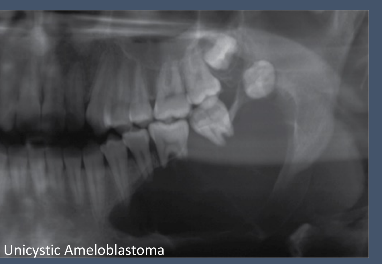
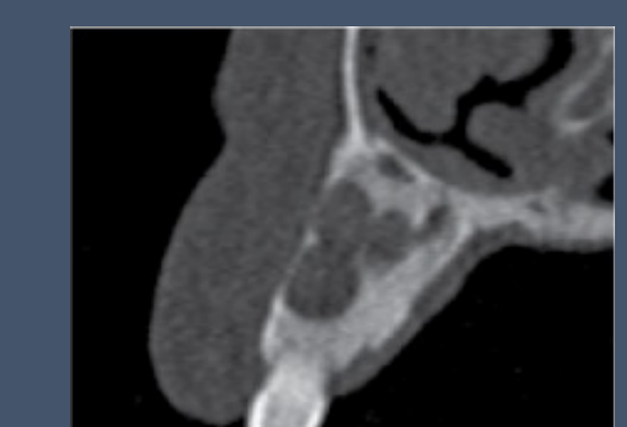
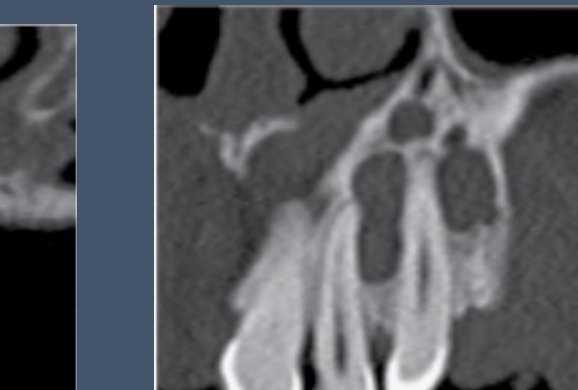
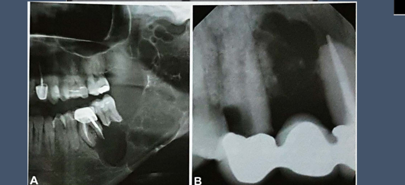
Well-defined, expansile, multilocular lucency with thick, curved internal septa resulting in “soap bubble” appearance
-
MDCT/MRI Review
-
Refer to Surgeon
 |  |  |
 |  |  |
 |
Benign Non-odontogenic Tumours2324
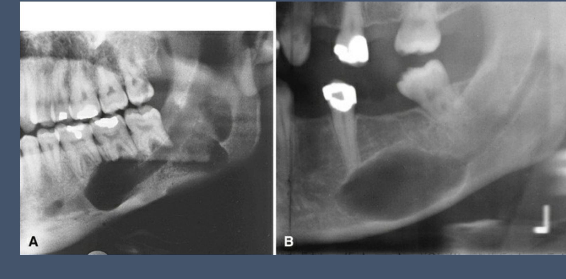
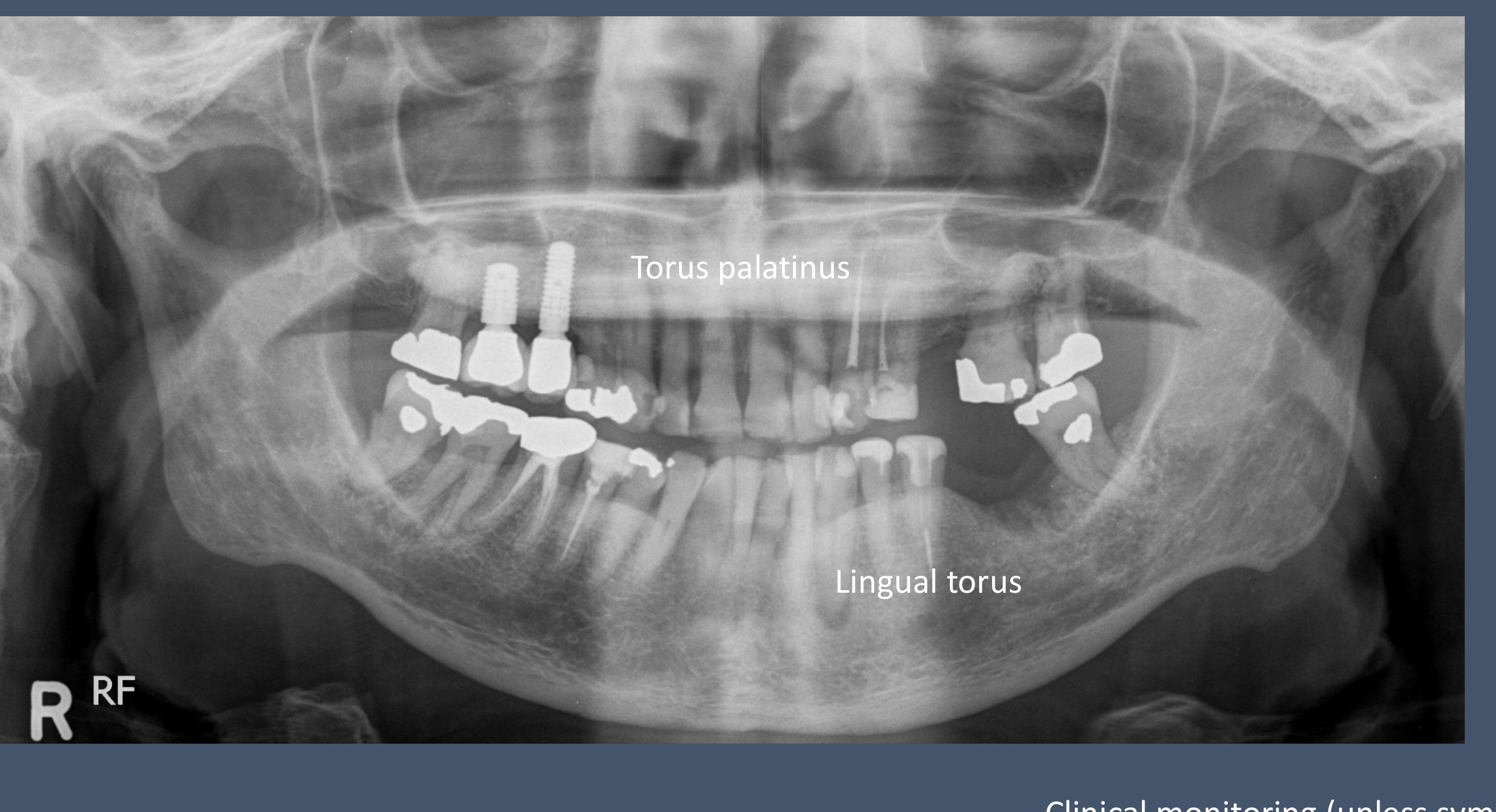
- Fusiform, expansile lucency associated with mandibular canal
- Neurofibroma (strong association with Neurofibromatosis type 1 (NF1))
- MDCT/MRI Review
- Refer to Surgeon
 |  |
Idiopathic Lesions and Bone Islands25
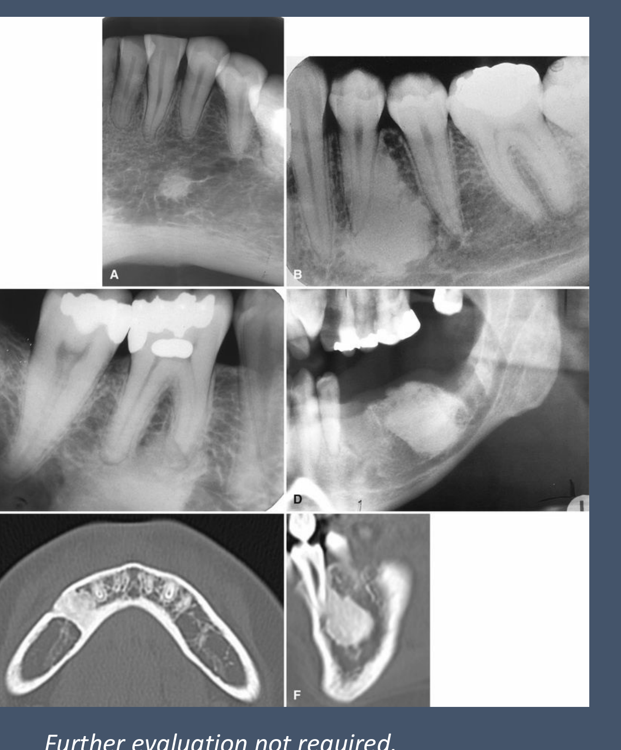
- Bone islands are also considered as hamartomas.
- Localised dense bone (often homogeneous cortical density).
- Can occur anywhere.
- Most common site: mandibular premolar-molar region.
- Various morphologies.
- Seldom resorb teeth.
- Bone islands continuous with cortical plates are termed enostosis.
- Further evaluation not required.
- Monitor with I/O or OPG (unless symptomatic).
Fibro-osseous Lesions
Cemento-osseous Dysplasia26
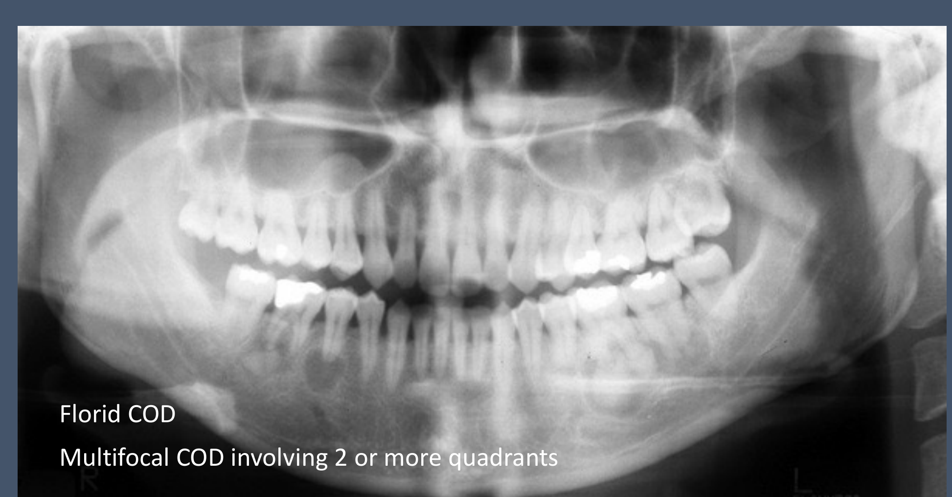
- Florid COD: Multifocal COD involving 2 or more quadrants
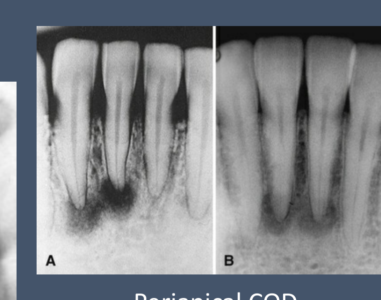
- Periapical COD
- Can consider CBCT review
- Cemento-osseous Dysplasia (unifocal or multifocal)
- Early, immature lesions are lucent
- Late, mature lesions are opaque with a lucent rim and sclerotic borders
- Fibro-osseous
 |  |
Fibrous Dysplasia27
- Fibro-osseous
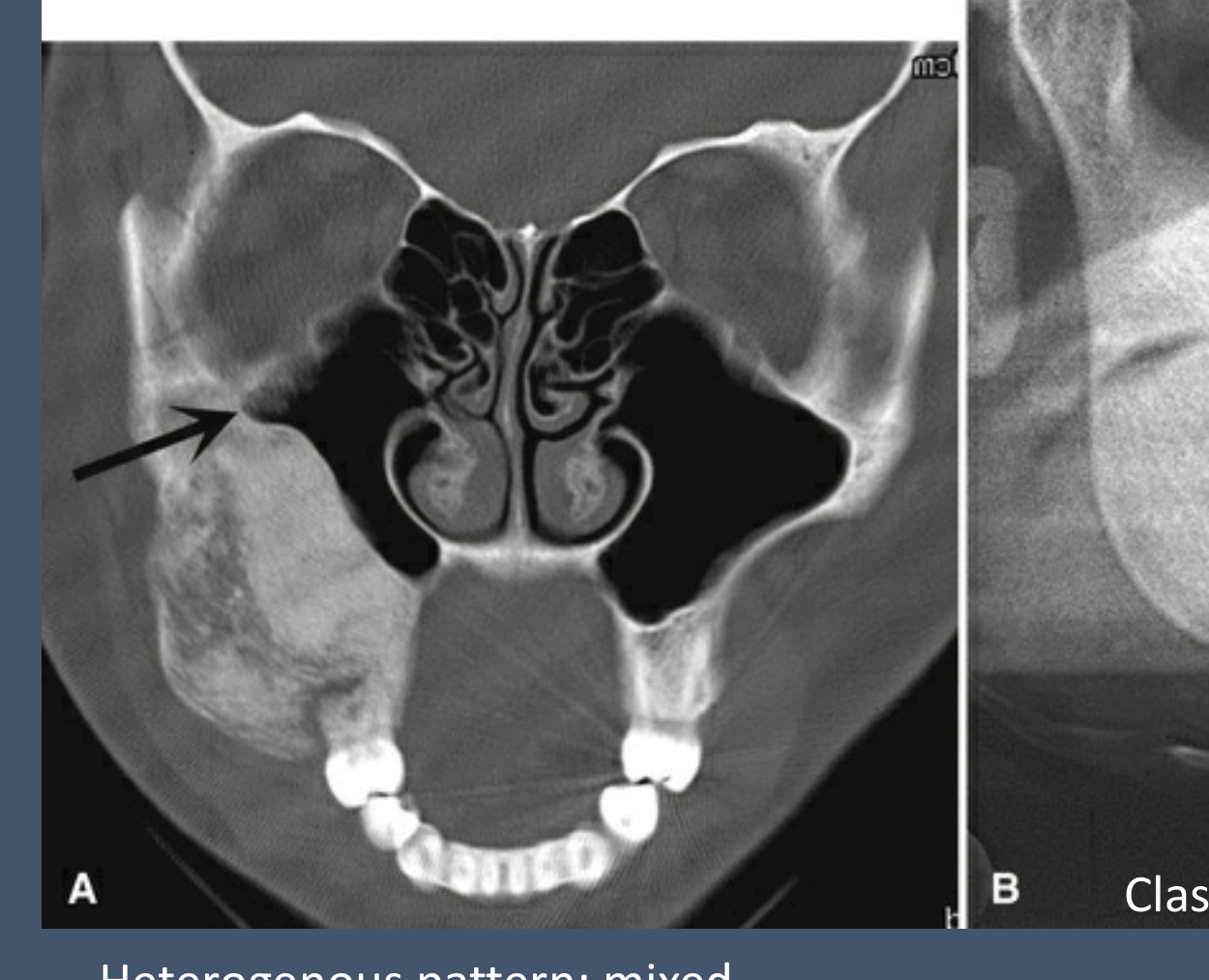
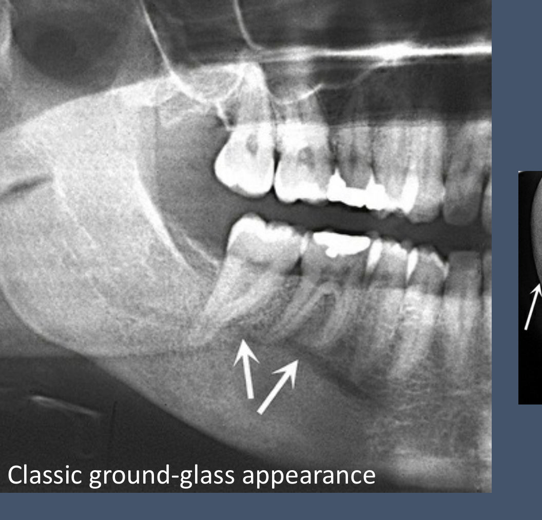
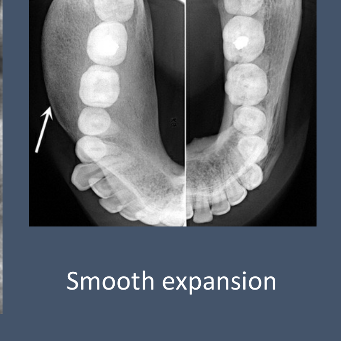
- Fibrous Dysplasia
- Classic ground-glass appearance
- Smooth expansion
- Heterogenous pattern; mixed hypodensity & ground-glass bone
- CBCT vs MDCT
 |  |
 |
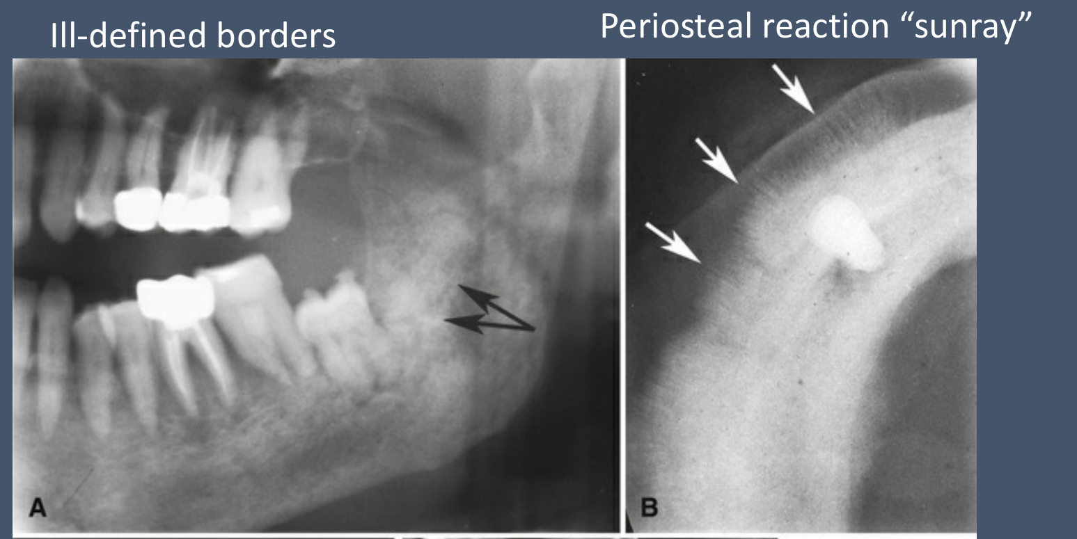
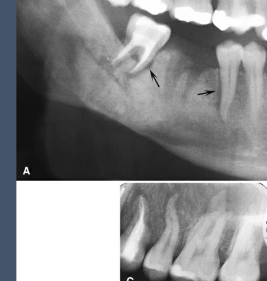
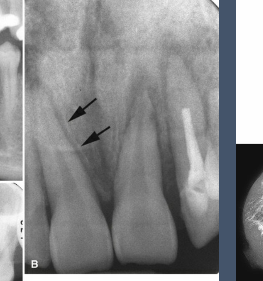
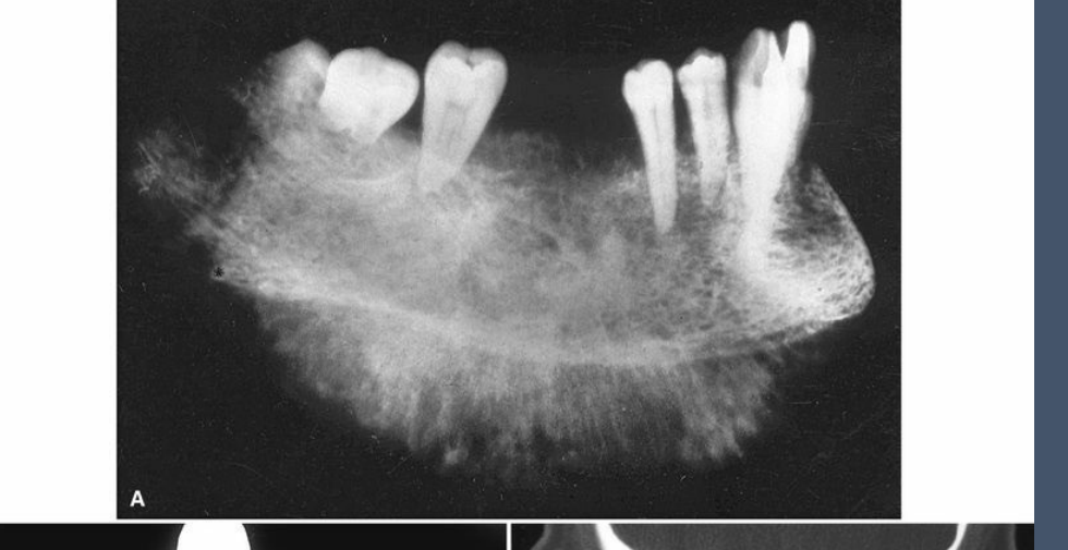
Malignant Neoplasms28
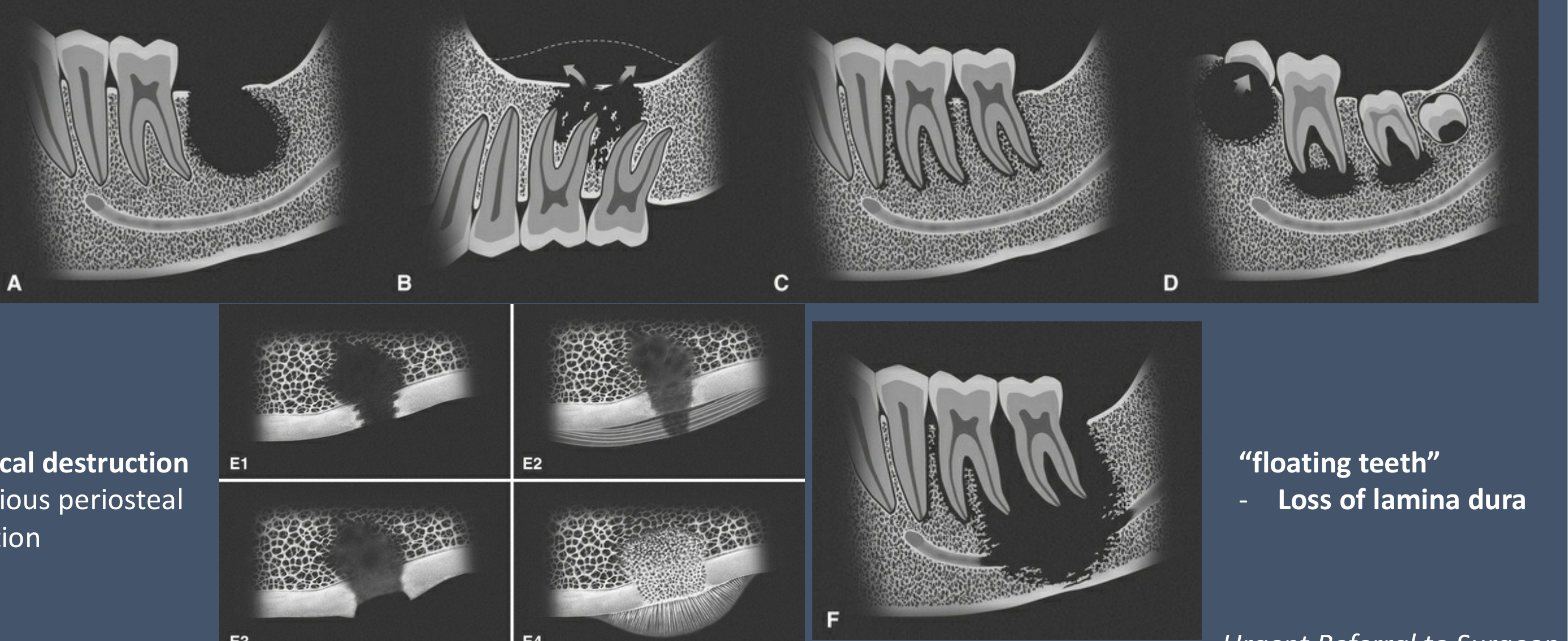
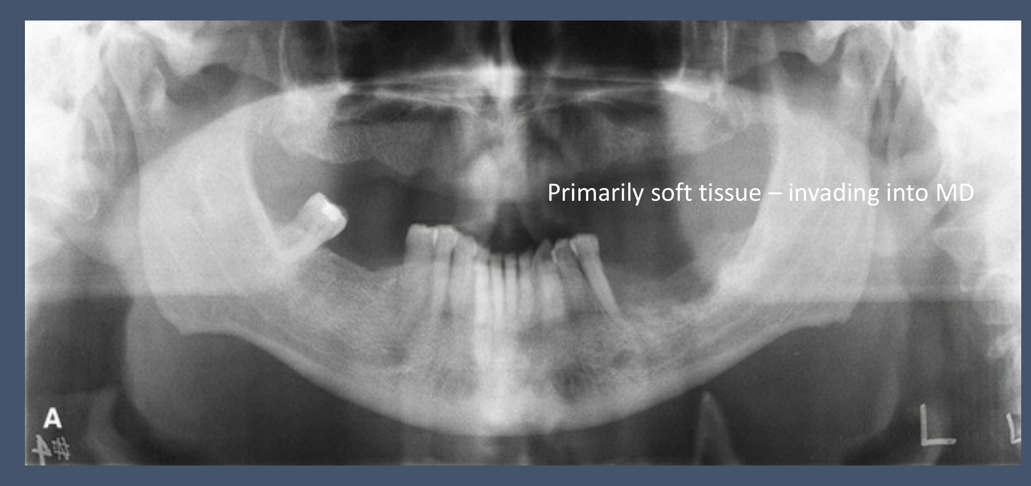
- Ill-defined borders, fast-growing, destructive, invasive (invade through path of least resistance)
- Cortical destruction + various periosteal reaction
- “Floating teeth” - loss of lamina dura
- Urgent Referral to Surgeon
Squamous Cell Carcinoma2930
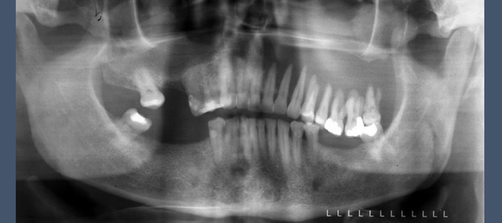
- Squamous cell carcinoma (left maxillary sinus)
- Refer to OMF Surgeons straight away
- Urgent Referral
 |  |
Metastatic and Primary Bone Cancer3132
-
Osteoblastic Cancer
-
Metastatic cancer – primary prostate cancer (some breast cancer also)
-
Urgent Referral
-
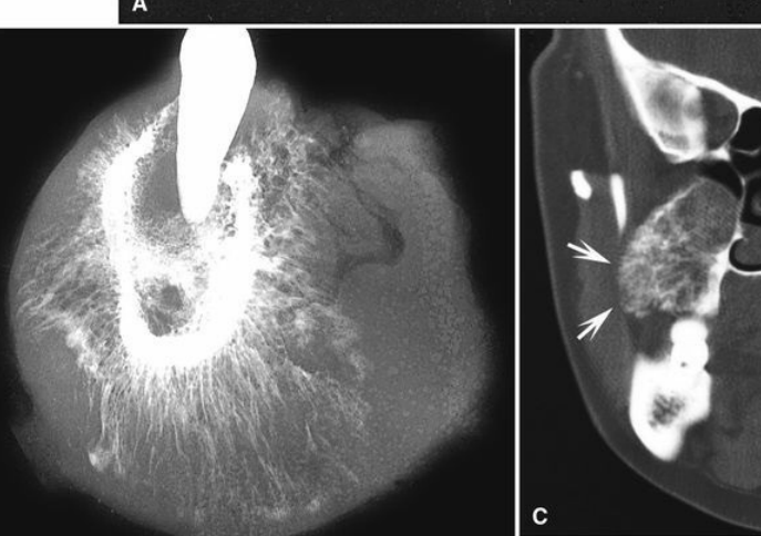
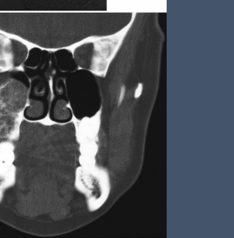
Primary Bone Cancer: Osteogenic sarcoma
-
Urgent Referral
 |  |  |
 |  |  |
Orthopantomogram Report Checklist33
- Identify any significant OPG error
- Check stage of dentition (deciduous / mixed / permanent)
- Identify any missing teeth (& supernumerary teeth, if present)
- Are there implants in edentulous sites?
- Comment on peri-implant crestal bone level.
- Are there root remnants?
- Are there implants in edentulous sites?
- Identify any impacted or ectopic teeth
- Location
- Angulation
- Morphology (both crown & root system)
- Relationship with adjacent structures eg. Adjacent molars, mx sinus, IDC, etc.
- Any dental anomalies? (eg. Dens invaginatus, supernumeraries, etc)
- Identify periodontal bone loss
- Perio-endo bony defects
- Horizontal bone loss
- Angular bony defects / vertical bone loss
- Furcation involvement
- Are there calculus deposits? Where?
- Is there existing restorative therapy?
- Identify any faulty restorations eg. overhang, insufficient margin
- Identify caries including recurrent caries
- Identify non-carious tooth loss (NCTL) eg. attrition
- Is there existing endodontic therapy?
- Identify periapical lucencies ie. periapical inflammatory lesions
- Any associated root resorption, antral floor remodelling, reactive sclerosis, reactive mucosal thickening in antrum?
- Identify any abnormal lucencies or opacities in the jaws
- Location
- Border and Morphology
- Well-defined? Ill-defined?
- Overall shape – oval, round, elongated, etc.
- Internal architecture
- Effects on adjacent structures
- Surgical Sieve - Differential diagnoses if can.
- Extragnathic structures
- Maxillary sinuses – Symmetrical? Size? Opacity? Floor and walls intact?
- Condyles – Symmetrical? Deformity? Remodelling? Sclerosis? Pitting? Beaking / lipping? Loose bodies?
- Airspace
- Cervical spine
- Any presence of soft tissue calcifications?
- Check submandibular region, tonsillar regions, carotid bulb region, etc.
- Pterygomaxillary fissure
- Orbits
- Summary (for long reports or reports with significant pathology)
- Further imaging required? If yes, which modality?
Further Reading and References34
- Essentials of dental radiography and radiology 5th edition by Whaites, Eric; Drage, Nicholas
- White and Pharoah’s Oral radiology: principles and interpretation 8th edition By Ernest Lam
- Atlas of Oral and Maxillofacial Radiology By Bernard Koong
Footnotes
-
Original PDF page 1: L13PR2, p.1 ↩
-
Original PDF page 2: L13PR2, p.2 ↩
-
Original PDF page 3: L13PR2, p.3 ↩
-
Original PDF page 4: L13PR2, p.4 ↩
-
Original PDF page 5: L13PR2, p.5 ↩
-
Original PDF page 6: L13PR2, p.6 ↩
-
Original PDF page 7: L13PR2, p.7 ↩
-
Original PDF page 8: L13PR2, p.8 ↩
-
Original PDF page 9: L13PR2, p.9 ↩
-
Original PDF page 10: L13PR2, p.10 ↩
-
Original PDF page 11: L13PR2, p.11 ↩
-
Original PDF page 12: L13PR2, p.12 ↩
-
Original PDF page 13: L13PR2, p.13 ↩
-
Original PDF page 14: L13PR2, p.14 ↩
-
Original PDF page 15: L13PR2, p.15 ↩
-
Original PDF page 16: L13PR2, p.16 ↩
-
Original PDF page 17: L13PR2, p.17 ↩
-
Original PDF page 18: L13PR2, p.18 ↩
-
Original PDF page 19: L13PR2, p.19 ↩
-
Original PDF page 20: L13PR2, p.20 ↩
-
Original PDF page 21: L13PR2, p.21 ↩
-
Original PDF page 22: L13PR2, p.22 ↩
-
Original PDF page 23: L13PR2, p.23 ↩
-
Original PDF page 24: L13PR2, p.24 ↩
-
Original PDF page 25: L13PR2, p.25 ↩
-
Original PDF page 26: L13PR2, p.26 ↩
-
Original PDF page 27: L13PR2, p.27 ↩
-
Original PDF page 28: L13PR2, p.28 ↩
-
Original PDF page 29: L13PR2, p.29 ↩
-
Original PDF page 30: L13PR2, p.30 ↩
-
Original PDF page 31: L13PR2, p.31 ↩
-
Original PDF page 32: L13PR2, p.32 ↩
-
Original PDF page 33: L13PR2, p.33 ↩
-
Original PDF page 34: L13PR2, p.34 ↩