Interpretation of OPGs Workshop1
DR MAY LAM OMF RADIOLOGIST
Instructional Guidelines
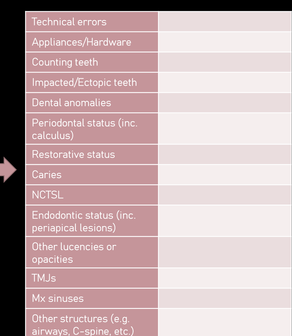
Technical errors2
- Appliances/Hardware
- Counting teeth
- Impacted/Ectopic teeth
- Dental anomalies
- Periodontal status (inc. calculus)
- Restorative status
- Caries
- NCTSL
- Endodontic status (inc. periapical lesions)
- Other lucencies or opacities
- TMJs
- Mx sinuses
- Other structures (e.g. airways, C-spine, etc.)
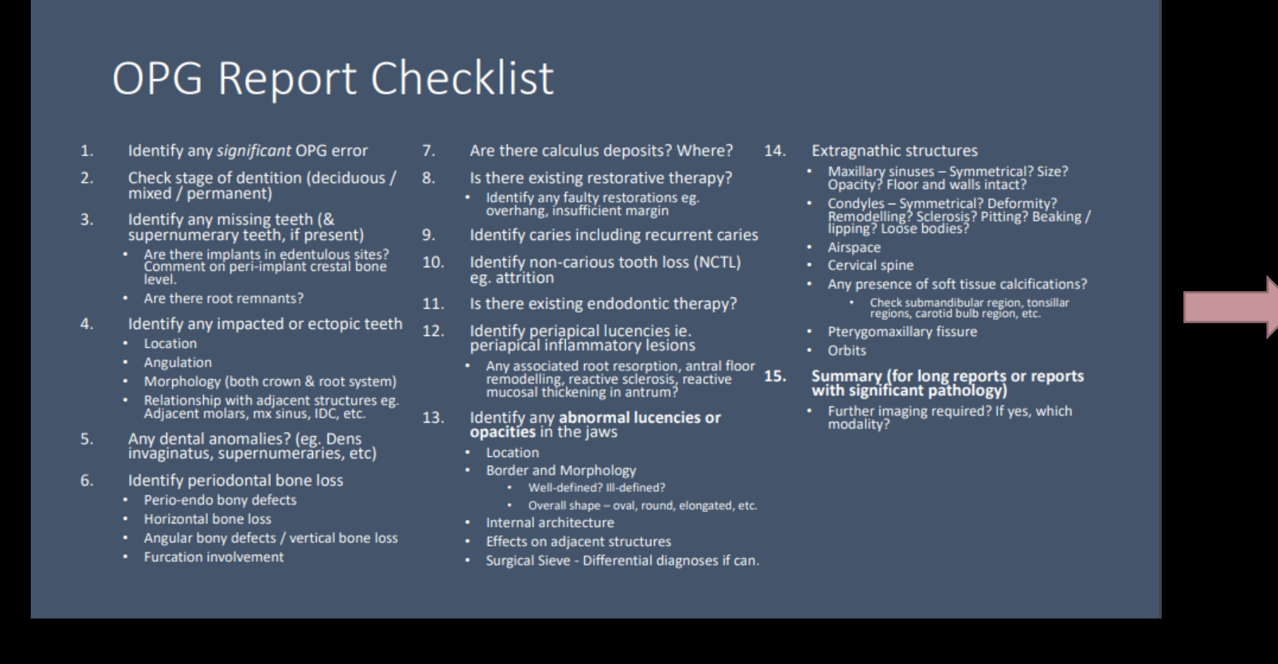 |  |
OPG Report Checklist
- Identify any significant OPG error
- Check stage of dentition (deciduous / mixed / permanent)
- Identify any missing teeth (& supernumerary teeth, if present)
- Are there implants in edentulous sites?
- Comment or peri-implant crestal bone level.
- Are there root remnants?
- Identify any impacted or ectopic teeth
- Location
- Angulation
- Morphology (both crown & root system)
- Relationship with adjacent structures e.g., adjacent molars, mx sinus, IDC, etc.
- Any dental anomalies? (e.g., Dens invaginatus, supernumeraries, etc.)
- Identify periodontal bone loss
- Perio-endo bony defects
- Horizontal bone loss
- Angular bony defects / vertical bone loss
- Furcation involvement
- Are there calculus deposits? Where?
- Is there existing restorative therapy?
- Identify any faulty restorations e.g., overhang, insufficient margin
- Identify caries including recurrent caries
- Identify non-carious tooth loss (NCTL)
- e.g., attrition
- Is there existing endodontic therapy?
- Identify periapical lucencies i.e., periapical inflammatory lesions
- Any associated root resorption, antral floor remodelling, reactive sclerosis, reactive mucosal thickening in antrum?
- Identify any abnormal lucencies or opacities in the jaws
- Location
- Border and Morphology
- Well-defined? Ill-defined?
- Overall shape - oval, round, elongated, etc.
- Internal architecture
- Effects on adjacent structures
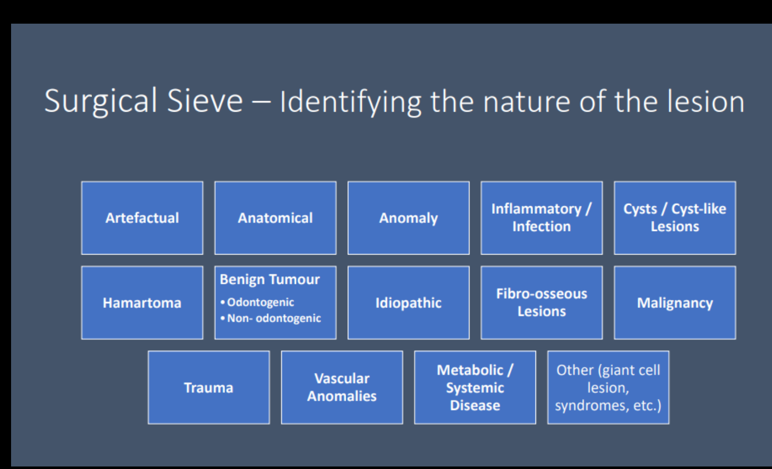
- Surgical Sieve - Differential diagnoses if can.
- Extragnathic structures
- Maxillary sinuses – Symmetrical? Size? Opacity? Floor and walls intact?
- Condyles – Symmetrical? Deformity? Remodelling? Sclerosis? Pitting? Beaking / lipping? Loose bodies?
- Airspace
- Cervical spine
- Any presence of soft tissue calcifications?
- Check submandibular region, tonsillar regions, carotid bulb region, etc.
- Pterygomaxillary fissure
- Orbits
- Summary (for long reports or reports with significant pathology)
- Further imaging required? If yes, which modality?
Analysis of Intraosseous Lesions
Step 1: Localize Abnormality3
- Anatomic position (epicenter)
- Localized or generalized
- Unilateral or bilateral
- Single or multifocal
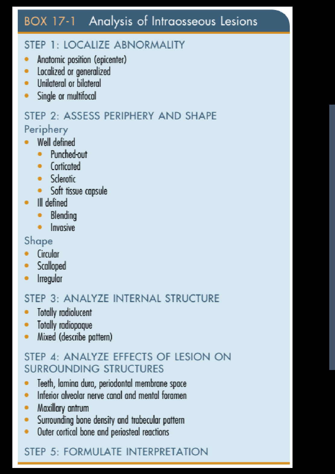 |  |
Step 2: Assess Periphery and Shape
Periphery
- Well defined
- Punched-out
- Corticated
- Sclerotic
- Soft tissue capsule
- Ill defined
- Blending
- Invasive
Shape
- Circular
- Scalloped
- Irregular
Step 3: Analyze Internal Structure
- Totally radiolucent
- Totally radiopaque
- Mixed (describe pattern)
Step 4: Analyze Effects of Lesion on Surrounding Structures
- Teeth, lamina dura, periodontal membrane space
- Inferior alveolar nerve canal and mental foramen
- Maxillary antrum
- Surrounding bone density and trabecular pattern
- Outer cortical bone and periosteal reactions
Step 5: Formulate Interpretation
Clinical Case Studies4
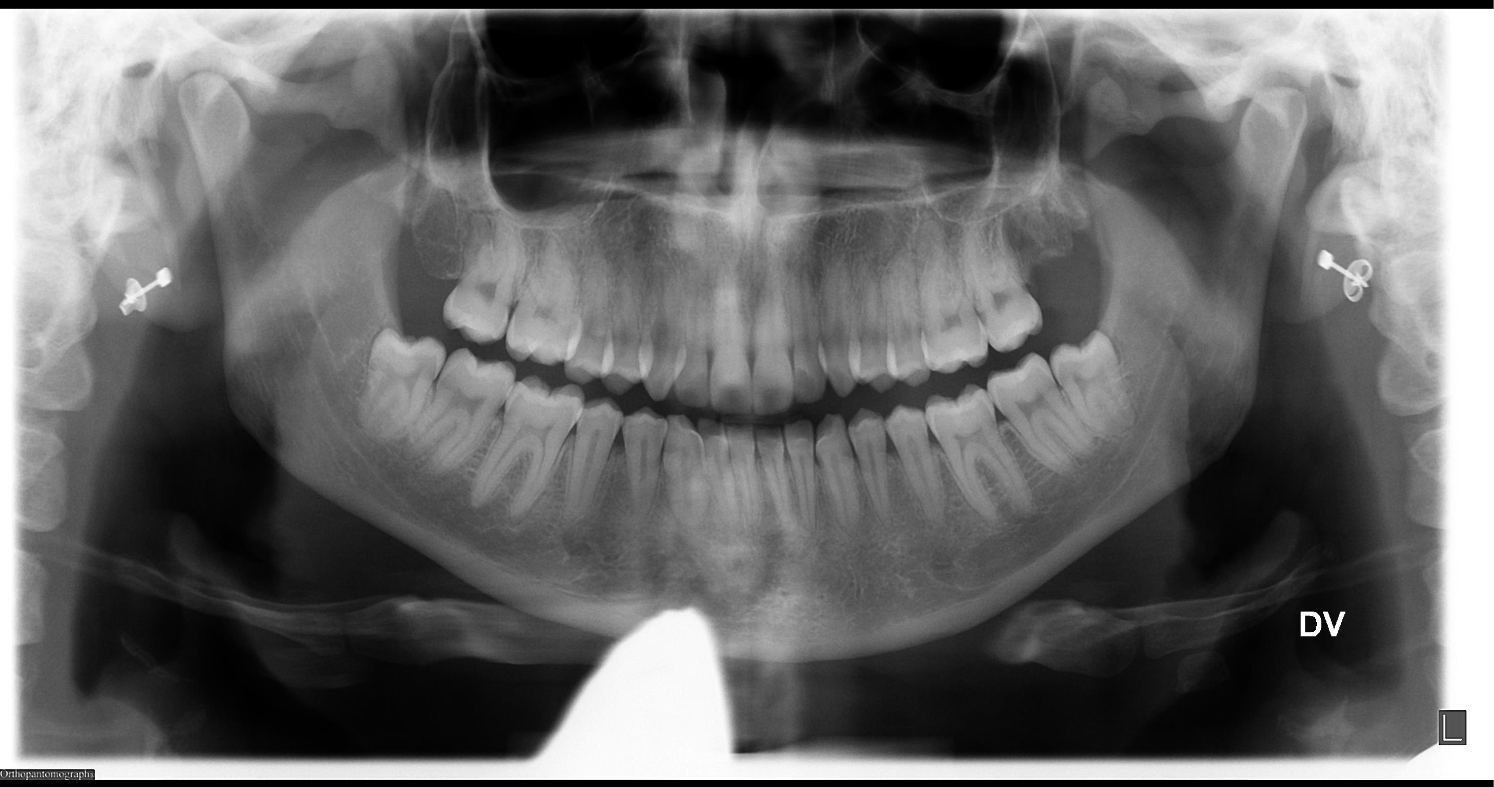
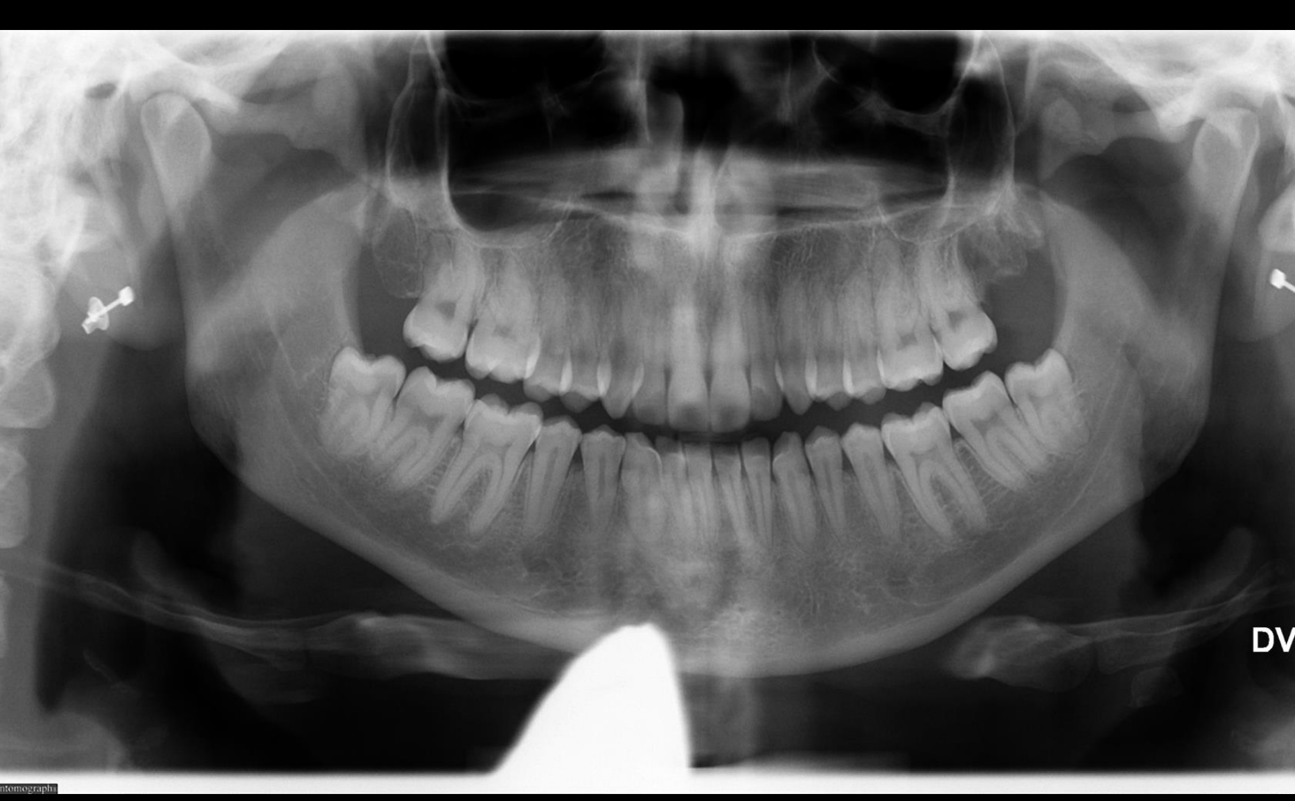
Case Study 1: Impacted Third Molars56
-
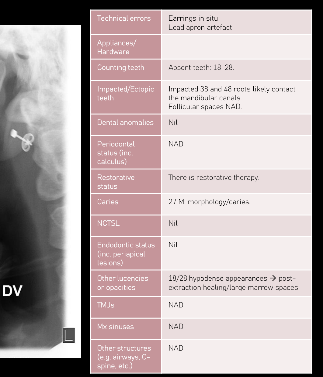
Technical errors: Earrings in situ, Lead apron artefact
-
Appliances/Hardware: (none)
-
Counting teeth: Absent teeth: 18, 28.
-
Impacted/Ectopic teeth: Impacted 38 and 48 roots likely contact the mandibular canals. Follicular spaces NAD.
-
Dental anomalies: Nil
-
Periodontal status (inc. calculus): NAD
-
Restorative status: There is restorative therapy.
-
Caries: 27 M: morphology/caries.
-
NCTSL: Nil
-
Endodontic status (inc. periapical lesions): Nil
-
Other lucencies or opacities: 18/28 hypodense appearances → post-extraction healing/large marrow spaces.
-
TMJs: NAD
-
Mx sinuses: NAD
-
Other structures (e.g. airways, C-spine, etc.): NAD
-
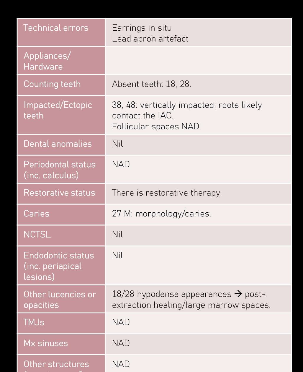
Technical errors: Earrings in situ, Lead apron artefact
-
Appliances/Hardware: (none)
-
Counting teeth: Absent teeth: 18, 28.
-
Impacted/Ectopic teeth: 38, 48: vertically impacted; roots likely contact the IAC. Follicular spaces NAD.
-
Dental anomalies: Nil
-
Periodontal status (inc. calculus): NAD
-
Restorative status: There is restorative therapy.
-
Caries: 27 M: morphology/caries.
-
NCTSL: Nil
-
Endodontic status (inc. periapical lesions): Nil
-
Other lucencies or opacities: 18/28 hypodense appearances → post-extraction healing/large marrow spaces.
-
TMJs: NAD
-
Mx sinuses: NAD
-
Other structures (e.g. airways, C-spine, etc.): NAD
 |  |
 |
Findings7
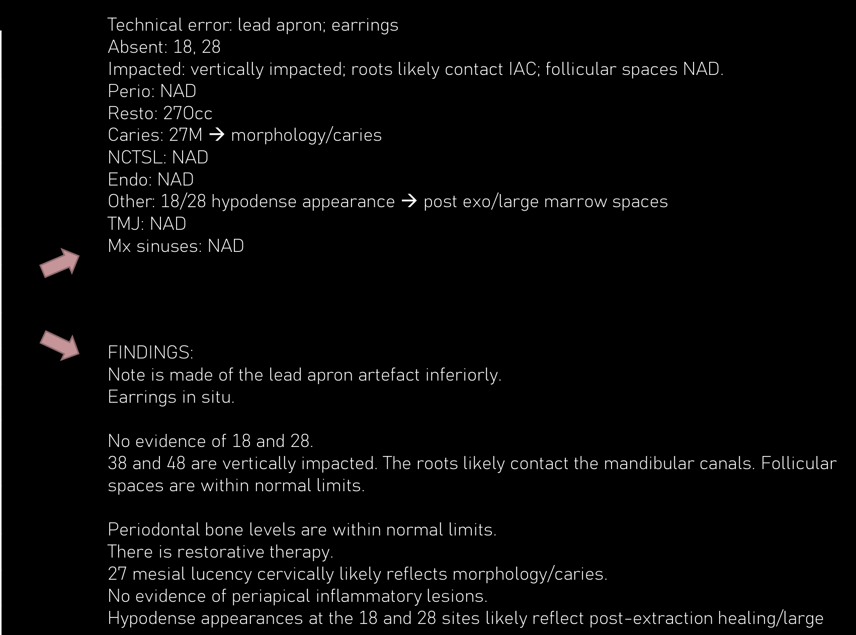
- No evidence of 18 and 28.
- 38 and 48 are vertically impacted. The roots likely contact the mandibular canals. Follicular spaces are within normal limits.
- Periodontal bone levels are within normal limits.
- There is restorative therapy.
- 27 mesial lucency cervically likely reflects morphology/caries.
- No evidence of periapical inflammatory lesions.
- Hypodense appearances at the 18 and 28 sites likely reflect post-extraction healing/large marrow spaces.
- Allowing for the oblique view, condylar appearances are within normal limits.
- The maxillary sinuses are clear.
- Technical error: lead apron; earrings.
 |  |
Summary
- Absent: 18, 28
- Impacted: vertically impacted; roots likely contact IAC; follicular spaces NAD.
- Perio: NAD
- Resto: 270cc
- Caries: 27M → morphology/caries
- NCTSL: NAD
- Endo: NAD
- Other: 18/28 hypodense appearance → post exo/large marrow spaces
- TMJ: NAD
- Mx sinuses: NAD
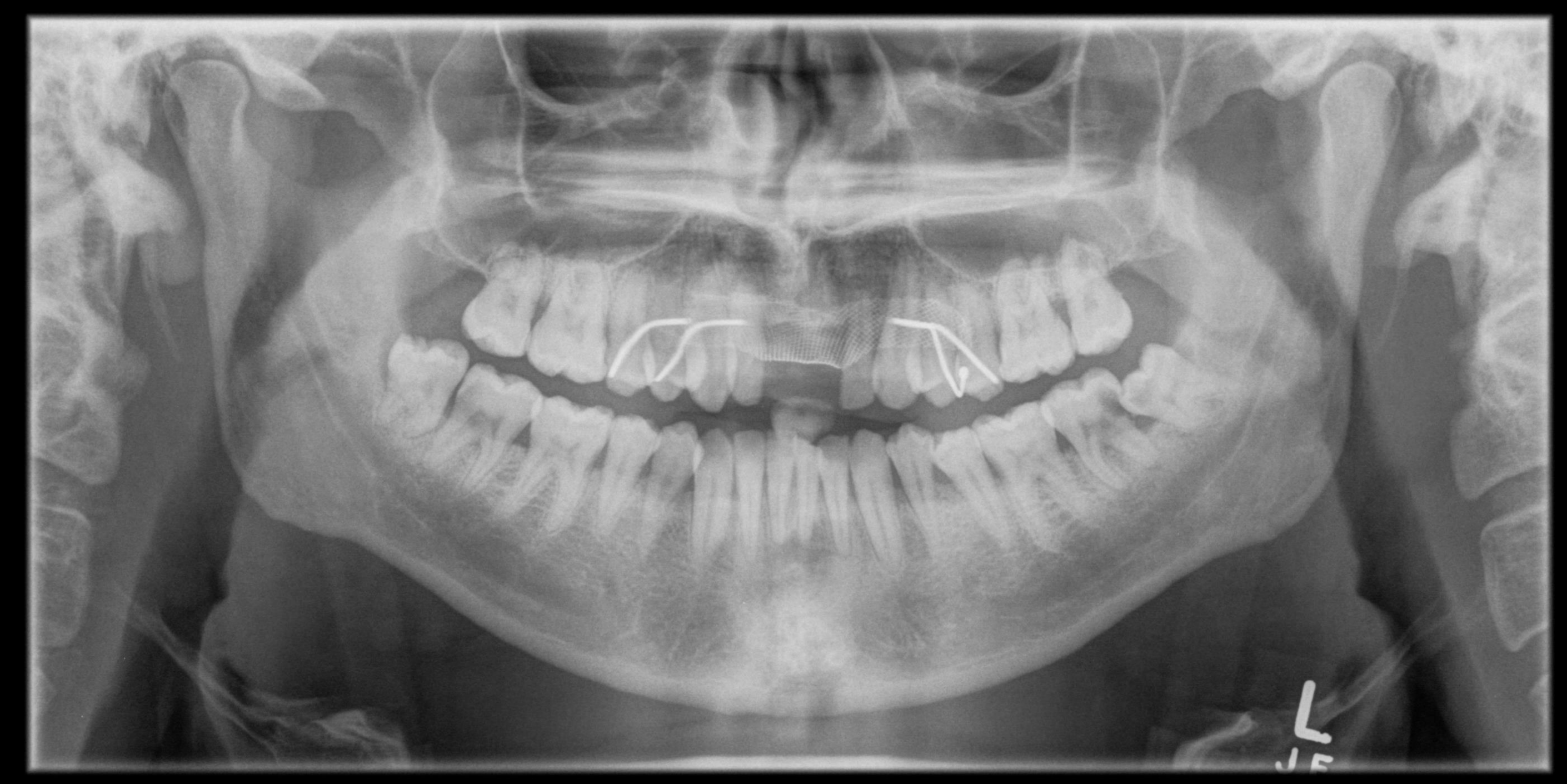
Case Study 2: Chronic Pericoronitis89
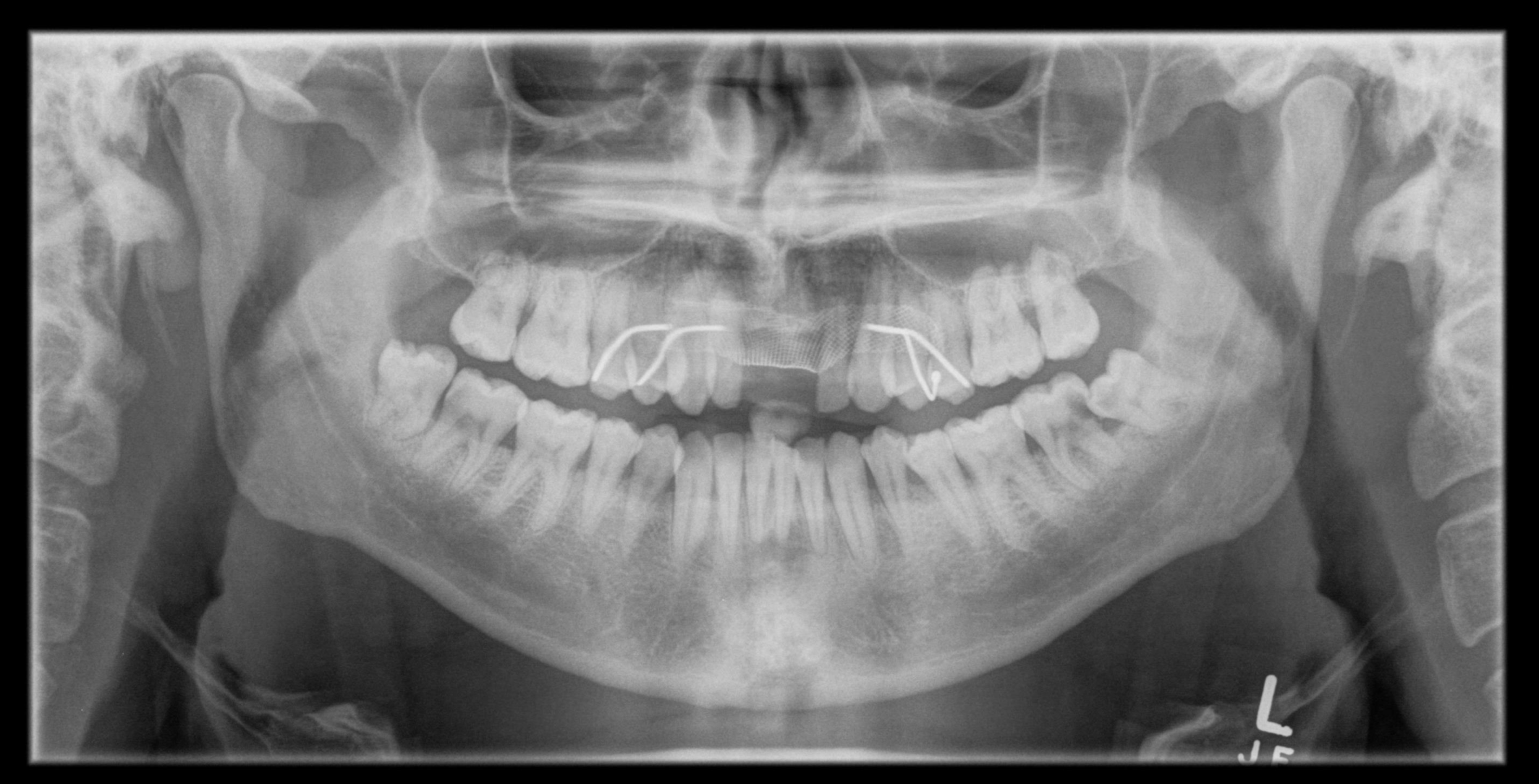
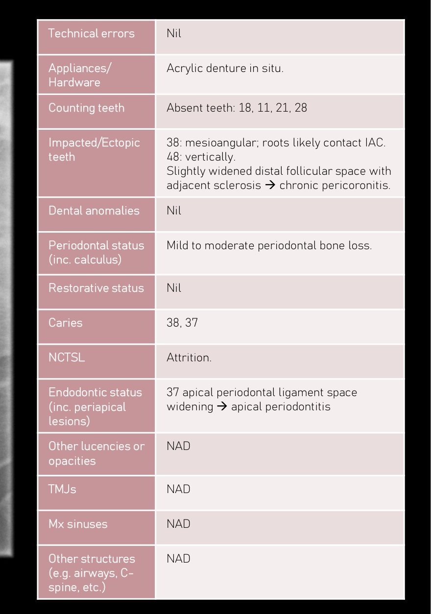
- Technical errors: Nil
- Appliances/Hardware: Acrylic denture in situ.
- Counting teeth: Absent teeth: 18, 11, 21, 28
- Impacted/Ectopic teeth: 38: mesioangular; roots likely contact IAC. 48: vertically. Slightly widened distal follicular space with adjacent sclerosis → chronic pericoronitis.
- Dental anomalies: Nil
- Periodontal status (inc. calculus): Mild to moderate periodontal bone loss.
- Restorative status: Nil
- Caries: 38, 37
- NCTSL: Attrition.
- Endodontic status (inc. periapical lesions): 37 apical periodontal ligament space widening → apical periodontitis
- Other lucencies or opacities: NAD
- TMJs: NAD
- Mx sinuses: NAD
- Other structures (e.g. airways, C-spine, etc.): NAD
 |  |
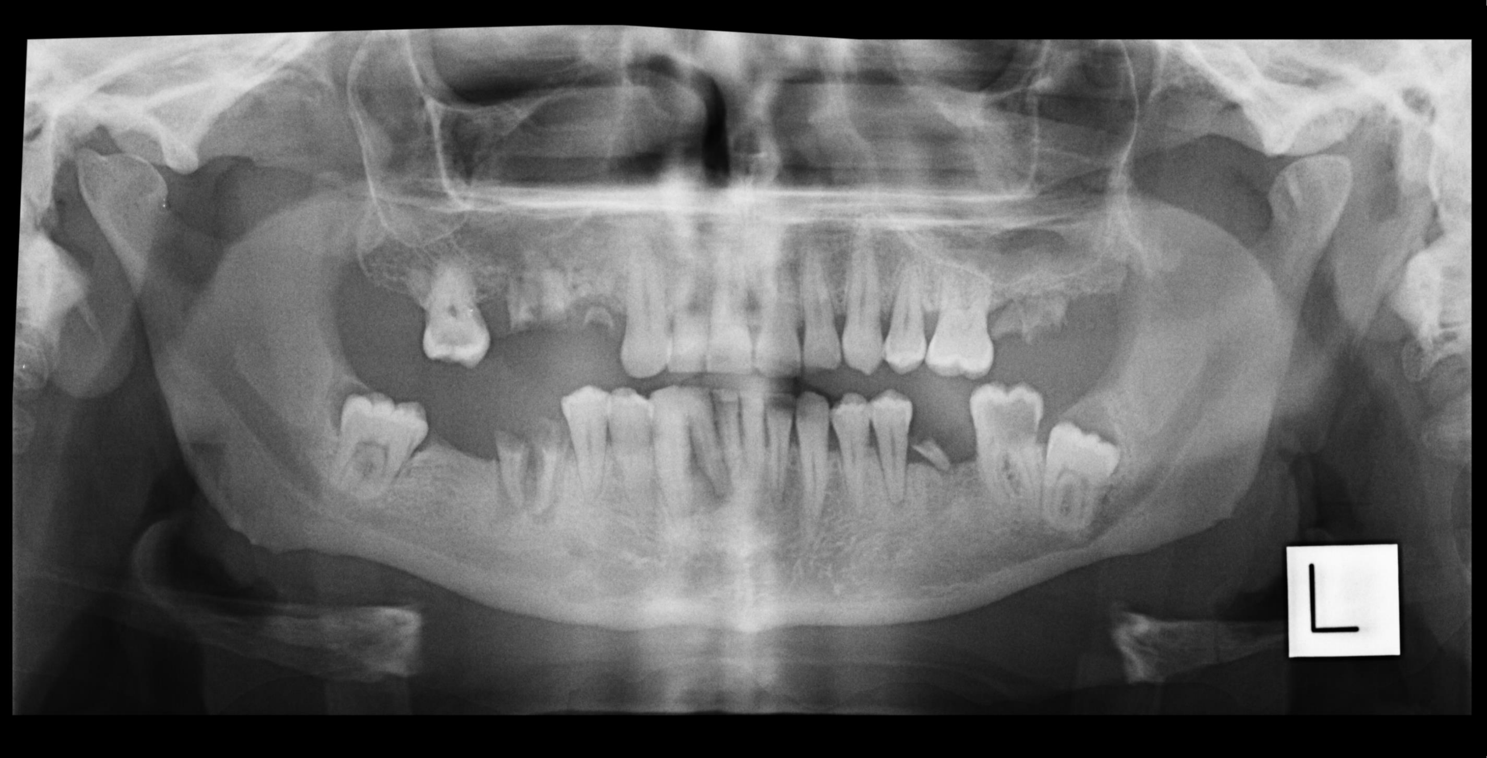 |
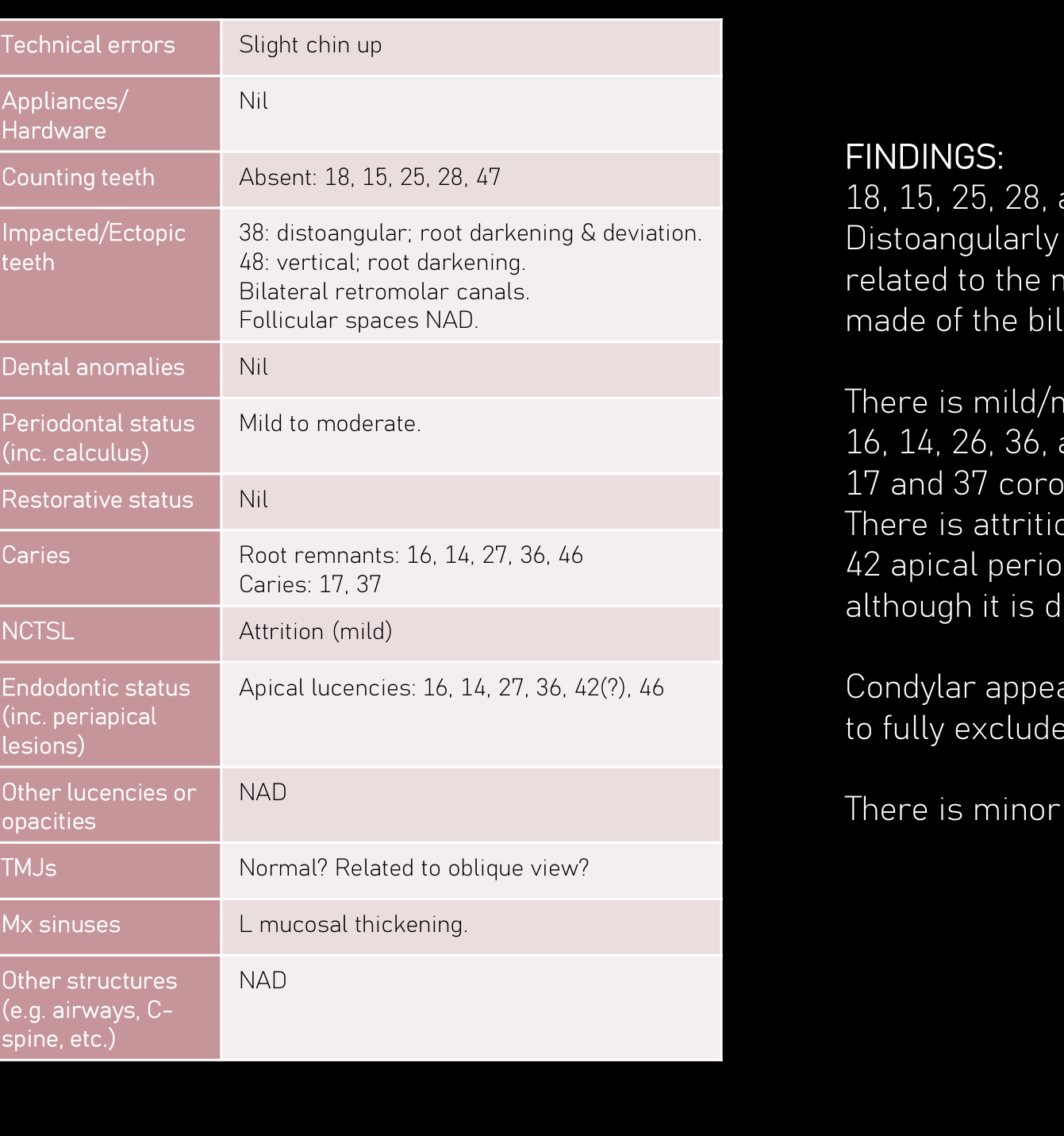
Case Study 3: Retromolar Canals1011
- Technical errors: Slight chin up
- Appliances/Hardware: Nil
- Counting teeth: Absent: 18, 15, 25, 28, 47
- Impacted/Ectopic teeth: 38: distoangular; root darkening & deviation. 48: vertical; root darkening. Bilateral retromolar canals. Follicular spaces NAD.
- Dental anomalies: Nil
- **Periodontal status (inc. calc
 | 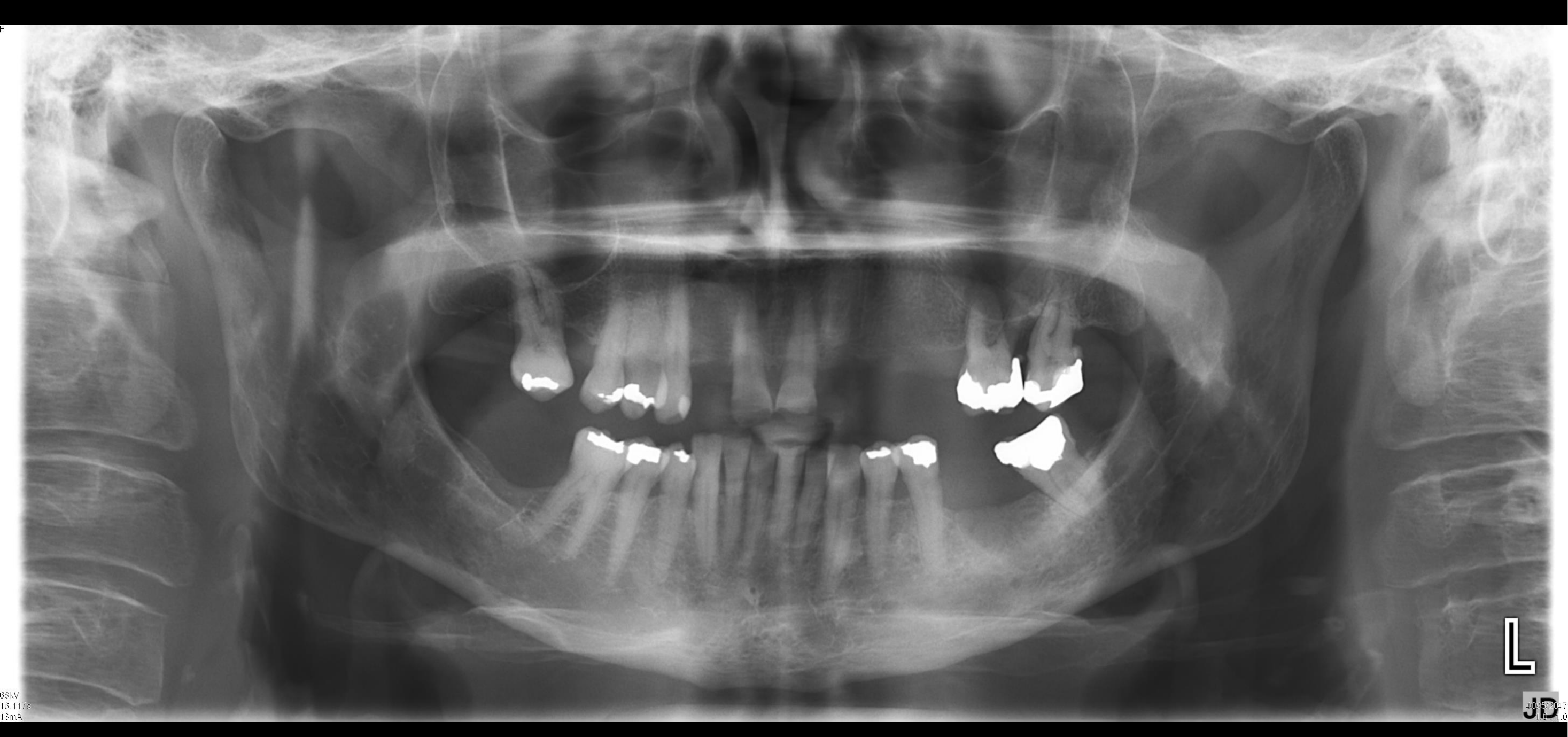 |
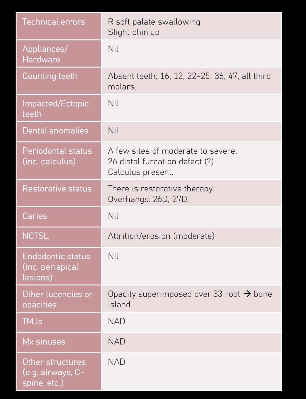
Case Study 4: Periodontal Bone Loss1213
| Technical errors | R soft palate swallowing Slight chin up |
| Appliances/ Hardware | Nil |
| Counting teeth | Absent teeth: 16, 12, 22-25, 36, 47, all third molars. |
| Impacted/Ectopic teeth | Nil |
| Dental anomalies | Nil |
| Periodontal status (inc. calculus) | A few sites of moderate to severe. 26 distal furcation defect (?) Calculus present. |
| Restorative status | There is restorative therapy. Overhangs: 26D, 27D. |
| Caries | Nil |
| NCTSL | Attrition/erosion (moderate) |
| Endodontic status (inc. periapical lesions) | Nil |
| Other lucencies or opacities | Opacity superimposed over 33 root → bone island |
| TMJs | NAD |
| Mx sinuses | NAD |
| Other structures (e.g. airways, C- spine, etc.) | NAD |
FINDINGS: 16, 12, 22-25, 36, 47, and all third molars are absent.
There a few sides of moderate to severe periodontal bone loss, with likely 26 distal bony furcation defect. Dental calculus deposit is noted There is restorative therapy. Note is made of the 26/27 distal overhangs. No definite caries is seen although it cannot be ruled out with this technique. There is moderate attrition/erosion. No evidence of periapical inflammatory lesion. Opacity superimposed over the 33 root likely reflects a bone island.
Appearances of remaining osseous structures in this view are within normal limits.
 | 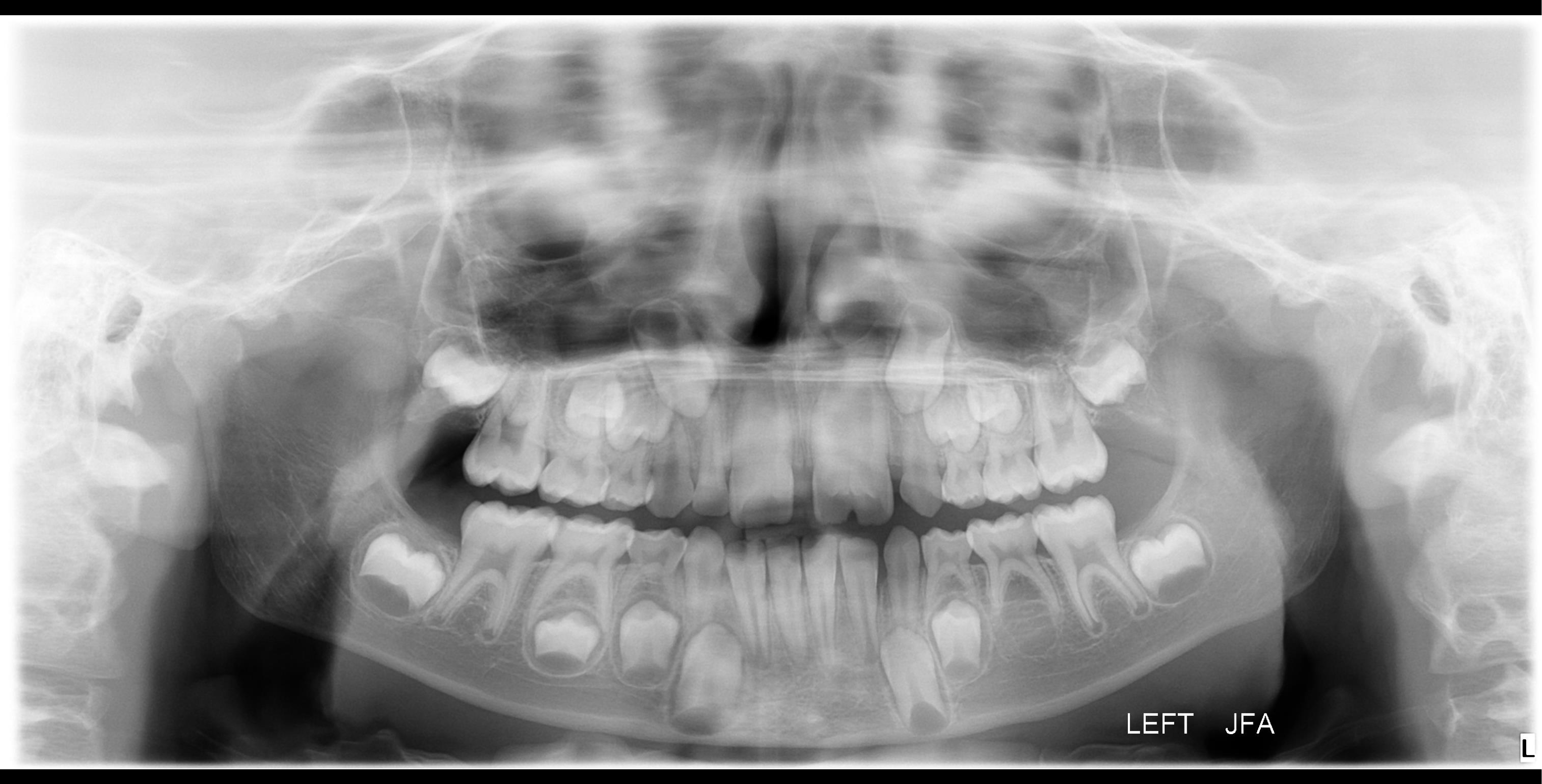 |
Case Study 5: Dental Fusion and Caries1415
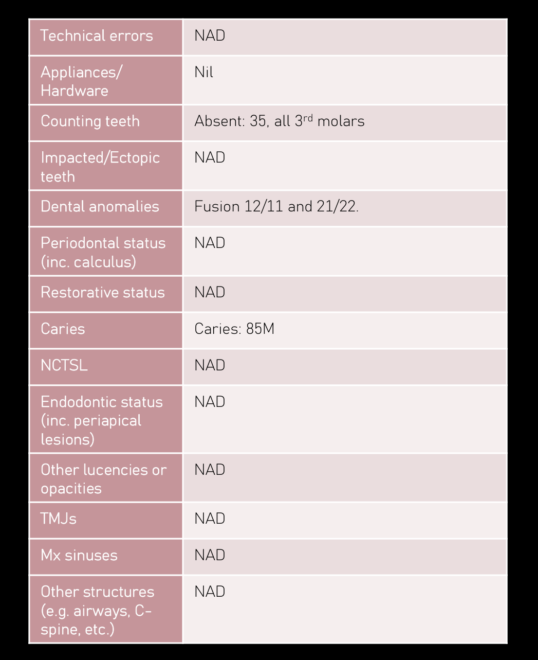
| Technical errors | NAD |
|---|---|
| Appliances/Hardware | Nil |
| Counting teeth | Absent: 35, all 3rd molars |
| Impacted/Ectopic teeth | NAD |
| Dental anomalies | Fusion 12/11 and 21/22. |
| Periodontal status (inc. calculus) | NAD |
| Restorative status | NAD |
| Caries | Caries: 85M |
| NCTSL | NAD |
| Endodontic status (inc. periapical lesions) | NAD |
| Other lucencies or opacities | NAD |
| TMJs | NAD |
| Mx sinuses | NAD |
| Other structures (e.g. airways, C-spine, etc.) | NAD |
Appearances of the teeth in the maxillary central incisor positions likely reflect fusion of 12/11 and 21/22. The tooth in the right maxillary lateral incisor position is presumably the 52. There is no evidence of 35 and all third molars at this stage. Remaining permanent teeth are present.
Periodontal bone levels of this mixed dentition are within normal limits. 85 mesial coronal lucency likely reflects caries. No periapical inflammatory lesions.
Appearances are remaining osseous structures in this are within normal limits.
 | 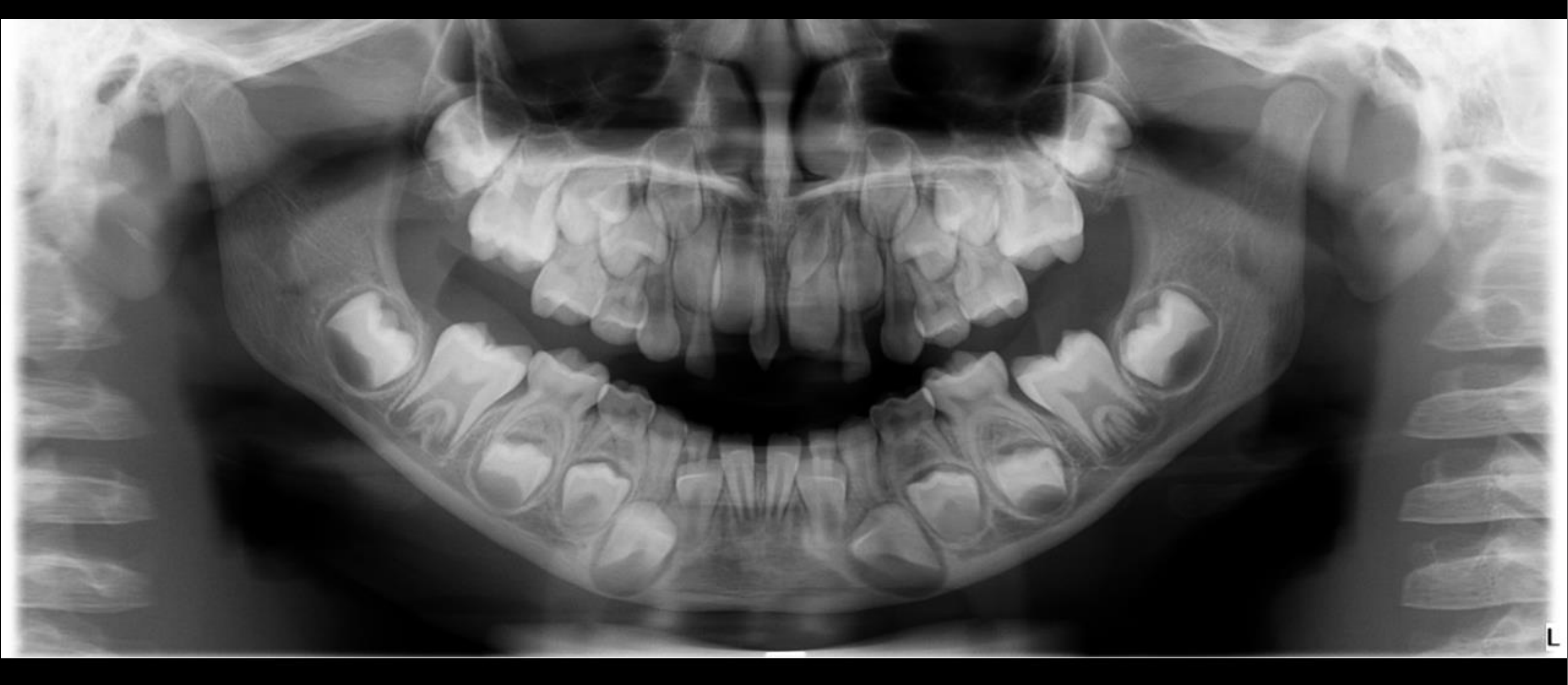 |
Case Study 6: Supernumerary Teeth1617
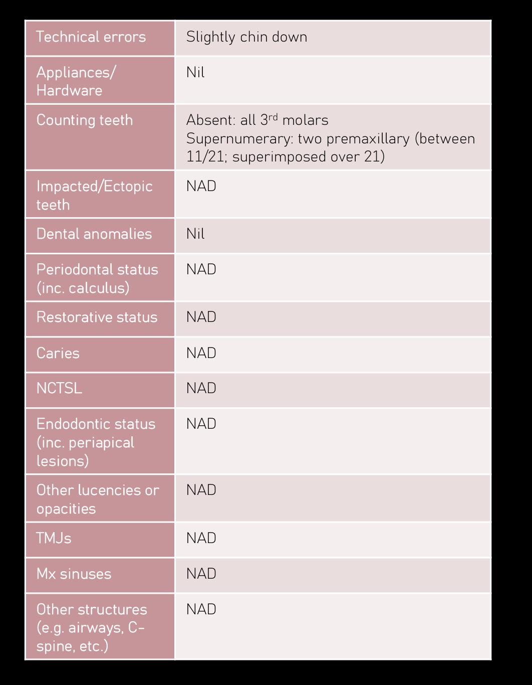
| Technical errors | Slightly chin down |
|---|---|
| Appliances/ Hardware | Nil |
| Counting teeth | Absent: all 3rd molars Supernumerary: two premaxillary (between 11/21; superimposed over 21) |
| Impacted/Ectopic teeth | NAD |
| Dental anomalies | Nil |
| Periodontal status (inc. calculus) | NAD |
| Restorative status | NAD |
| Caries | NAD |
| NCTSL | NAD |
| Endodontic status (inc. periapical lesions) | NAD |
| Other lucencies or opacities | NAD |
| TMJs | NAD |
| Mx sinuses | NAD |
| Other structures (e.g. airways, C-spine, etc.) | NAD |
FINDINGS:
No evidence of third molars at this stage. Remaining permanent teeth are present, at various stages of development and eruption.
There are two premaxillary supernumerary teeth. The right supernumerary tooth is erupted, likely impeding the eruption of 11. The left supernumerary tooth crown is superimposed over the 21 root.
Periodontal bone levels of this mixed dentition are within normal limits. No definite caries seen although it cannot be ruled out with this technique. No evidence of periapical inflammatory lesions. No evidence of external root resorption of permanent teeth. Intraoral radiographs or Cone Beam CT could be considered in further evaluation.
Appearances of remaining osseous structures in this view are within normal limits.
 | 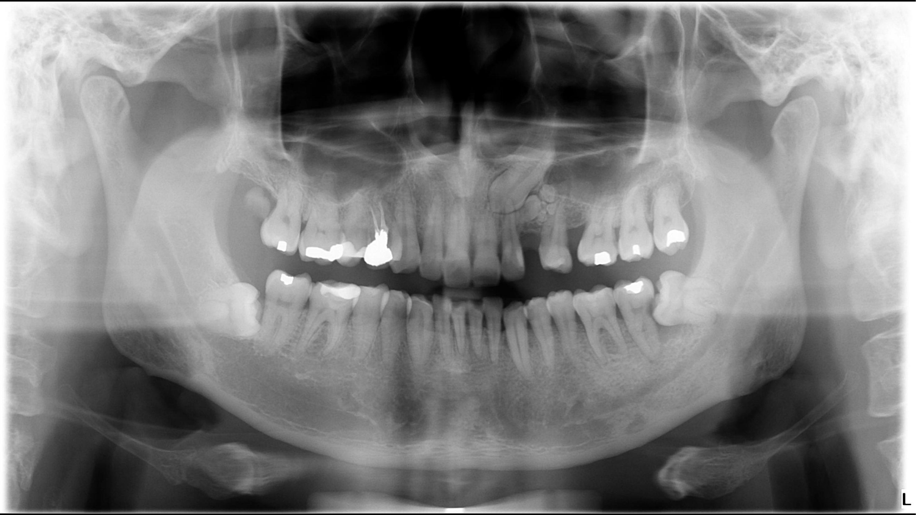 |
Case Study 7: Ectopic Eruption and Resorption1819
| Technical errors | Nil |
|---|---|
| Appliances/Hardware | Nil |
| Counting teeth | Absent: 25 |
| Impacted/Ectopic teeth | 23: ectopically impacted; crown superimposed over 21 → resorption? 38, 48: horizontal; likely contact IAC. 38 follicular space widening with adjacent sclerosis → chronic pericoronitis + periodontal bone loss. |
| Dental anomalies | Location: 23 site Periphery: well-defined, lucent rim with corticated borders Internal: odontoid opacities Surrounding: superimposed over 24 DDx: compound odontoma Tx: depends at this age... |
| Periodontal status (inc. calculus) | Minor to moderate |
| Restorative status | There is restorative therapy. |
| Caries | Root remnant: 18 Caries: 37D |
| NCTSL | Attrition |
| Endodontic status (inc. periapical lesions) | 37: apical PDL widening → inflammatory lesion Endodontically treated: 14 |
| Other lucencies or opacities | Opacities at apical aspects of 41/42 & 46 → bone islands |
| TMJs | NAD |
| Mx sinuses | Mild L Mx sinus |
| Other structures (e.g. airways, C-spine, etc.) | NAD |
FINDINGS: Ectopic 23 crown is projected over the apical aspects of the 21 and 22 where appearances of 21 raise the possibility of associated root resorption. The lesion at the 23 region, projected over the 24 with an estimated overall dimension of approximately 12mm demonstrates internal odontoid appearances, likely related to an odontome. Opacity at 18 site likely reflects a carious root remnant, essentially located within the mucosa. 28 is erupted. Impacted mandibular third molar roots likely contact the mandibular canals. There is likely associated distal caries and severe periodontal bone loss of the 37. Sclerosis adjacent to this second molar and 38 is likely reactive in nature, related to the periodontal bone loss as well as chronic pericoronitis. Widened apical periodontal space of 37 may be related to increased mobility/loading but an inflammatory lesion is included in the differential diagnosis. A few sites of minor and moderate periodontal bone loss noted elsewhere. There is restorative therapy. 26 and 36 lucencies reflect morphology or caries. There is attrition. 14 is endodontically treated. Opacities at the apical aspect of 41/42 and inferior to 46 likely reflect bone islands. There is likely minimal mucosal thickening at the left maxillary sinus floor. COMMENT: Opaque lesion at 23 region described likely reflects an odontome. The impacted ectopic 23 crown is projected over the apical aspect of 21/22 where appearances raise the possibility of root resorption associated with the 21. In view of these appearances and the other dentoalveolar findings described, Cone Beam CT could be considered in further evaluation.
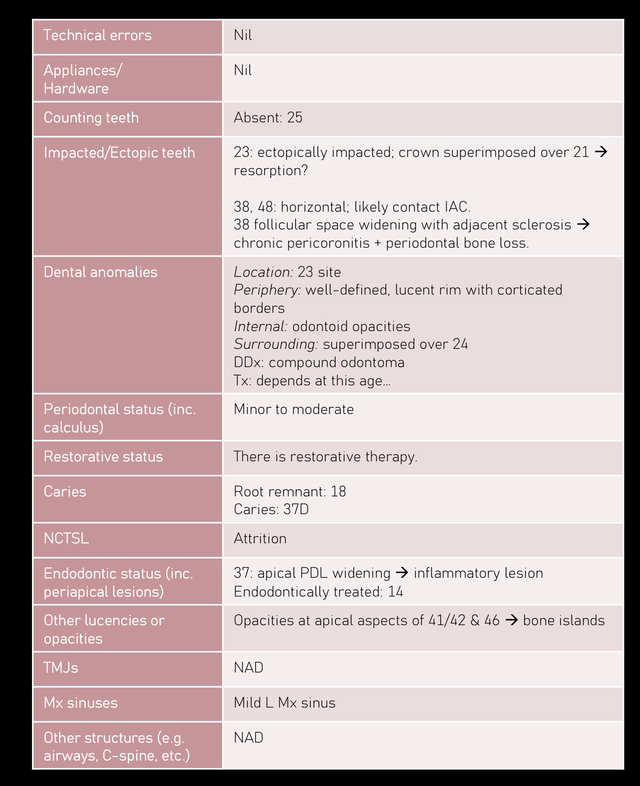 | 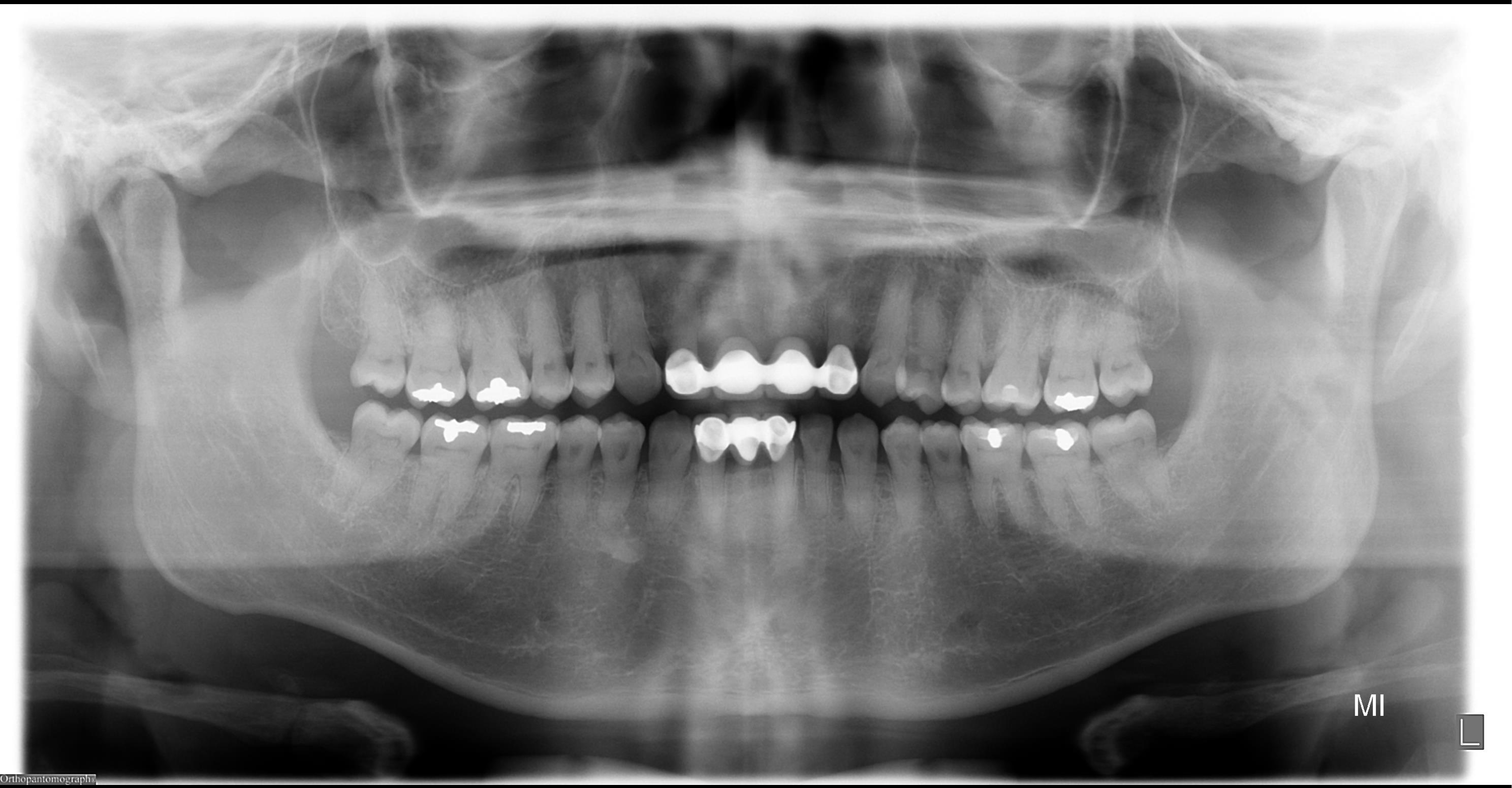 |
Case Study 8: Dentine Dysplasia Type II20
| Technical errors | Slightly chin up |
|---|---|
| Appliances/ Hardware | Nil |
| Counting teeth | Absent: 11, 21, 41 |
| Impacted/Ectopic teeth | Nil |
| Dental anomalies | Location: generalised, all teeth Shape: thistle-shaped pulp chambers; narrow root canals DDx: dentine dysplasia type II |
| Periodontal status (inc. calculus) | NAD |
| Restorative status | NAD |
| Caries | NAD |
| NCTSL | NAD |
| Endodontic status (inc. periapical lesions) | NAD |
| Other lucencies or opacities | Opacity apical aspect of 44 → bone island |
| TMJs | NAD |
| Mx sinuses | NAD |
| Other structures (e.g. airways, C-spine, etc.) | NAD |
FINDINGS:
11, 21, and 41 are absent.
There is generalised unusual morphology of the root canal system of the visualised teeth, with thistle-shaped pulp chambers, and narrow root canal systems radicularly. Appearances likely reflect dentine dysplasia type II.
Periodontal bone levels are within normal limits. No definite caries is seen on cannot be ruled out with this technique. No evidence of periapical inflammatory lesions. Opacity at the apical aspect of 44 likely reflects a bone island.
Appearances remaining osseous structures in this view are within normal limits.
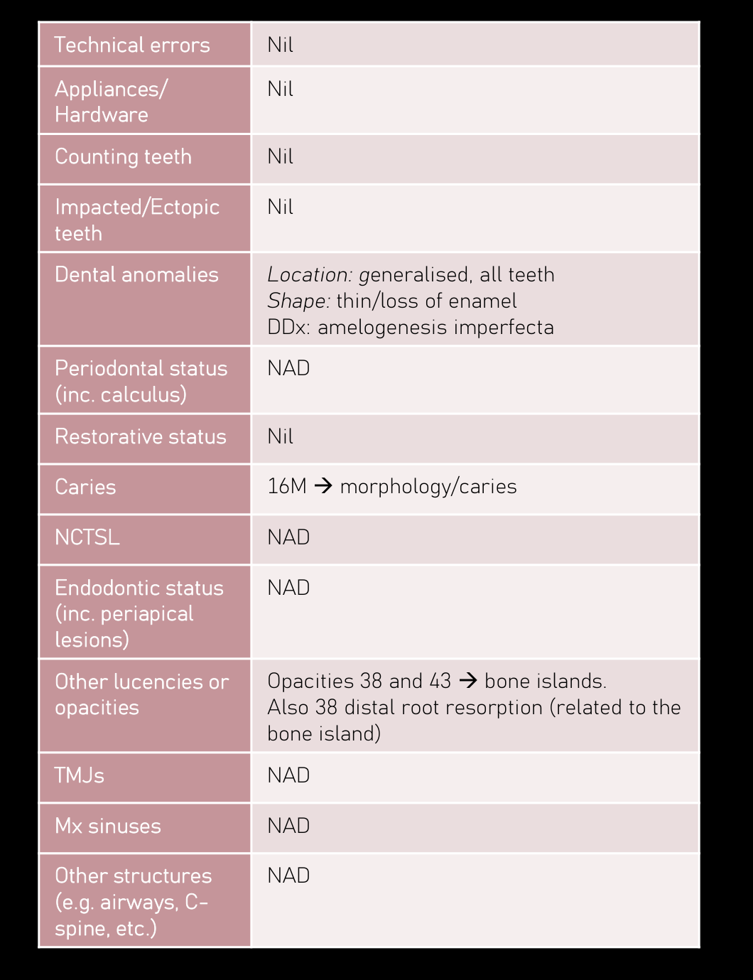
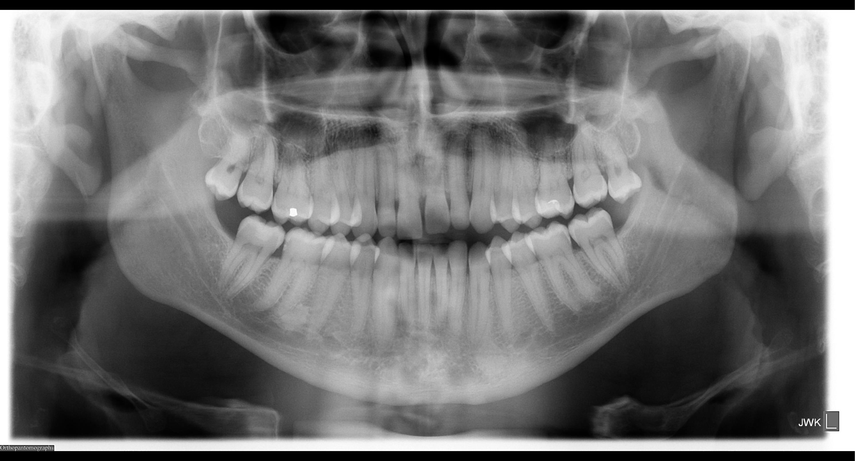
Case Study 9: Amelogenesis Imperfecta2122
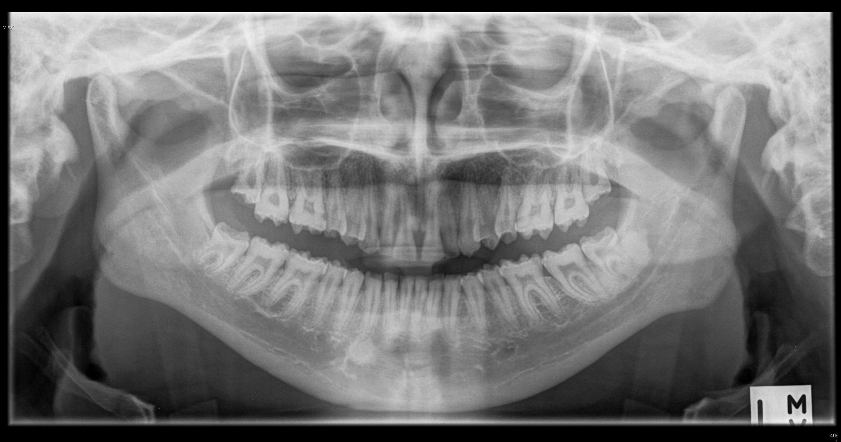
| Technical errors | Nil |
|---|---|
| Appliances/Hardware | Nil |
| Counting teeth | Nil |
| Impacted/Ectopic teeth | Nil |
| Dental anomalies | Location: generalised, all teeth Shape: thin/loss of enamel DDx: amelogenesis imperfecta |
| Periodontal status (inc. calculus) | NAD |
| Restorative status | Nil |
| Caries | 16M → morphology/caries |
| NCTSL | NAD |
| Endodontic status (inc. periapical lesions) | NAD |
| Other lucencies or opacities | Opacities 38 and 43 → bone islands. Also 38 distal root resorption (related to the bone island) |
| TMJs | NAD |
| Mx sinuses | NAD |
| Other structures (e.g. airways, C-spine, etc.) | NAD |
FINDINGS:
All permanent teeth are present. There is generalised thinning/loss of enamel, consistent with amelogenesis imperfecta.
Periodontal bone levels are within normal limits.
16 mesio-cervical lucency likely reflects morphology/caries.
No evidence of periapical inflammatory lesions.
Opacities surrounding the 38 roots, and at the apical aspect of 43 likely reflect bone islands. Note is made of the 38 distal root resorption, sometimes seen associated with bone island.
Appearances of the other osseous structures in this view are within normal limits.
 |  |
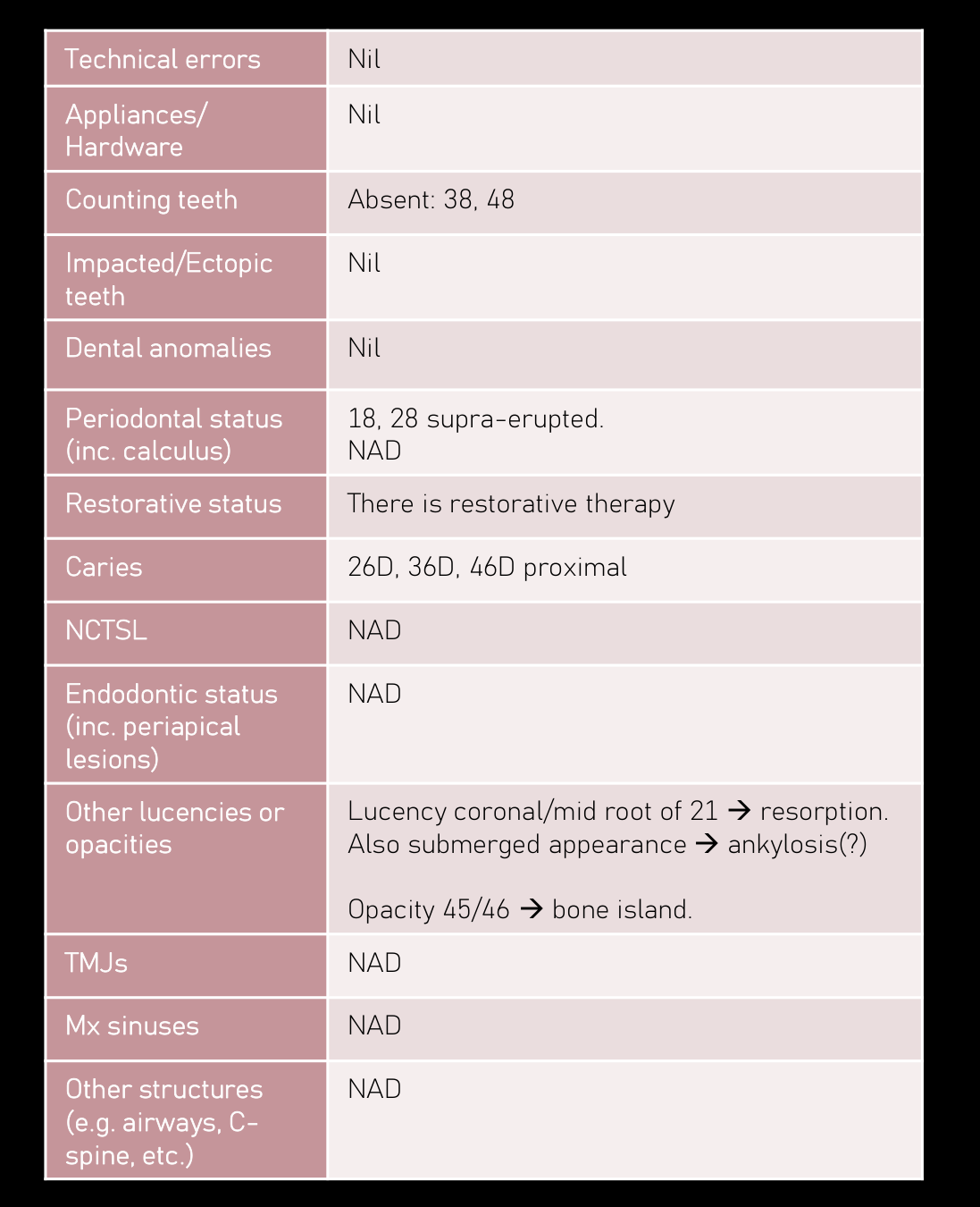
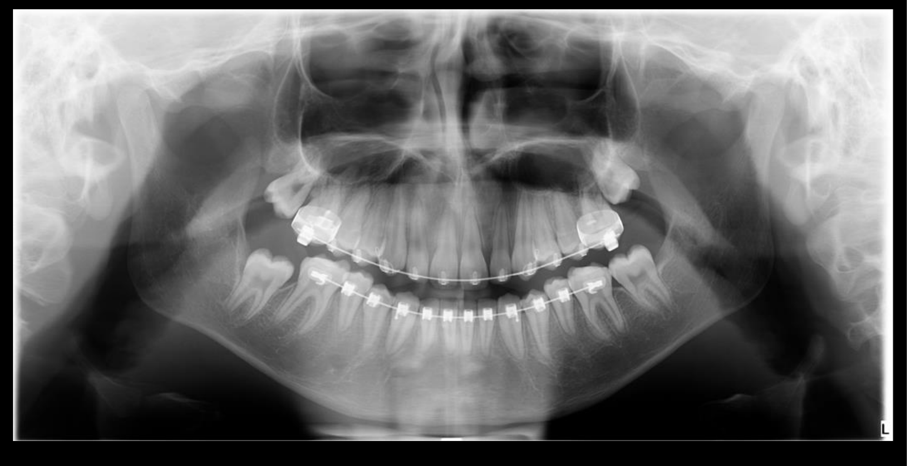
Case Study 10: Periapical Lesions2324
| Technical errors | Nil |
| Appliances/ Hardware | Nil |
| Counting teeth | Absent: 38, 48 |
| Impacted/Ectopic teeth | Nil |
| Dental anomalies | Nil |
| Periodontal status (inc. calculus) | 18, 28 supra-erupted. NAD |
| Restorative status | There is restorative therapy |
| Caries | 26D, 36D, 46D proximal |
| NCTSL | NAD |
| Endodontic status (inc. periapical lesions) | NAD |
| Other lucencies or opacities | Lucency coronal/mid root of 21 → resorption. Also submerged appearance → ankylosis(?) Opacity 45/46 → bone island. |
| TMJs | NAD |
| Mx sinuses | NAD |
| Other structures (e.g. airways, C- spine, etc.) | NAD |
No evidence of 38 and 48. 18 and 28 are overerupted.
Periodontal bone levels are within normal limits. There is restorative therapy. 26 distal, 36 distal, and 46 distal proximal lucencies likely reflect caries. No evidence of periapical inflammatory lesions. 21 radicular lucency at the coronal/mid root level with submerged appearances likely reflect resorption and ankylosis. This region is unavoidably not well demonstrated on account of superimposition of anatomical structures. Opacity between 45/46 likely reflects a bone island.
Appearances of the remaining osseous structures in this view are within normal limits.
 |  |
Case Study 11: Orthodontic Hardware and Eruption Cyst2526
<table border="1">
	<tr>
		<th>Technical errors</th>
		<td>Nil</td>
	</tr>
	<tr>
		<th>Appliances/ Hardware</th>
		<td>Orthodontic hardware (FFA)</td>
	</tr>
	<tr>
		<th>Counting teeth</th>
		<td>Absent: 3rd molars</td>
	</tr>
	<tr>
		<th>Impacted/Ectopic teeth</th>
		<td>27 & 47 delayed eruption.<br>47 soft tissue density with prominent follicular space $\rightarrow$ eruption cyst?</td>
	</tr>
	<tr>
		<th>Dental anomalies</th>
		<td>Nil</td>
	</tr>
	<tr>
		<th>Periodontal status (inc. calculus)</th>
		<td>NAD.</td>
	</tr>
	<tr>
		<th>Restorative status</th>
		<td>Nil</td>
	</tr>
	<tr>
		<th>Caries</th>
		<td>Nil</td>
	</tr>
	<tr>
		<th>NCTSL</th>
		<td>Nil</td>
	</tr>
	<tr>
		<th>Endodontic status (inc. periapical lesions)</th>
		<td>Widened PDL spaces $\rightarrow$ ortho tx</td>
	</tr>
	<tr>
		<th>Other lucencies or opacities</th>
		<td>Nil</td>
	</tr>
	<tr>
		<th>TMJs</th>
		<td>NAD</td>
	</tr>
	<tr>
		<th>Mx sinuses</th>
		<td>NAD</td>
	</tr>
	<tr>
		<th>Other structures (e.g. airways, C-spine, etc.)</th>
		<td>NAD</td>
	</tr>
</table>

**FINDINGS:**
Orthodontic hardware present.

No evidence of 3rd molars at this stage.

Soft tissue density overlying the unerupted 47 crown, with prominent residual follicular spaces are noted. The possibility of an eruption cyst is considered. Clinical correlation may clarify.

Periodontal bone levels are within normal limits.

No definite caries seen although it cannot be ruled out with this technique. Widened periodontal ligament spaces are likely related to the orthodontic therapy.

No evidence of external root resorption of permanent teeth. Intraoral imaging could be considered in further evaluation.

Appearances of remaining osseous structures in this view are within normal limits.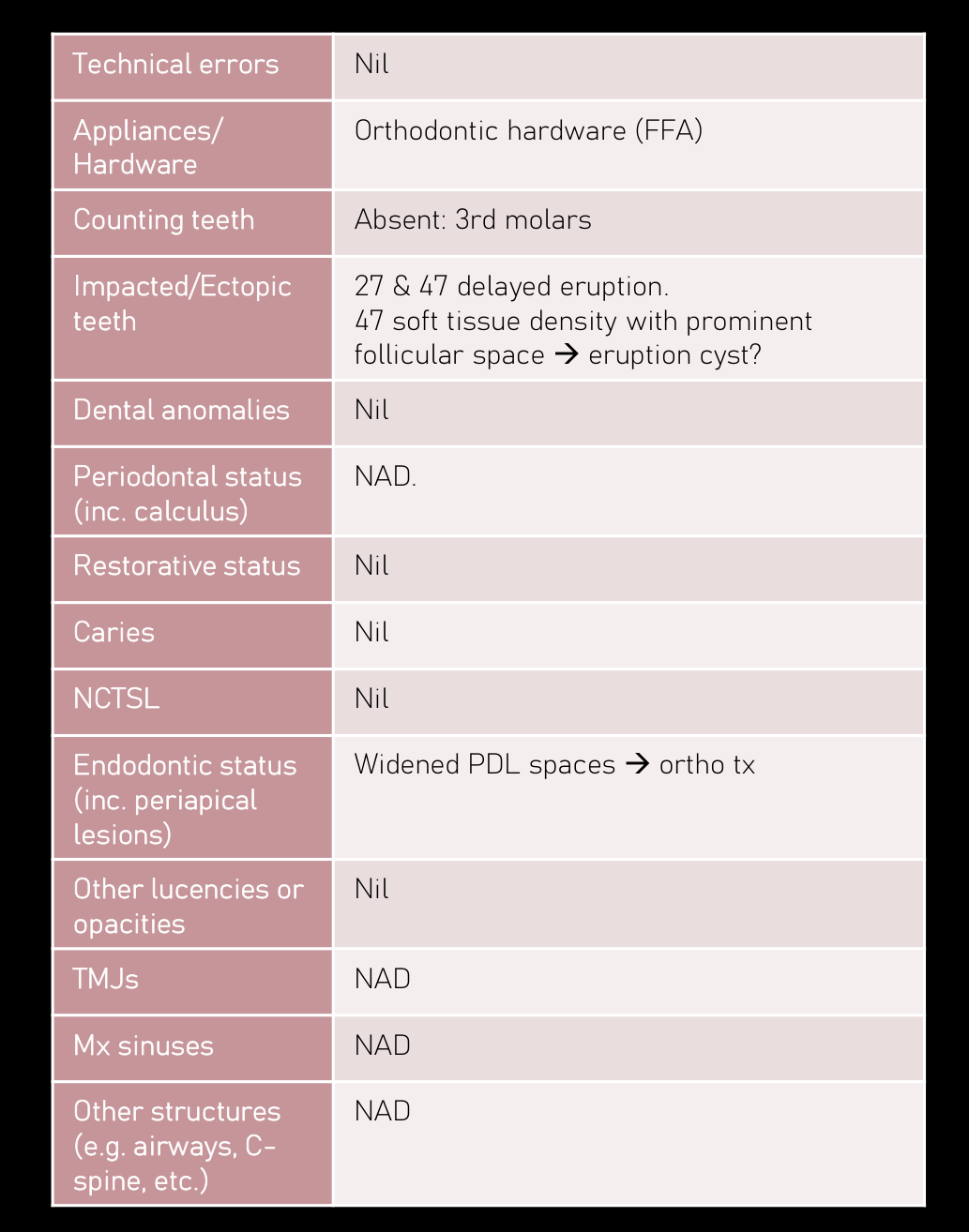 | 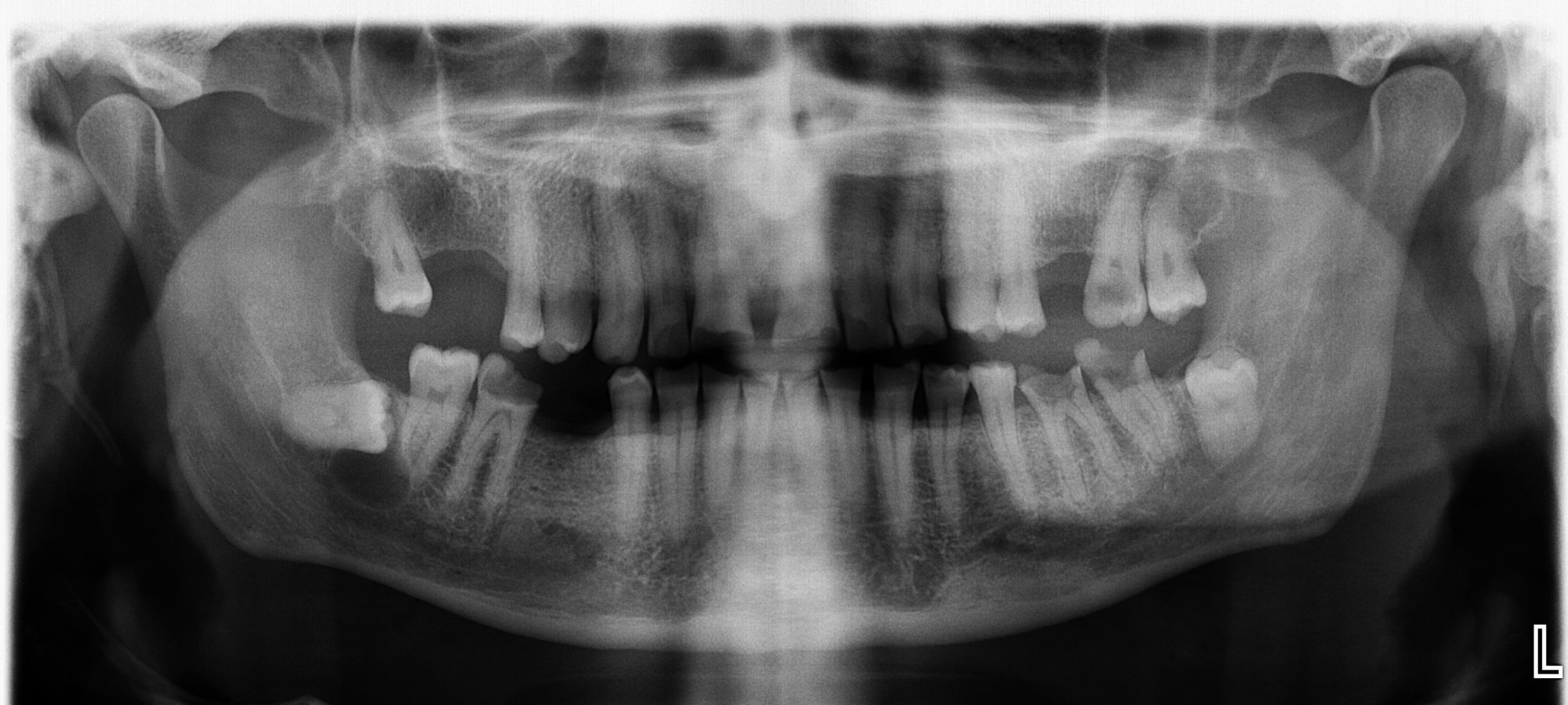 |
Case Study 12: Mandibular Canal Proximity2728
<table border="1" dir="ltr">
 <tbody>
  <tr>
   <th> Technical errors </th>
   <td> Slightly chin up </td>
  </tr>
  <tr>
   <th> Appliances/ Hardware </th>
   <td> Nil </td>
  </tr>
  <tr>
   <th> Counting teeth </th>
   <td> 17, 16, 26, 45 </td>
  </tr>
  <tr>
   <th> Impacted/Ectopic teeth </th>
   <td> 38: vertically; roots likely contact IAC; follicular space NAD.<br/>48: horizontally; root darkening; hypercementosis.<br/><br/>Location: centred inferior to 48 crown. Involves follicular space.<br/>Periphery: well-defined; corticated.<br/>Internal: lucent<br/>Surrounding: inferior displacement of IAC. 47 root resorption?<br/>DDx: cyst (dentigerous cyst or OKC).<br/>Tx: OMFS referral </td>
  </tr>
  <tr>
   <th> Dental anomalies </th>
   <td> Nil </td>
  </tr>
  <tr>
   <th> Periodontal status (inc. calculus) </th>
   <td> Mild to moderate </td>
  </tr>
  <tr>
   <th> Restorative status </th>
   <td> Nil </td>
  </tr>
  <tr>
   <th> Caries </th>
   <td> 27, 37, 36, 46 </td>
  </tr>
  <tr>
   <th> NCTSL </th>
   <td> Attrition/erosion (severe) </td>
  </tr>
  <tr>
   <th> Endodontic status (inc. periapical lesions) </th>
   <td> 27 apical lucency.<br/>37, 36 widened PDL spaces + adjacent sclerosis $\rightarrow$ inflammatory lesions </td>
  </tr>
  <tr>
   <th> Other lucencies or opacities </th>
   <td> Nil </td>
  </tr>
  <tr>
   <th> TMJs </th>
   <td> NAD </td>
  </tr>
  <tr>
   <th> Mx sinuses </th>
   <td> NAD </td>
  </tr>
  <tr>
   <th> Other structures (e.g. airways, C-spine, etc.) </th>
   <td> NAD </td>
  </tr>
 </tbody>
</table>

<div> FINDINGS:<br/>There is a well-defined corticated lucency within the right posterior mandible, centred inferior to the 48 crown, in continuity with the follicular space. This lucency is superimposed over the 47 distal root apex where it is difficult to exclude root resorption. There is mild displacement of the right mandibular canal inferiorly.<br/><br/>Horizontally impacted 48 roots are likely intimately related to the right mandibular canal. Increased periradicular density likely reflects hypercementosis.<br/><br/> OTHER FINDINGS:<br/>Impacted 38 roots likely contact the left mandibular canal. Follicular space is within normal limits.<br/>There are a few sites of mild to moderate periodontal bone loss.<br/>27, 37, 36, and 46 coronal lucencies likely reflect caries/lost restoration.<br/>There is substantial attrition/erosion.<br/> 27 apical lucency likely reflects an inflammatory lesion.<br/>37 and 36 widened apical periodontal ligament spaces with adjacent sclerosis also likely reflects inflammatory lesions.<br/>There is mild mucosal thickening within the visualised left maxillary sinus.<br/><br/> COMMENT:<br/>1. Well-defined right posterior mandibular lucency described likely reflects an odontogenic keratocyst. A dentigerous cyst is less likely. Further evaluation with Multi-slice CT +/- MRI is recommended.<br/>2. Other dentoalveolar findings, including caries and inflammatory lesions, are described. </div>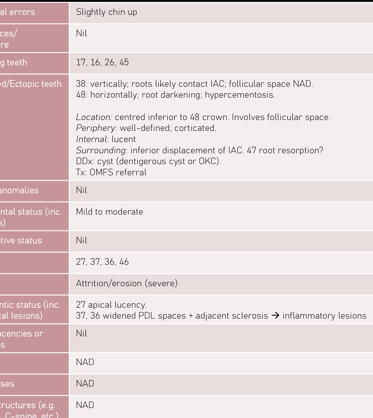 | 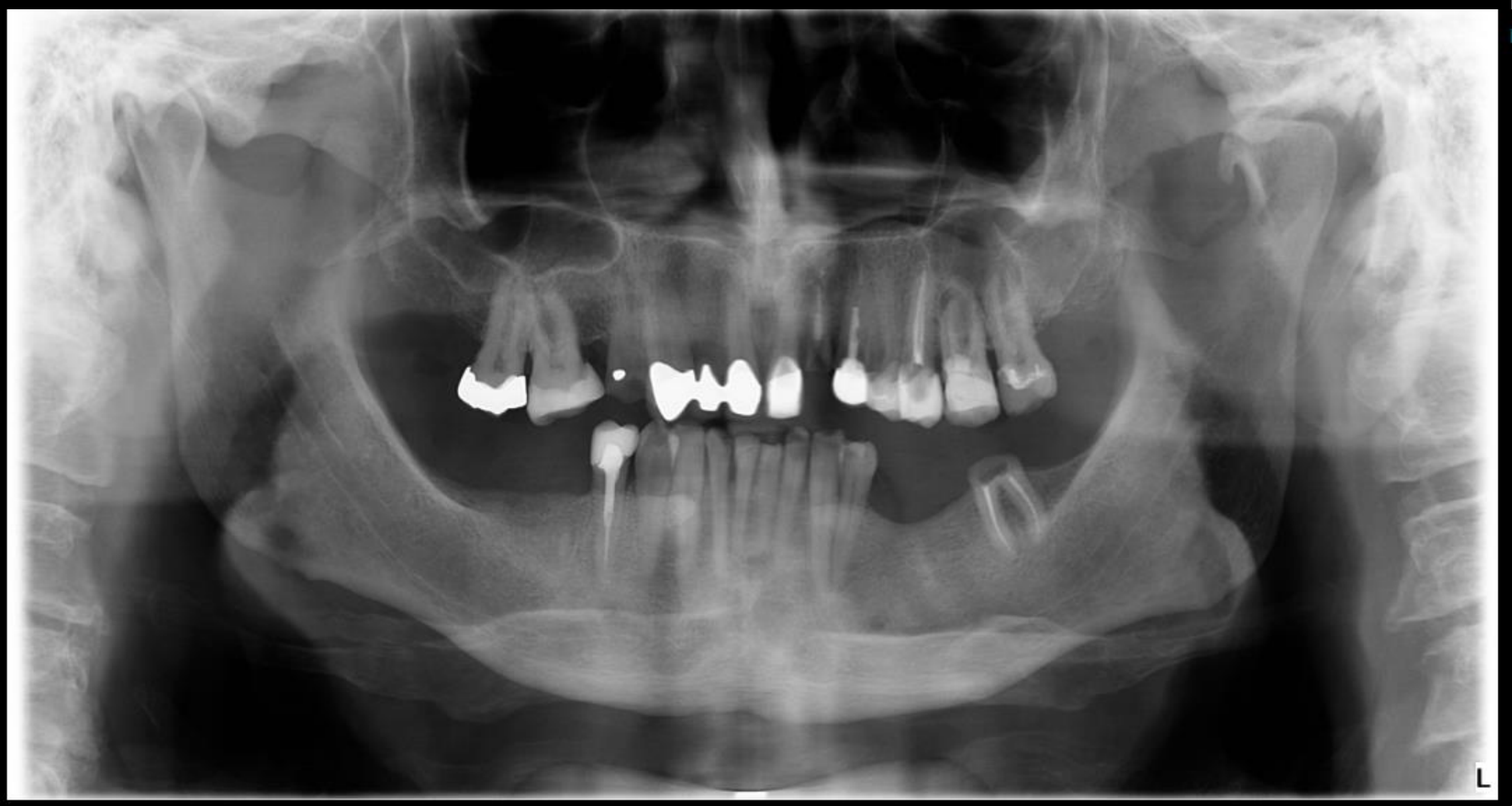 |
Case Study 13: Extensive Tooth Absence2930
| Technical errors | Nil |
| Appliances/Hardware | Nil |
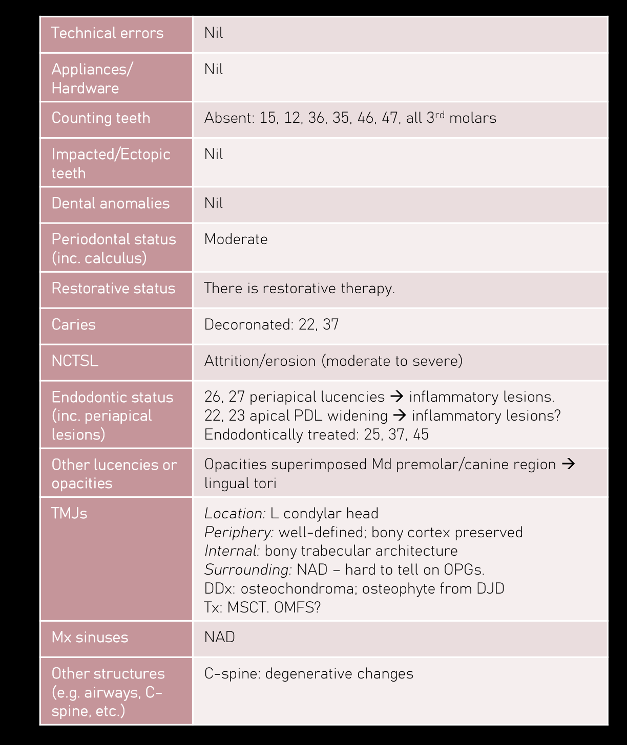
| Counting teeth | Absent: 15, 12, 36, 35, 46, 47, all 3rd molars |
| Impacted/Ectopic teeth | Nil |
| Dental anomalies | Nil |
| Periodontal status (inc. calculus) | Moderate |
| Restorative status | There is restorative therapy. |
| Caries | Decoronated: 22, 37 |
| NCTSL | Attrition/erosion (moderate to severe) |
| Endodontic status (inc. periapical lesions) | 26, 27 periapical lucencies $\rightarrow$ inflammatory lesions. 22, 23 apical PDL widening $\rightarrow$ inflammatory lesions? Endodontically treated: 25, 37, 45 |
| Other lucencies or opacities | Opacities superimposed Md premolar/canine region $\rightarrow$ lingual tori |
| TMJs | Location: L condylar head Periphery: well-defined; bony cortex preserved Internal: bony trabecular architecture Surrounding: NAD – hard to tell on OPGs. DDx: osteochondroma; osteophyte from DJD Tx: MSCT. OMFS? |
| Mx sinuses | NAD |
| Other structures (e.g. airways, C-spine, etc.) | C-spine: degenerative changes |
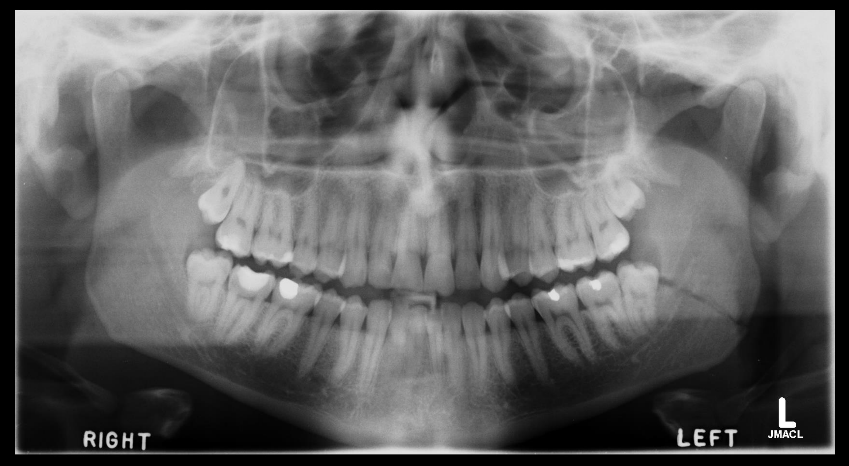
FINDINGS: There are a few sites of moderate periodontal bone loss. There is restorative therapy. 22 and 37 are decoronated, likely related to lost restoration. There is attrition/erosion. 26 and 27 periapical lucencies likely reflect inflammatory pathology. 22 and 23 apical periodontal ligament space widening likely reflects fibrous healing although it is difficult to fully exclude early inflammatory lesion. 25, 37, and 45 are also endodontically treated. Opacity superimposed over the 21 apical aspect raises the possibility of an impacted supernumerary tooth. Bilateral mandibular lingual tori noted. Intraoral radiographs or Cone Beam CT could be considered in further evaluation. There is mucosal thickening within the left maxillary sinus, likely reactive in nature. Allowing for the oblique view, left condylar appearances likely reflect a bony prominence. Degenerative changes of the visualised cervical spine are noted.
COMMENT: The dentoalveolar findings, including inflammatory lesions, are described. Allowing for the oblique view, left condylar appearances likely reflect arthropathy and suggest the presence of a bony prominence. An osteochondroma would be included in the differential diagnosis. Multislice CT could be considered for further evaluation.
 |  |
Case Study 14: Maxillary Sinus Approximation3132
<table border="1">
	<tr>
		<th>Technical errors</th>
		<td>Nil</td>
	</tr>
	<tr>
		<th>Appliances/</th>
		<td>Nil</td>
	</tr>
	<tr>
		<th>Hardware</th>
		<td></td>
	</tr>
	<tr>
		<th>Counting teeth</th>
		<td>NAD</td>
	</tr>
	<tr>
		<th>Impacted/Ectopic</th>
		<td>18/28: impacted; roots approximate sinus floor. Follicles NAD.</td>
	</tr>
	<tr>
		<th>teeth</th>
		<td>38/48: vertically; roots contact IAC. Widened distal follicular</td>
	</tr>
	<tr>
		<td></td>
		<td>spaces + adjacent sclerosis → chronic pericoronitis.</td>
	</tr>
	<tr>
		<th>Dental anomalies</th>
		<td>Nil</td>
	</tr>
	<tr>
		<th>Periodontal status</th>
		<td>NAD</td>
	</tr>
	<tr>
		<th>(inc. calculus)</th>
		<td></td>
	</tr>
	<tr>
		<th>Restorative status</th>
		<td>There is restorative therapy.</td>
	</tr>
	<tr>
		<th>Caries</th>
		<td>NAD</td>
	</tr>
	<tr>
		<th>NCTSL</th>
		<td>Attrition (mild to moderate)</td>
	</tr>
	<tr>
		<th>Endodontic status</th>
		<td>NAD</td>
	</tr>
	<tr>
		<th>(inc. periapical</th>
		<td></td>
	</tr>
	<tr>
		<th>lesions)</th>
		<td></td>
	</tr>
	<tr>
		<th>Other lucencies or</th>
		<td>Linear lucency from 38 distal follicular space to L Md angle →</td>
	</tr>
	<tr>
		<th>opacities</th>
		<td>displaced oblique fracture.</td>
	</tr>
	<tr>
		<th>TMJs</th>
		<td>NAD</td>
	</tr>
	<tr>
		<th>Mx sinuses</th>
		<td>NAD</td>
	</tr>
	<tr>
		<th>Other structures</th>
		<td>NAD</td>
	</tr>
	<tr>
		<th>(e.g. airways, C-</th>
		<td></td>
	</tr>
	<tr>
		<th>spine, etc.)</th>
		<td></td>
	</tr>
</table>
FINDINGS:
There is an oblique fracture of the left mandibular angle, involving the 38 distal
follicular space, with separation and displacement.
The condylar necks are intact.
No evidence of maxillary or zygomatic fracture.
The antral margins and orbital rims appear intact.
OTHER FINDINGS:
Impacted 18 and 28 roots approximate the sinus floor. Residual follicular spaces are
within normal limits.
Vertically impacted 38 and 48 roots likely contact the mandibular canals. Widened
distal follicular spaces with adjacent sclerosis likely reflect chronic pericoronitis.
Periodontal bone levels are within normal limits.
There is restorative therapy.
No definite caries seen although cannot be ruled out with this technique.
There is attrition.
No evidence of periapical inflammatory lesions.
Appearance of remaining osseous structures in this view are within normal limits.
COMMENT:
Displaced compound fracture extending from the 38 distal follicular space to the left
mandibular angle.
Other dentoalveolar findings are described.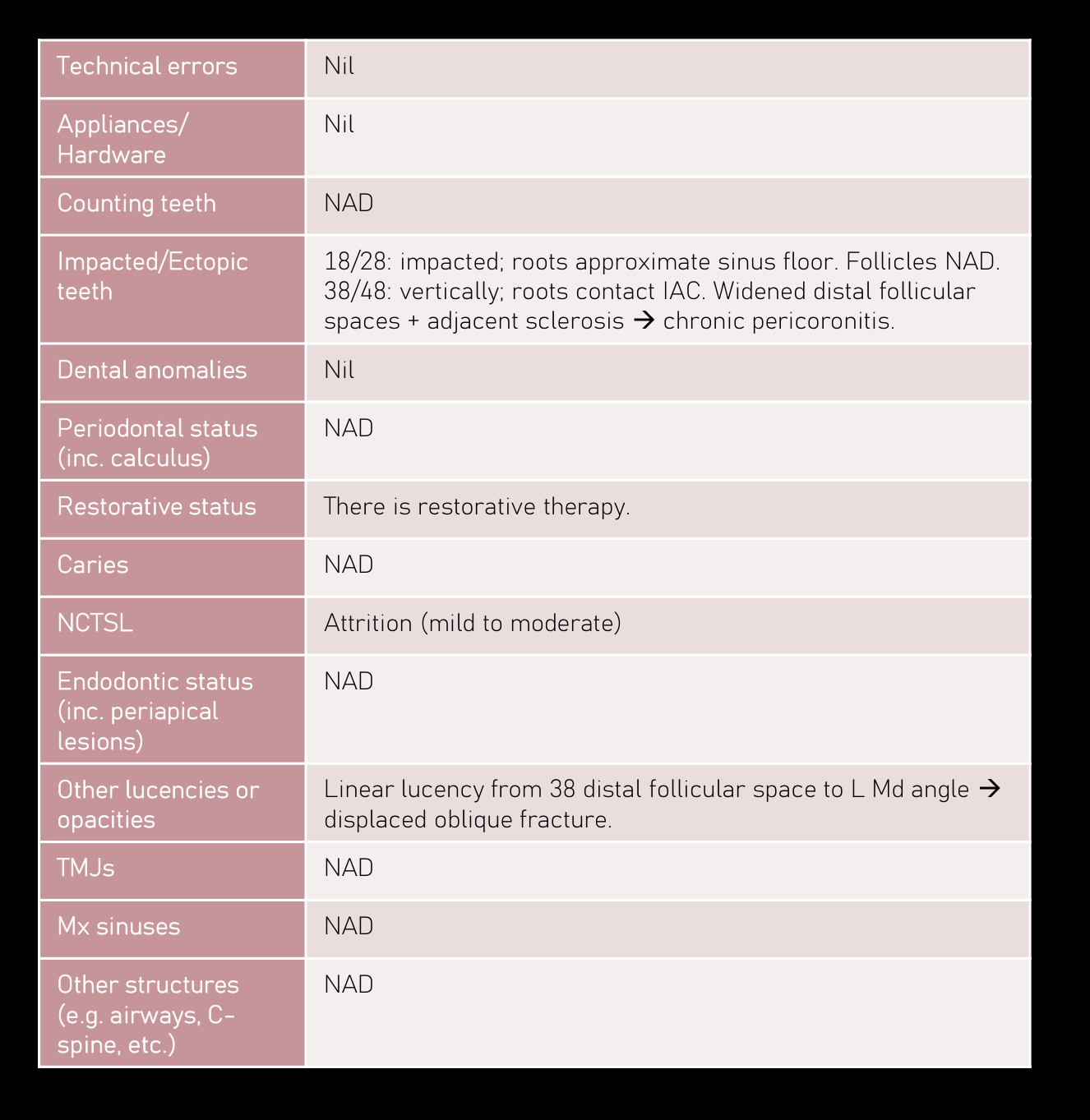 | 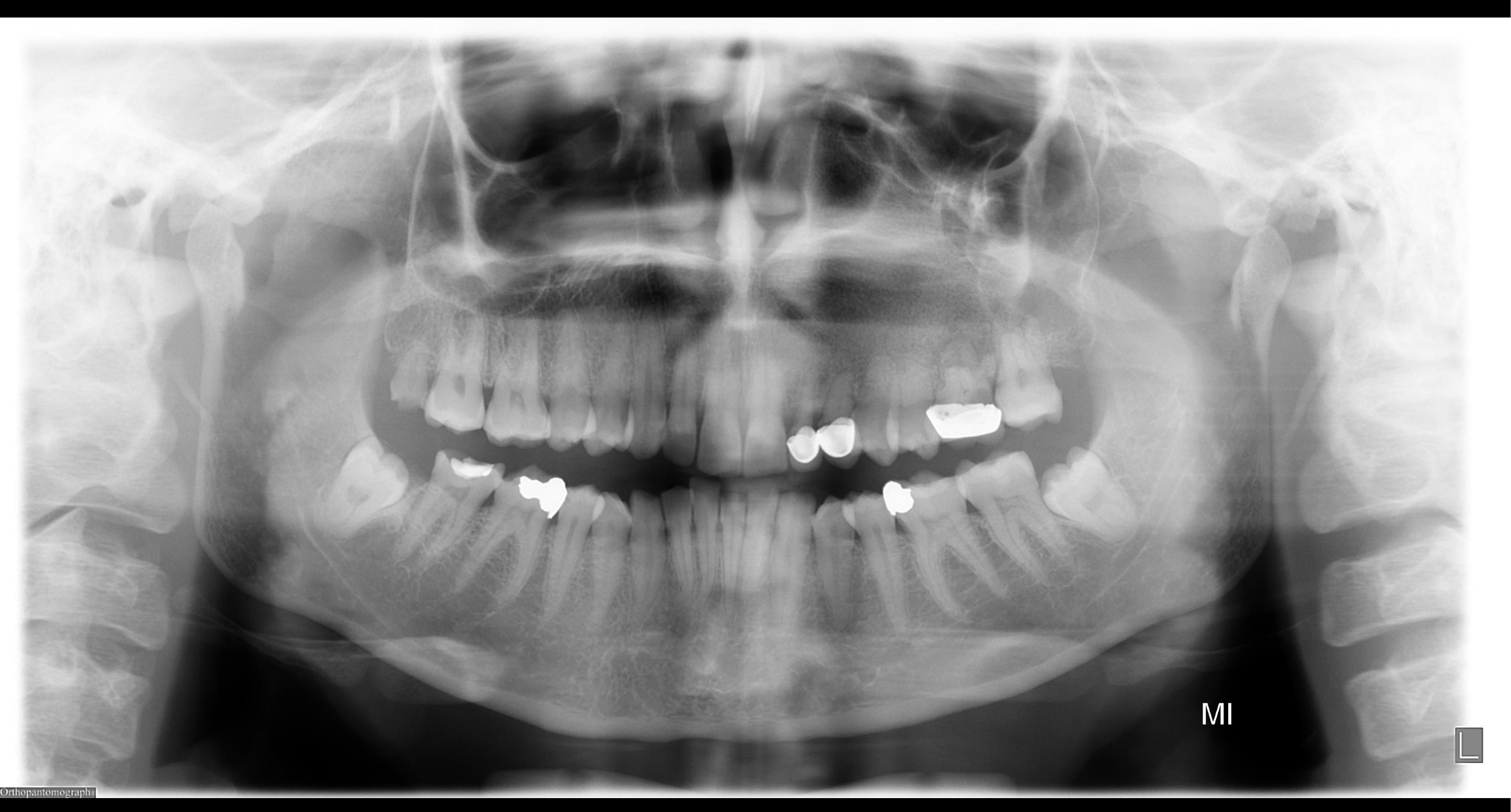 |
Case Study 15: Microdontia and Enamel Hypoplasia3334
<table border="1" cellspacing="0">
    <tr> <th> Technical errors </th> <td> Nil </td> </tr>
    <tr> <th> Appliances/ Hardware </th> <td> Nil </td> </tr>
    <tr> <th> Counting teeth </th> <td> Absent: 28 </td> </tr>
    <tr> <th> Impacted/Ectopic teeth </th> <td> 38, 48: mesioangular; roots contact IAC; follicular spaces NAD </td> </tr>
    <tr> <th> Dental anomalies </th> <td> 18: microdontic. Loss of enamel $\rightarrow$ hypoplasia? </td> </tr>
    <tr> <th> Periodontal status (inc. calculus) </th> <td> Mild </td> </tr>
    <tr> <th> Restorative status </th> <td> There is restorative therapy. </td> </tr>
    <tr> <th> Caries </th> <td> 26D, 23M(?), 47Occ(?) </td> </tr>
    <tr> <th> NCTSL </th> <td> Attrition </td> </tr>
    <tr> <th> Endodontic status (inc. periapical lesions) </th> <td> 26 apical PDL widening $\rightarrow$ inflammatory lesion </td> </tr>
    <tr> <th> Other lucencies or opacities </th> <td> Location: L Mx (22-26 region)<br>Periphery: ill-defined<br>Internal: ground-glass bony architecture<br>Surrounding: elevation of L Mx sinus floor; 22-26 root resorption.<br>DDx: fibrous dysplasia<br>Tx plan: if known; already being monitored by OMFS; if unknown $\rightarrow$ MSCT/OMFS? </td> </tr>
    <tr> <th> TMJs </th> <td> NAD </td> </tr>
    <tr> <th> Mx sinuses </th> <td> L Mx sinus opacification? </td> </tr>
    <tr> <th> Other structures (e.g. airways, C-spine, etc.) </th> <td> NAD </td> </tr>
</table>

No evidence of 28.

Microdontic 18 coronal appearances likely relate to hypoplasia.

Mesioangularly impacted 38 and 48 roots likely contact the mandibular canals. Follicular spaces are within normal limits.

There are a few sites of mild periodontal bone loss.

There is restorative therapy.

Lucency adjacent to the 26 restoration likely reflects secondary caries. Lucencies adjacent to several restorations, notably 23 and 47, may be artefactual though it is difficult to fully exclude deficiency/caries from OPGs.

There is attrition.

26 apical periodontal ligament space widening likely reflects an inflammatory lesion.

There is ill-defined, ground glass bony architecture within the left maxilla, extending from the maxillary midline to the 26 region. Superior elevation of the left antral floor is noted. There is blunting of the 22-26 roots. The periodontal ligament spaces remain preserved. Appearances likely reflect fibrous dysplasia.

Condylar appearances likely relate to the oblique view.

Opacification of the remaining left maxillary sinus may relate to the projection, although it is difficult to fully exclude sinus pathology from OPGs.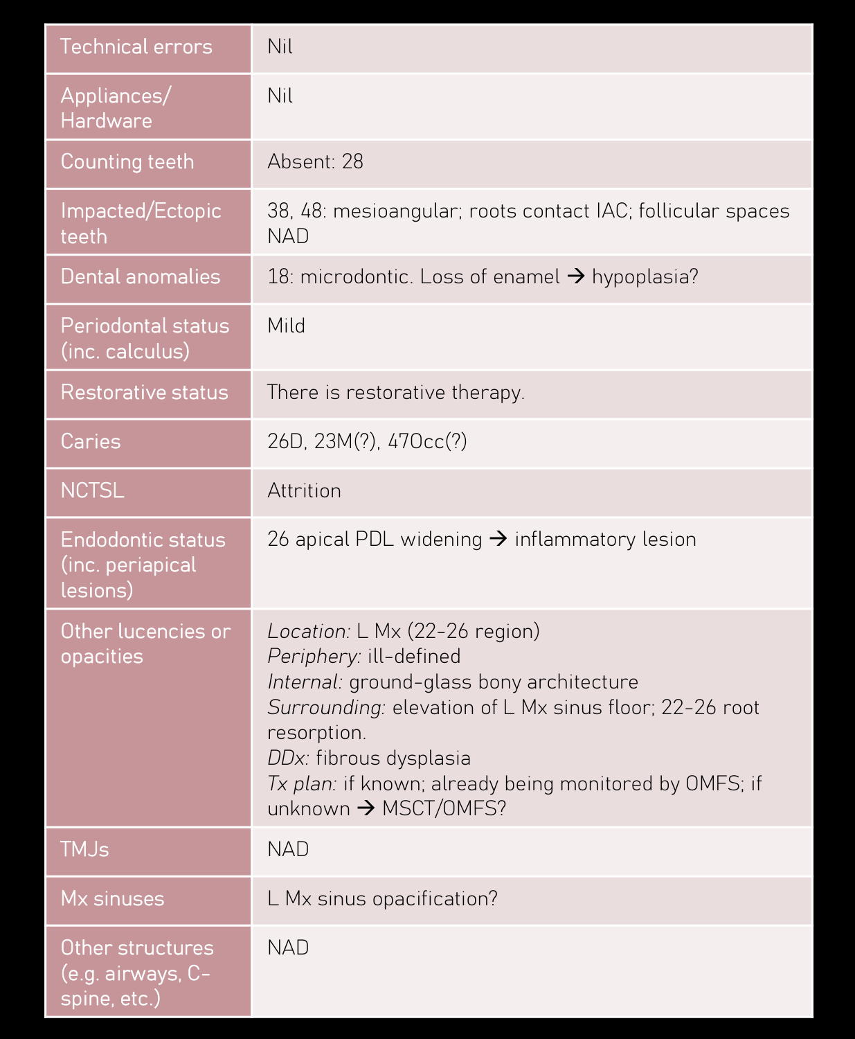 | 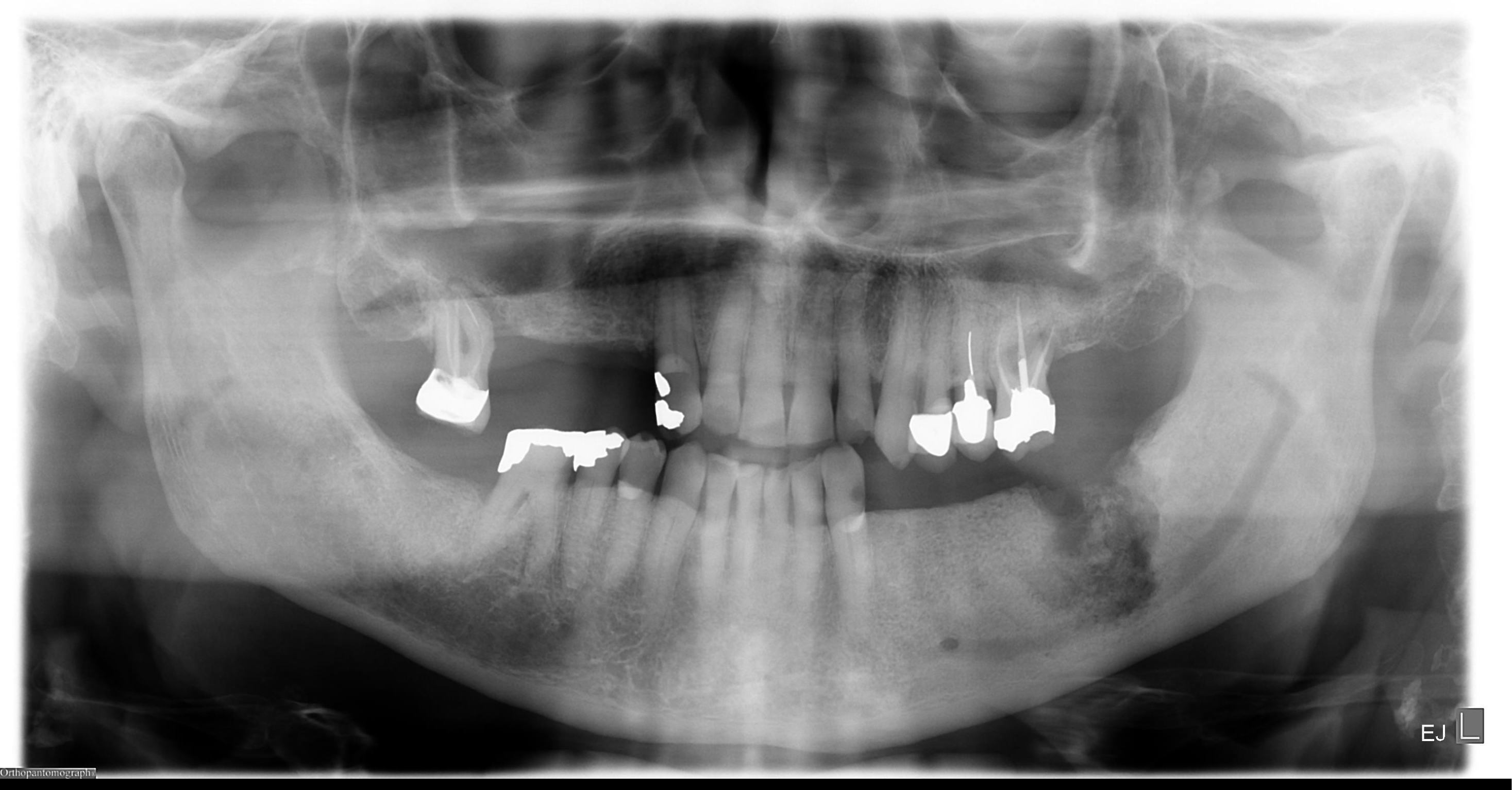 |
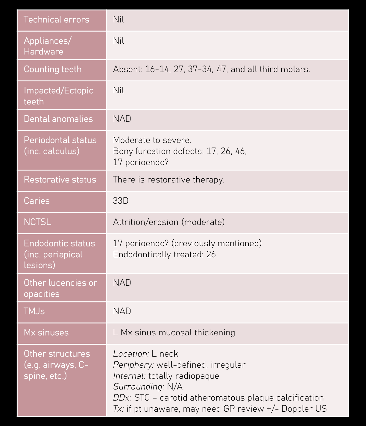
Case Study 16: Perio-Endo Defects35363738
| Technical errors | Nil |
| Appliances/Hardware | Nil |
| Counting teeth | Absent: 16-14, 27, 37-34, 47, and all third molars. |
| Impacted/Ectopic teeth | Nil |
| Dental anomalies | NAD |
| Periodontal status (inc. calculus) | Moderate to severe. Bony furcation defects: 17, 26, 46, 17 perioendo? |
| Restorative status | There is restorative therapy. |
| Caries | 33D |
| NCTSL | Attrition/erosion (moderate) |
| Endodontic status (inc. periapical lesions) | 17 perioendo? (previously mentioned) Endodontically treated: 26 |
| Other lucencies or opacities | NAD |
| TMJs | NAD |
| Mx sinuses | L Mx sinus mucosal thickening |
| Other structures (e.g. airways, C-spine, etc.) | Location:L neck Periphery:well-defined, irregular Internal:totally radiopaque Surrounding:N/A DDx:STC – carotid atheromatous plaque calcification Tx:if pt unaware, may need GP review +/- Doppler US |
FINDINGS:
There is ill-defined bone loss in the 37 region, extending inferiorly and involving the left mandibular canal. Extensive adjacent sclerosis in the mandibular body, with loss of definition of the inferior cortical-medullary border. Appearances likely reflect infected osteonecrosis.
OTHER FINDINGS:
16-14, 27, 37-34, 47, and all third molars are absent.
There are a few sites of moderate to severe periodontal bone loss, with likely 17, 26 and 46 bony furcation defects. Bone loss extends to the 17 mesiobuccal root apex, in continuity with the widened apical periodontal ligament space, where early development of a perioendo lesion can be fully excluded.
There is restorative therapy.
33 disto-cervical lucency likely reflects caries.
There is attrition/erosion.
26 is endodontically treated.
Condylar appearances likely relate to the oblique view. Arthropathy is less likely.
There is likely mucosal thickening within the left maxillary sinus.
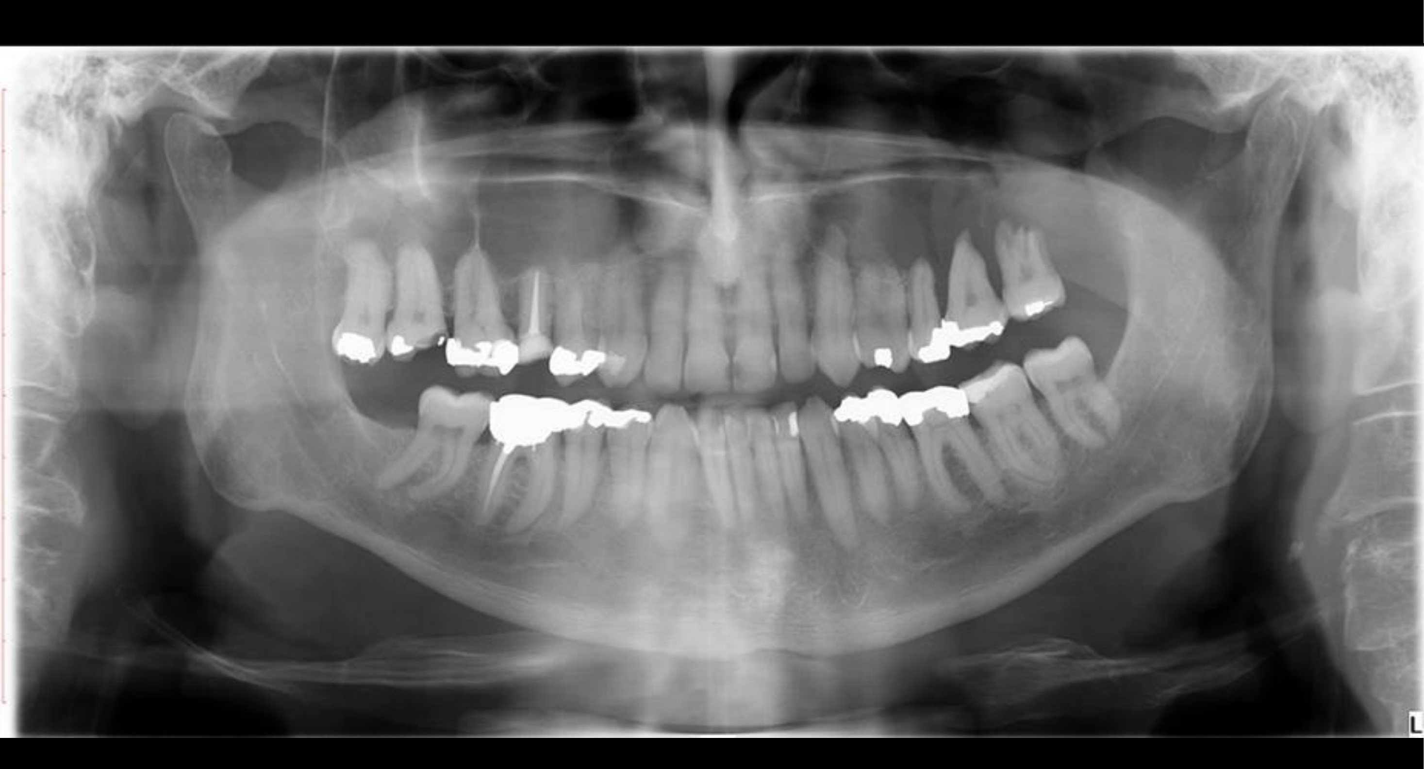
Curvilinear opacity within the left neck likely reflects carotid atheromatous plaque calcification. If clinically indicated, Doppler ultrasound could be considered for further evaluation.
There is a perio-endo defect associated with 37. Appearances likely reflect incomplete endodontic therapy. Shortened distal root appearances likely relate to previous resorption.
There is moderate to severe periodontal bone loss elsewhere with likely 17 and 27 distal bony furcation defects. Dental calculus deposits noted.
There is restorative therapy.
No definite caries seen although it is difficult to exclude with this technique.
There is attrition.
Hypodense appearances at the 16 apical aspect likely reflect inflammatory lesions.
Premaxillary midline appearances likely reflect the incisive canal/foramen.
Appearances of the condyles likely relate to the oblique view, although arthropathy cannot be fully excluded from OPGs.
There is mild mucosal thickening at the floor of the maxillary sinuses.
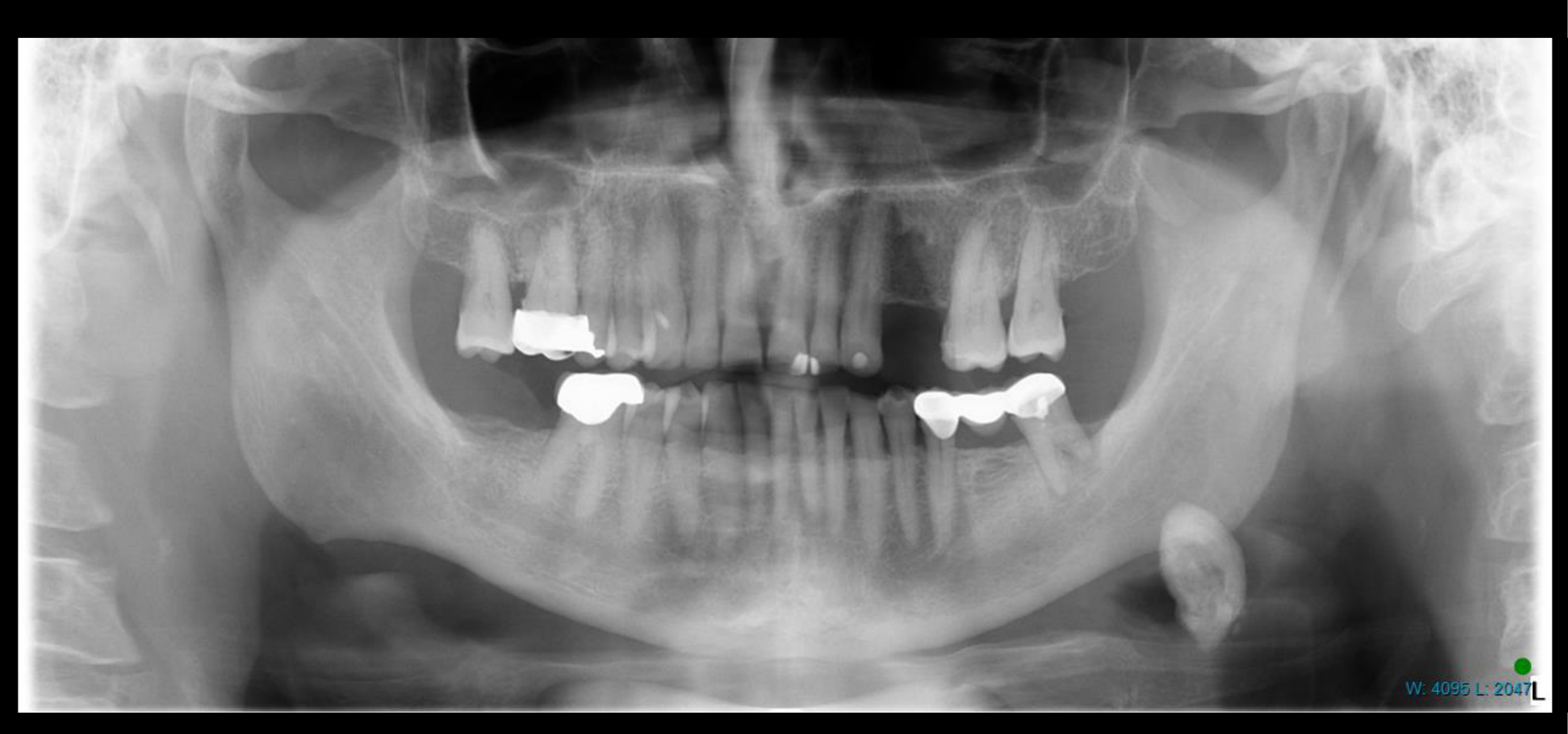
Opacity projected over the inferior aspect of the left posterior body of mandible, with an estimated maximal dimension of approximately 22mm, likely reflects a sialolith.
COMMENT:
- 37 perio-endo lesion and the other dentoalveolar findings are described.
- Premaxillary midline appearances likely relate to magnification of the incisive canal/foramen related to the limitations of tomography. If there is clinical concern or suspicion for a nasopalatine duct cyst, multislice CT could be considered in further evaluation.
- Opacity projected over the posterior aspect of the left mandible described likely reflects a sialolith within the proximal duct of the left submandibular salivary gland. Clinical correlation is suggested. If indicated, Ultrasound or Multislice CT could be considered for further evaluation.
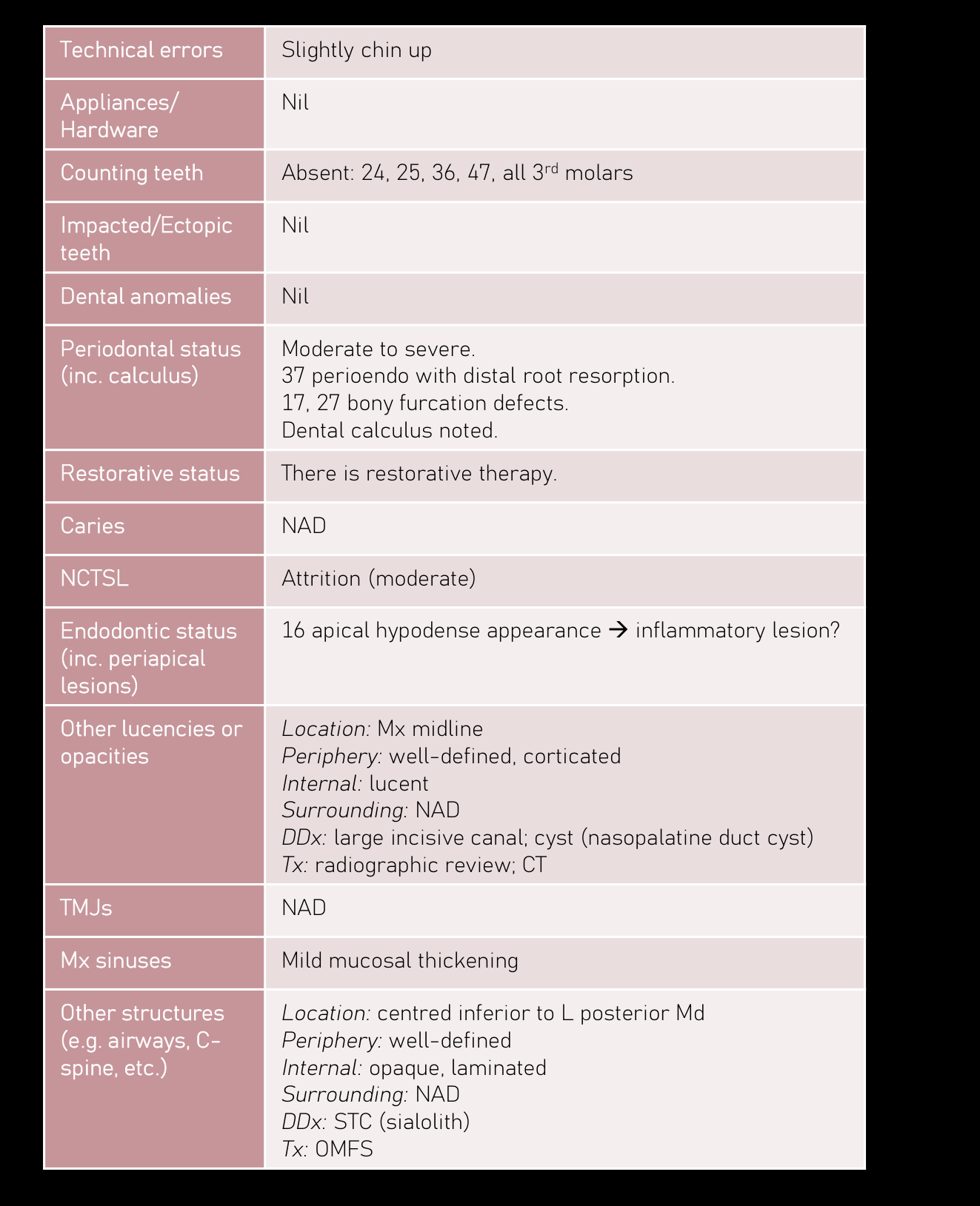
| Technical errors | Slightly chin up |
| Appliances/Hardware | Nil |
| Counting teeth | Absent: 24, 25, 36, 47, all 3rd molars |
| Impacted/Ectopic teeth | Nil |
| Dental anomalies | Nil |
| Periodontal status (inc. calculus) | Moderate to severe. 37 perioendo with distal root resorption. 17, 27 bony furcation defects. Dental calculus noted. |
| Restorative status | There is restorative therapy. |
| Caries | NAD |
| NCTSL | Attrition (moderate) |
| Endodontic status (inc. periapical lesions) | 16 apical hypodense appearance inflammatory lesion? |
| Other lucencies or opacities | Location: Mx midline Periphery: well-defined, corticated Internal: lucent Surrounding: NAD DDx: large incisive canal; cyst (nasopalatine duct cyst) Tx: radiographic review; CT |
| TMJs | NAD |
| Mx sinuses | Mild mucosal thickening |
| Other structures (e.g. airways, C-spine, etc.) | Location: centred inferior to L posterior Md Periphery: well-defined Internal: opaque, laminated Surrounding: NAD DDx: STC (sialolith) Tx: OMFS |
 |  |
 |  |
Case Study 17: Furcation Defects and Caries3940
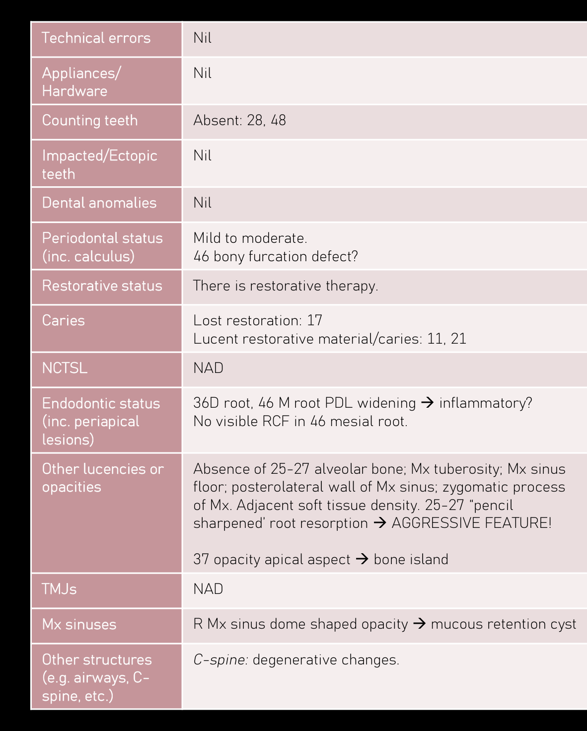
| Technical errors | Nil |
|---|---|
| Appliances/Hardware | Nil |
| Counting teeth | Absent: 28, 48 |
| Impacted/Ectopic teeth | Nil |
| Dental anomalies | Nil |
| Periodontal status (inc. calculus) | Mild to moderate. 46 bony furcation defect? |
| Restorative status | There is restorative therapy. |
| Caries | Lost restoration: 17 Lucent restorative material/caries: 11, 21 |
| NCTSL | NAD |
| Endodontic status (inc. periapical lesions) | 36D root, 46 M root PDL widening inflammatory? No visible RCF in 46 mesial root. |
| Other lucencies or opacities | Absence of 25-27 alveolar bone; Mx tuberosity; Mx sinus floor; posterolateral wall of Mx sinus; zygomatic process of Mx. Adjacent soft tissue density. 25-27 “pencil sharpened’ root resorption AGGRESSIVE FEATURE! 37 opacity apical aspect bone island |
| TMJs | NAD |
| Mx sinuses | R Mx sinus dome shaped opacity mucous retention cyst |
| Other structures (e.g., airways, C-spine, etc.) | C-spine: degenerative changes. |
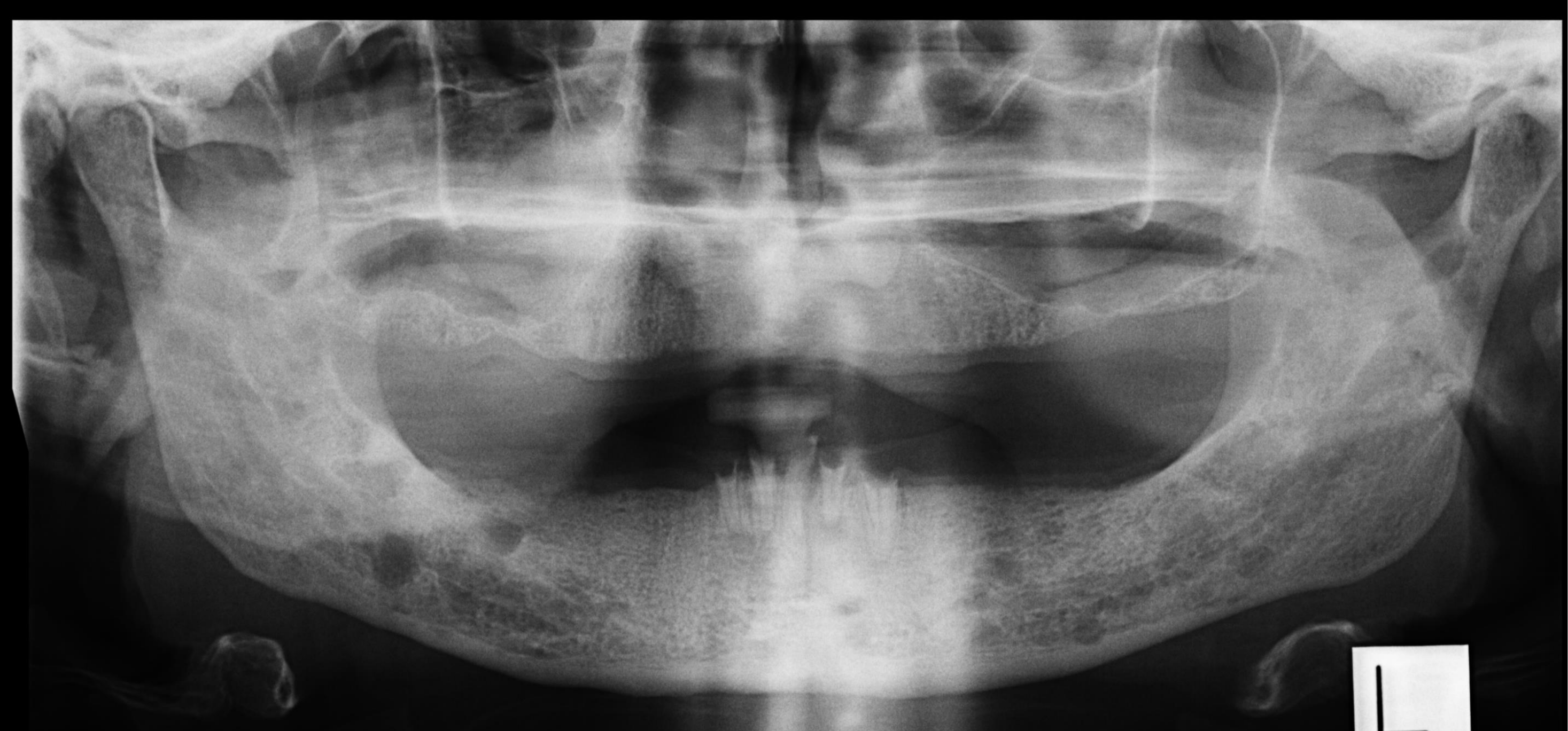
28 and 48 are absent.
There a few sites of mild to moderate periodontal bone loss, although 46 appearances likely reflect bony furcation defects.
There is restorative therapy.
17 coronal lucency likely reflects loss restoration.
11/21 mesial lucencies likely reflect lucent restorative material/caries.
36 distal root and 46 mesial root widened apical periodontal ligament spaces raise the possibility of inflammatory lesions. Endodontic material within the 46 mesial root is not visualised.
Opacity at the apical aspect of 37 likely reflects a bone island.
There is absence of the alveolar bone and left maxillary sinus floor in the 25-27 region, as well as absence of the maxillary tuberosity, posterior lateral wall of the maxillary sinus, and process of maxilla. There is potential sharpening root resorption associated with 25-27. Soft tissue density within the left maxillary sinus is noted.
Large dome shaped soft tissue density within the right maxillary sinus likely reflects a mucus retention cyst.
Condylar appearances likely relate to the oblique view. Arthropathy is less likely.
Degenerative changes of the visualised cervical spine are noted.
COMMENT:
There is absence of the left alveolar bone and maxillary sinus floor in the 25-27 region, as well as absence of the tuberosity, posterolateral wall of the maxillary sinus and zygomatic process of maxilla, with adjacent soft tissue density. “Pencil-sharpened” root resorption pattern noted. These features are highly suspicious for an aggressive lesion.
Prompt surgical opinion and MSCT is recommended.
 |  |
Case Study 18: Final Clinical Assessment41
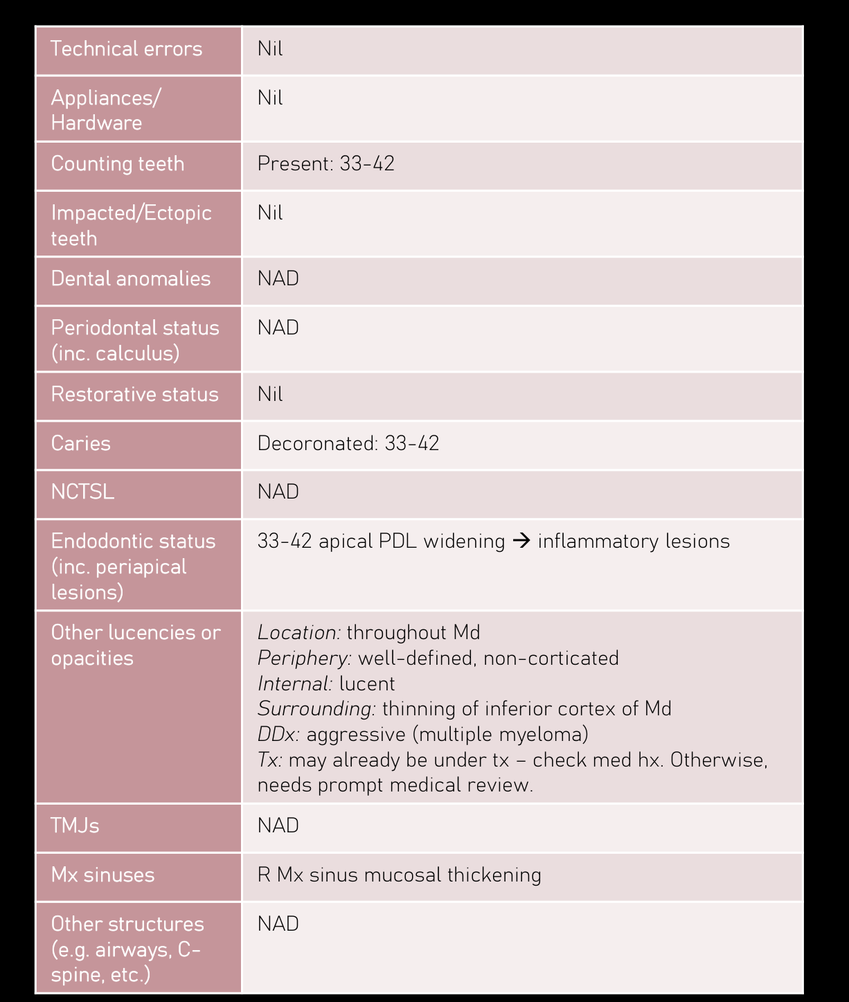
<table>
    <tr>
        <th>Technical errors</th>
        <td>Nil</td>
    </tr>
    <tr>
        <th>Appliances/<br>Hardware</th>
        <td>Nil</td>
    </tr>
    <tr>
        <th>Counting teeth</th>
        <td>Present: 33-42</td>
    </tr>
    <tr>
        <th>Impacted/Ectopic<br>teeth</th>
        <td>Nil</td>
    </tr>
    <tr>
        <th>Dental anomalies</th>
        <td>NAD</td>
    </tr>
    <tr>
        <th>Periodontal status<br>(inc. calculus)</th>
        <td>NAD</td>
    </tr>
    <tr>
        <th>Restorative status</th>
        <td>Nil</td>
    </tr>
    <tr>
        <th>Caries</th>
        <td>Decoronated: 33-42</td>
    </tr>
    <tr>
        <th>NCTSL</th>
        <td>NAD</td>
    </tr>
    <tr>
        <th>Endodontic status<br>(inc. periapical<br>lesions)</th>
        <td>33-42 apical PDL widening $\rightarrow$ inflammatory lesions</td>
    </tr>
    <tr>
        <th>Other lucencies or<br>opacities</th>
        <td><i>Location</i>: throughout Md<br>
            <i>Periphery</i>: well-defined, non-corticated<br>
            <i>Internal</i>: lucent<br>
            <i>Surrounding</i>: thinning of inferior cortex of Md<br>
            <b>DDx</b>: aggressive (multiple myeloma)<br>
            <b>Tx</b>: may already be under tx – check med hx. Otherwise, needs prompt medical review.
        </td>
    </tr>
    <tr>
        <th>TMJs</th>
        <td>NAD</td>
    </tr>
    <tr>
        <th>Mx sinuses</th>
        <td>R Mx sinus mucosal thickening</td>
    </tr>
    <tr>
        <th>Other structures<br>(e.g. airways, C-<br>spine, etc.)</th>
        <td>NAD</td>
    </tr>
</table>

<h2>FINDINGS:</h2>
<p>There are multifocal well-defined non-corticated lucencies within the mandible. There is thinning of the inferior cortex of mandible where these lesions contact this cortical border. These lesions demonstrate aggressive features and multiple myeloma is suspected. Correlation with the medical history and if indicated, medical review is suggested.</p>

<h2>OTHER FINDINGS:</h2>
<p>Decoronated 33-42 are present. Apical periodontal ligament space widening likely reflects inflammatory lesions.</p>

<p>Allowing for the oblique view, condylar appearances are within normal limits.</p>

<p>There is likely mucosal thickening within the right maxillary sinus.</p>
Conclusion42
Thank you
Footnotes
-
Original PDF page 1: L15 OPGT, p.1 ↩
-
Original PDF page 2: L15 OPGT, p.2 ↩
-
Original PDF page 3: L15 OPGT, p.3 ↩
-
Original PDF page 4: L15 OPGT, p.4 ↩
-
Original PDF page 5: L15 OPGT, p.5 ↩
-
Original PDF page 7: L15 OPGT, p.7 ↩
-
Original PDF page 6: L15 OPGT, p.6 ↩
-
Original PDF page 8: L15 OPGT, p.8 ↩
-
Original PDF page 10: L15 OPGT, p.10 ↩
-
Original PDF page 11: L15 OPGT, p.11 ↩
-
Original PDF page 12: L15 OPGT, p.12 ↩
-
Original PDF page 13: L15 OPGT, p.13 ↩
-
Original PDF page 14: L15 OPGT, p.14 ↩
-
Original PDF page 15: L15 OPGT, p.15 ↩
-
Original PDF page 16: L15 OPGT, p.16 ↩
-
Original PDF page 17: L15 OPGT, p.17 ↩
-
Original PDF page 18: L15 OPGT, p.18 ↩
-
Original PDF page 19: L15 OPGT, p.19 ↩
-
Original PDF page 20: L15 OPGT, p.20 ↩
-
Original PDF page 22: L15 OPGT, p.22 ↩
-
Original PDF page 23: L15 OPGT, p.23 ↩
-
Original PDF page 24: L15 OPGT, p.24 ↩
-
Original PDF page 25: L15 OPGT, p.25 ↩
-
Original PDF page 26: L15 OPGT, p.26 ↩
-
Original PDF page 27: L15 OPGT, p.27 ↩
-
Original PDF page 28: L15 OPGT, p.28 ↩
-
Original PDF page 29: L15 OPGT, p.29 ↩
-
Original PDF page 30: L15 OPGT, p.30 ↩
-
Original PDF page 31: L15 OPGT, p.31 ↩
-
Original PDF page 32: L15 OPGT, p.32 ↩
-
Original PDF page 33: L15 OPGT, p.33 ↩
-
Original PDF page 34: L15 OPGT, p.34 ↩
-
Original PDF page 35: L15 OPGT, p.35 ↩
-
Original PDF page 36: L15 OPGT, p.36 ↩
-
Original PDF page 37: L15 OPGT, p.37 ↩
-
Original PDF page 38: L15 OPGT, p.38 ↩
-
Original PDF page 39: L15 OPGT, p.39 ↩
-
Original PDF page 40: L15 OPGT, p.40 ↩
-
Original PDF page 41: L15 OPGT, p.41 ↩
-
Original PDF page 42: L15 OPGT, p.42 ↩
-
Original PDF page 43: L15 OPGT, p.43 ↩
-
Original PDF page 44: L15 OPGT, p.44 ↩