Panoramic Radiographic Anatomy Interactive Lecture1
Dr May Lam
Lecture Instructions and Objectives
Instructions for this Interactive Lecture2
-
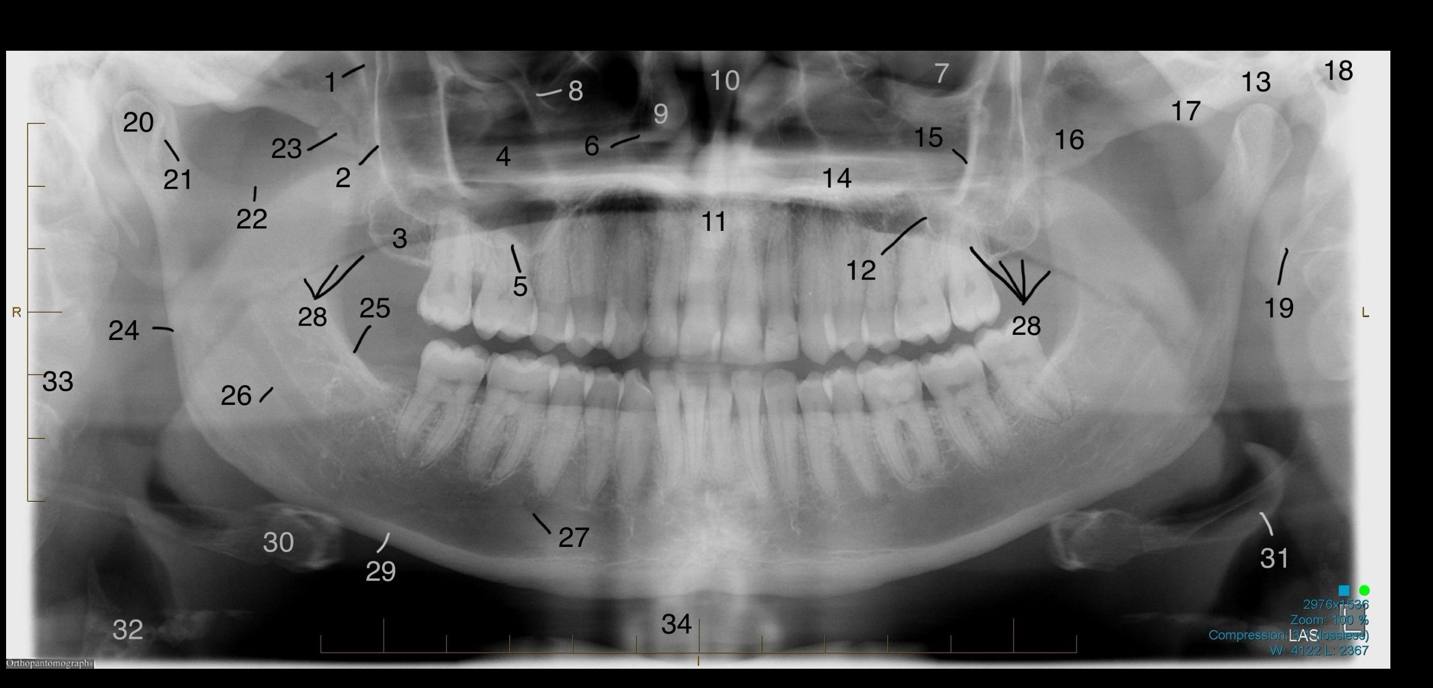
Label the anatomical structures outlined in the following panoramic radiographs.
-
Answers will be provided on LMS with Lecture 9.
Systematic Interpretation Approach
To master OPG interpretation, follow a consistent checklist to avoid missing critical findings. Use a five-step method for analyzing lesions: identify location, periphery, internal structure, and effects on surrounding structures, followed by a “surgical sieve” categorization. Always begin with a technical assessment (positioning, artifacts) as these influence the interpretation.
Case Studies in Radiographic Identification
Case One Anatomical Landmarks3
[!example] Case 1: Technical Errors and Dental Findings
- Technical Errors: Earrings left in situ; artifact from a lead apron visible as a large white section.
- Dental Findings: Absent 18/28; 38/48 vertically impacted with roots likely contacting mandibular canals; occlusal filling on 27.
- Caries: Potential lucency at the cervical region of 27 (requires clinical confirmation).
- Dentoalveolar: Hypodense bone in 18/28 regions representing post-extraction healing and large marrow spaces.
- Extra-dental: Normal TMJs, clear maxillary sinuses, and normal cervical spine/hyoid/orbital margins.
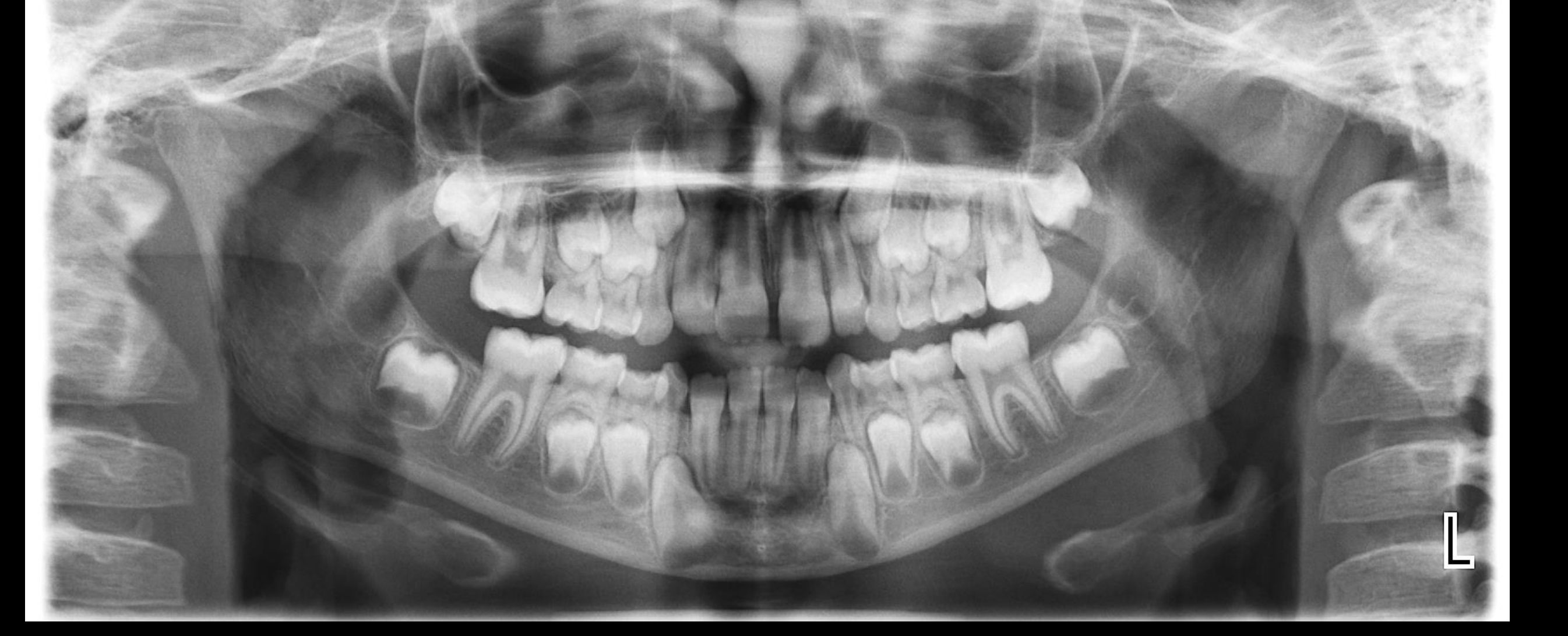
Case Two FDI Tooth Identification
Case 2 - Identify the Teeth using FDI numbers4
[!example] Case 2: Denture Artifacts and Pathology
- Technical Errors: Presence of an acrylic denture with four opaque ball clasps and wire mesh reinforcement.
- Dental Findings: Absent 18, 11, 21, 28; 38 mesio-angularly impacted (root touches canal); 48 vertically impacted.
- Pathology: Chronic pericoronitis (widened distal follicular space and reactive sclerosis behind 38/48).
- Endodontic/Periodontal: Apical periodontitis on 37 (widened PDL and sclerosis); mild to moderate bone loss; attrition on incisal edges.
- Caries: Lucencies on 37 (distal) and 38 (mesial).
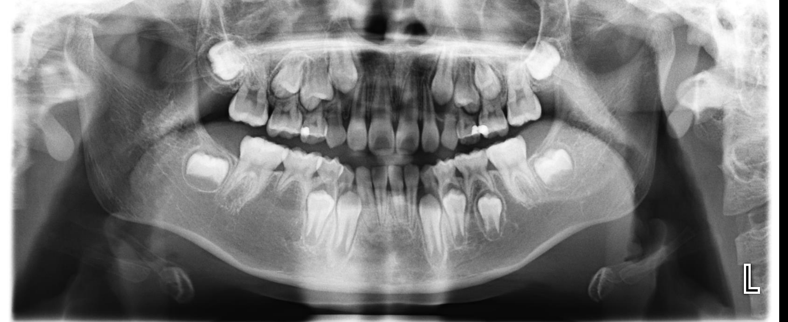
Case Three Anatomical Landmarks5
[!example] Case 3: Positioning Errors and Root Stumps
- Technical Errors: “Chin up” positioning resulting in a flat occlusal plane and hard palate superimposing over maxillary apices.
- Dental Findings: Absent 18, 15, 25, 28, 47; 38 disto-angularly impacted; 48 vertically impacted (both show root darkening/canal deviation).
- Normal Variants: Bifurcation of the mandibular canal (retromolar canals) visible.
- Pathology: Root stumps at 16, 14, 27, 36, 46 with associated periapical inflammatory lucencies; caries on 17 and 37.
- Extra-dental: Left sinus mucosal thickening; right condyle shows unusual morphology due to oblique angle.
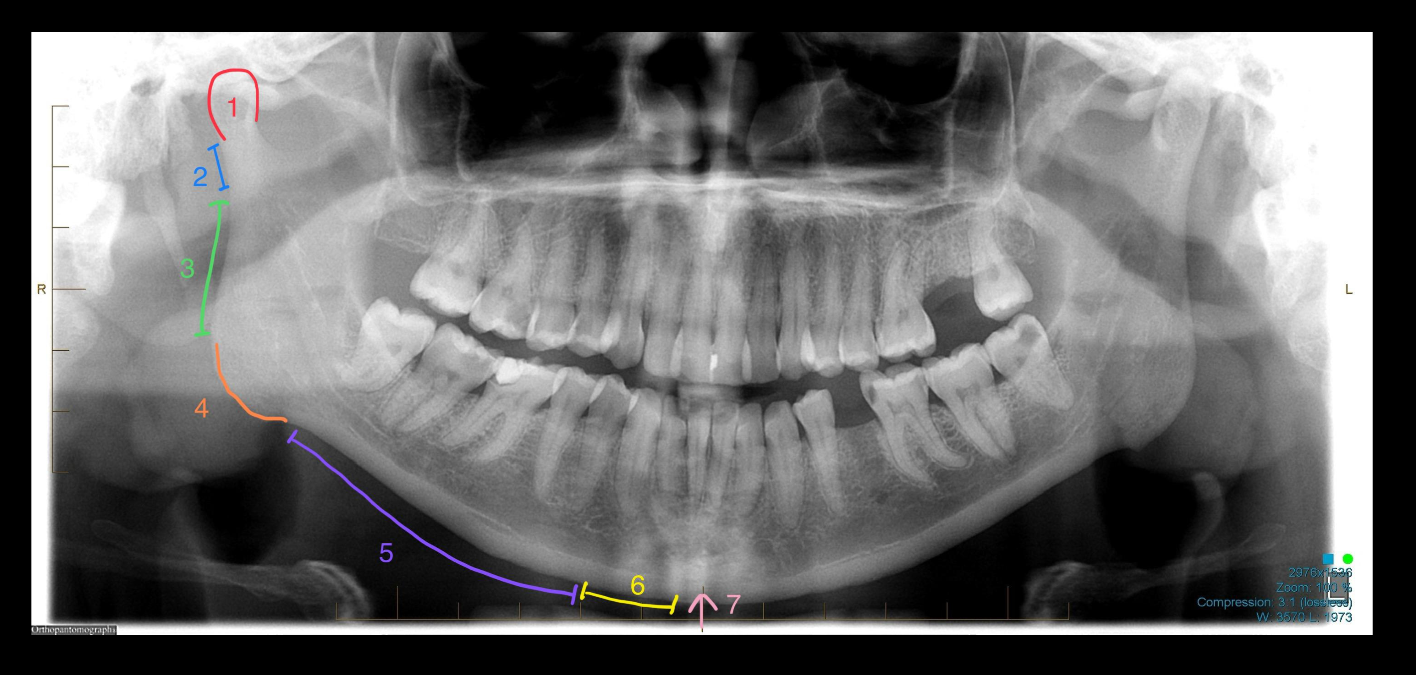
Case Four Anatomical Landmarks6
[!example] Case 4: Compound Odontoma
- Dental Findings: Absent 25; 23 ectopically impacted (crown over 21/22 apices); 38/48 horizontally impacted contacting the canal.
- Pathology: Chronic pericoronitis at 38; two bone islands (opacities) identified in the jaw.
- Compound Odontoma: Well-defined lesion at 23 site with a lucent rim, thin white margin, and multiple “odontoid density” denticles impeding eruption.
Case Five Midface Structures
Midface Structures Part A
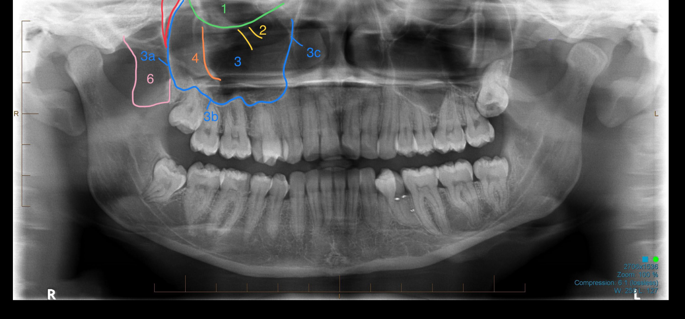
Case 5a – Midface Structures7
[!example] Case 5a: Mandibular Cyst and Erosion
- Technical Errors: “Chin up” positioning (flat occlusal plane).
- Dental Findings: Absent 18, 17, 16, 26, 45; 38 vertically impacted; 48 horizontally impacted with hypercementosis (bulbous root).
- Non-Caries Loss: Severe erosion (concave lucent appearances) and attrition.
- Mandibular Cyst (Dentigerous or OKC): Well-defined, corticated, totally lucent lesion centered below 48 crown, displacing the mandibular canal inferiorly.
Midface Structures Part B
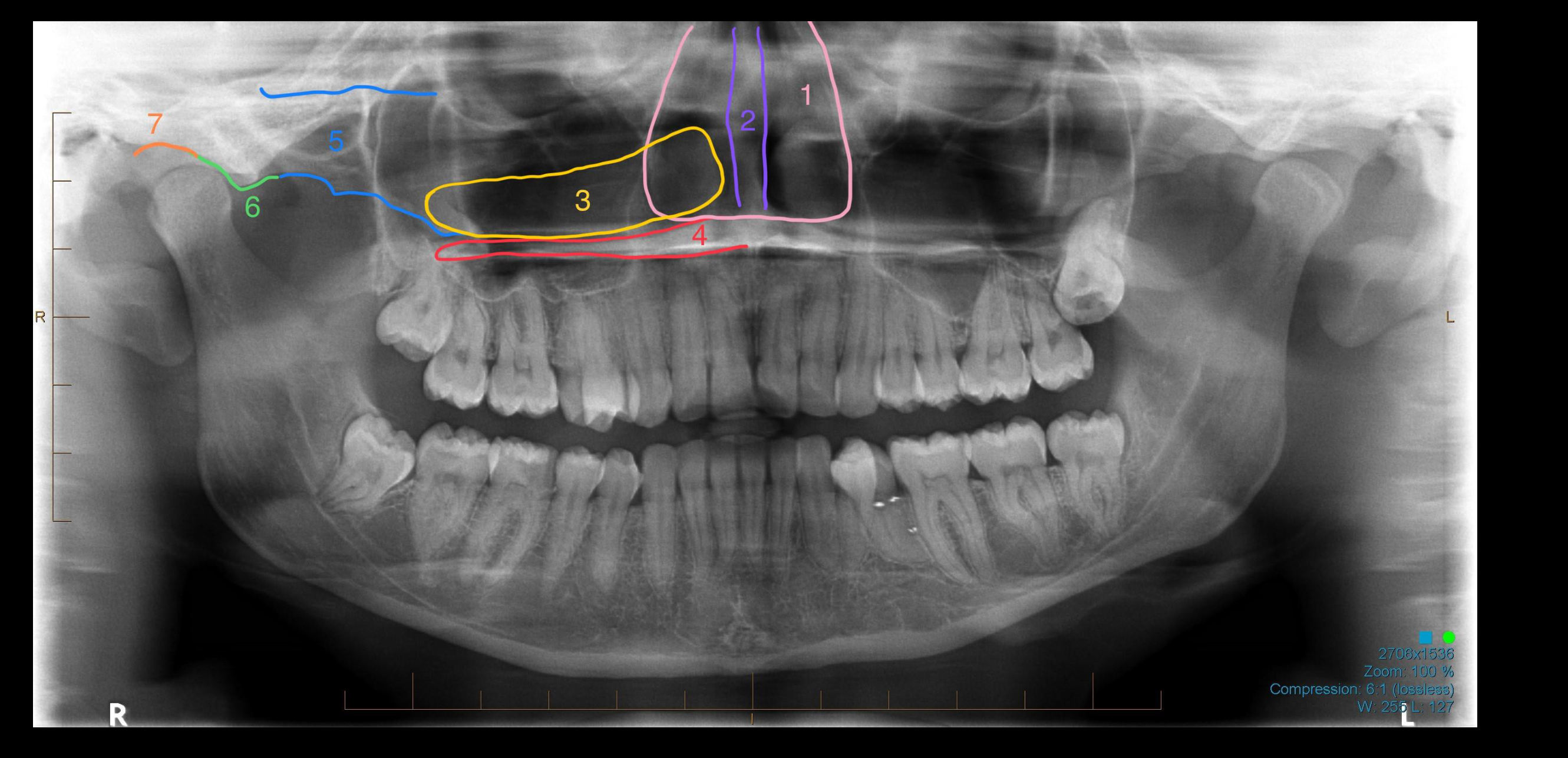
Case 5b – Midface Structures8
- Other Structures: Elongated styloid processes and stylohyoid calcification with pseudoarthrosis (normal variant).
Case Six Anatomical Landmarks9
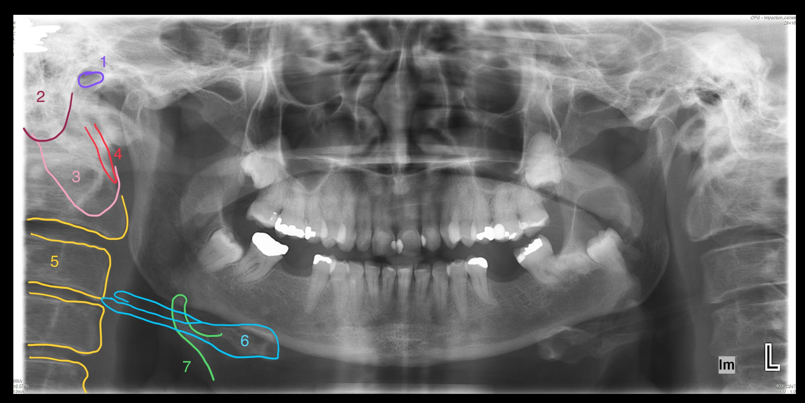
[!example] Case 6: Displaced Oblique Fracture
- Dental Findings: 18/28 unerupted near sinus floor; 38/48 vertically impacted contacting canal with chronic pericoronitis.
- Displaced Oblique Fracture: Located at left mandibular angle in continuity with 38 follicular space; signs include linear lucency and a “jump” in the canal cortex.
- Clinical Note: When one fracture is found, always look for a second (e.g., contralateral condyle).
Case Seven Anatomical Landmarks10
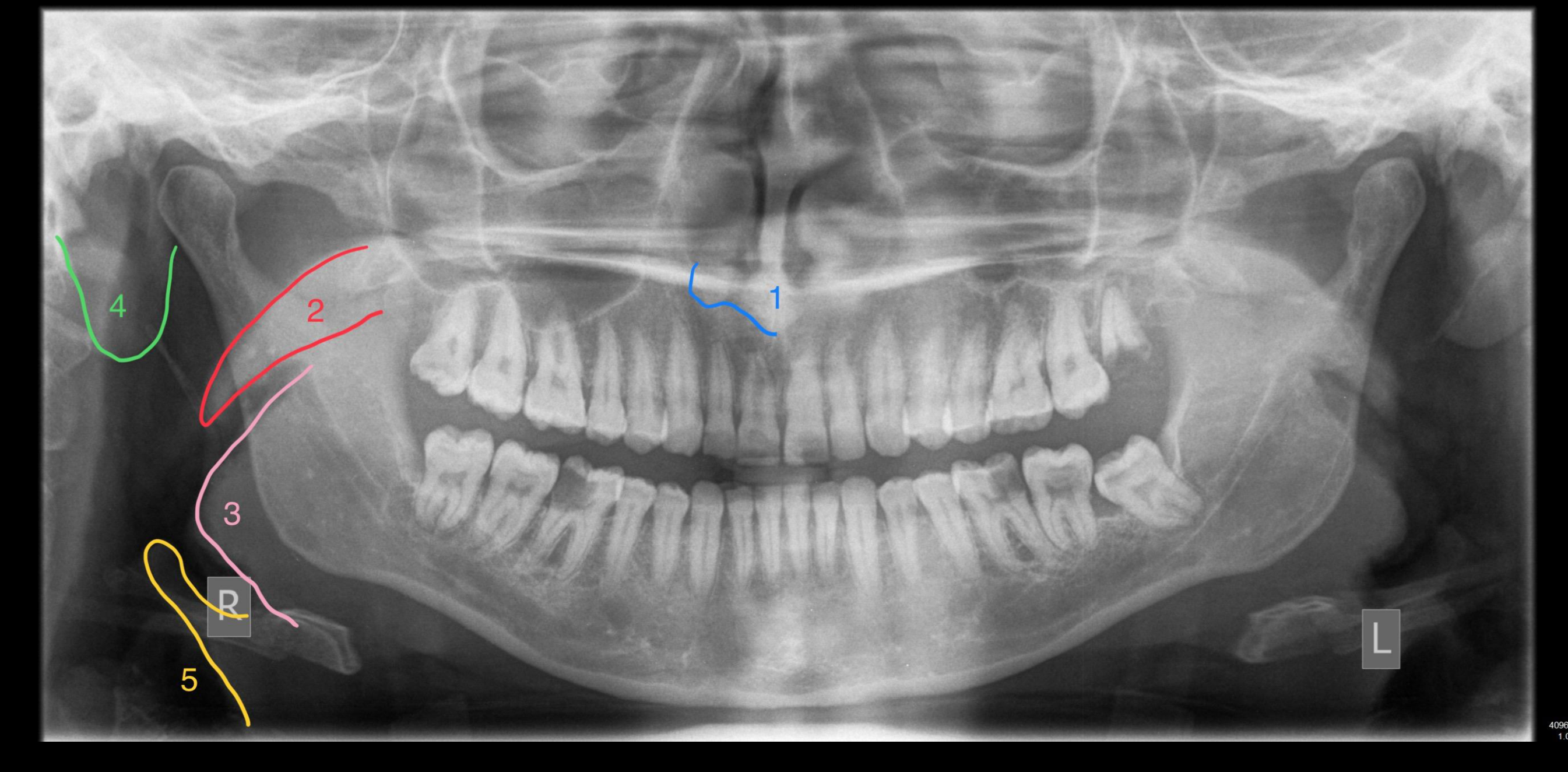
[!example] Case 7: Incisive Canal Cyst and Sialolith
- Dental Findings: Moderate bone loss; severe bone loss at 37 distal with a perio-endo configuration; crown and bridge work.
- Incisive Canal Cyst: Well-defined, circular, corticated lucency at maxillary midline superimposed over 11/21 apices.
- Sialolith (Salivary Stone): Well-defined opacity with a laminated appearance (tree rings) in the left submandibular gland region.
Case Eight Anatomical Landmarks11
[!example] Case 8: Malignant Neoplasia and Mucous Retention Cyst
- Dental Findings: Absent 28, 38, 48; bone island at 37 mesial; periapical lesion at 36.
- Malignant Neoplasia: Left maxilla showing missing alveolar bone, tuberosity, sinus floor, and posterior lateral wall.
- Root Findings: “Pencil sharpening” root resorption and lobulated soft tissue density (sinister features requiring surgical referral).
- Extra-dental: Right sinus contains a large, dome-shaped mucous retention cyst; degenerative changes in cervical spine (detached osteophyte).
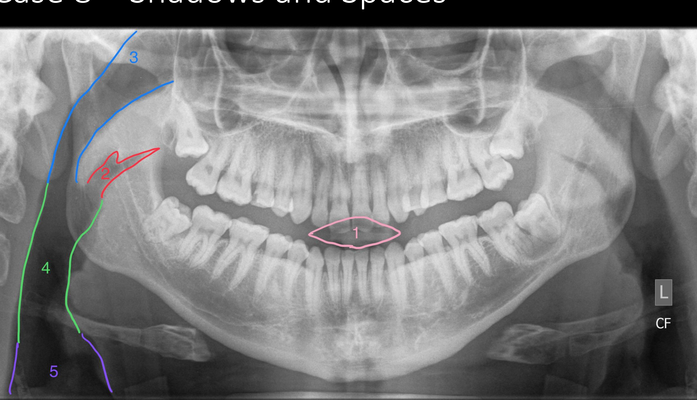
Case Nine Anatomical Landmarks12
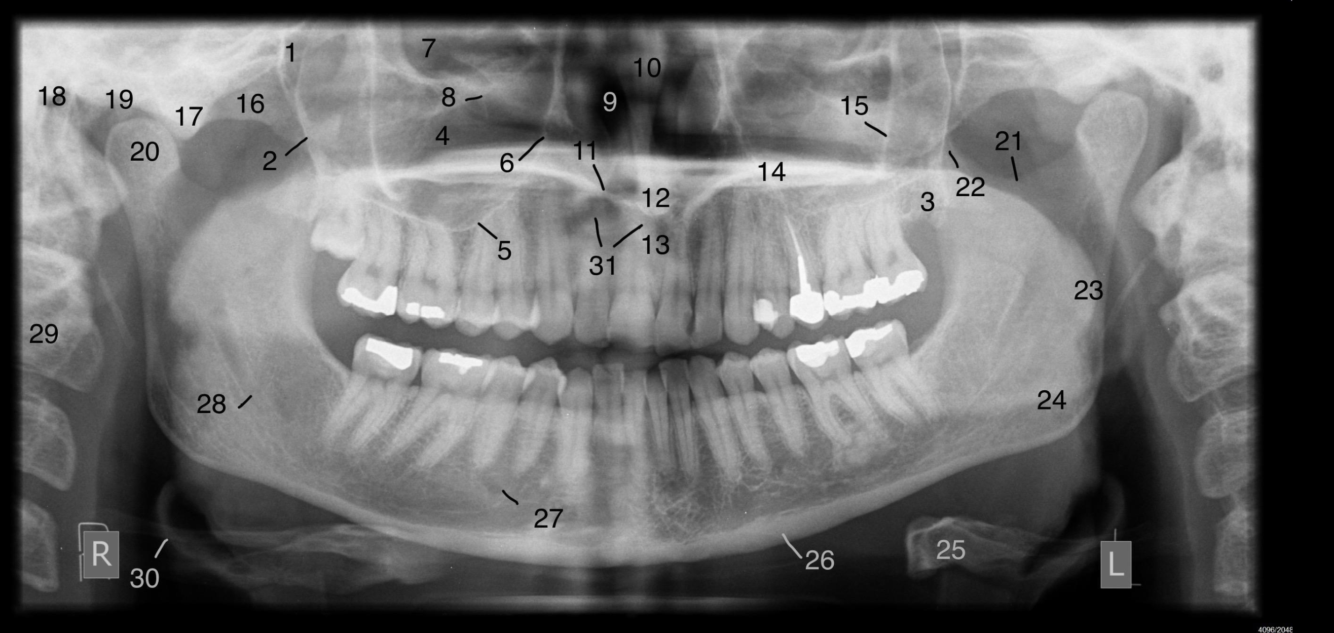
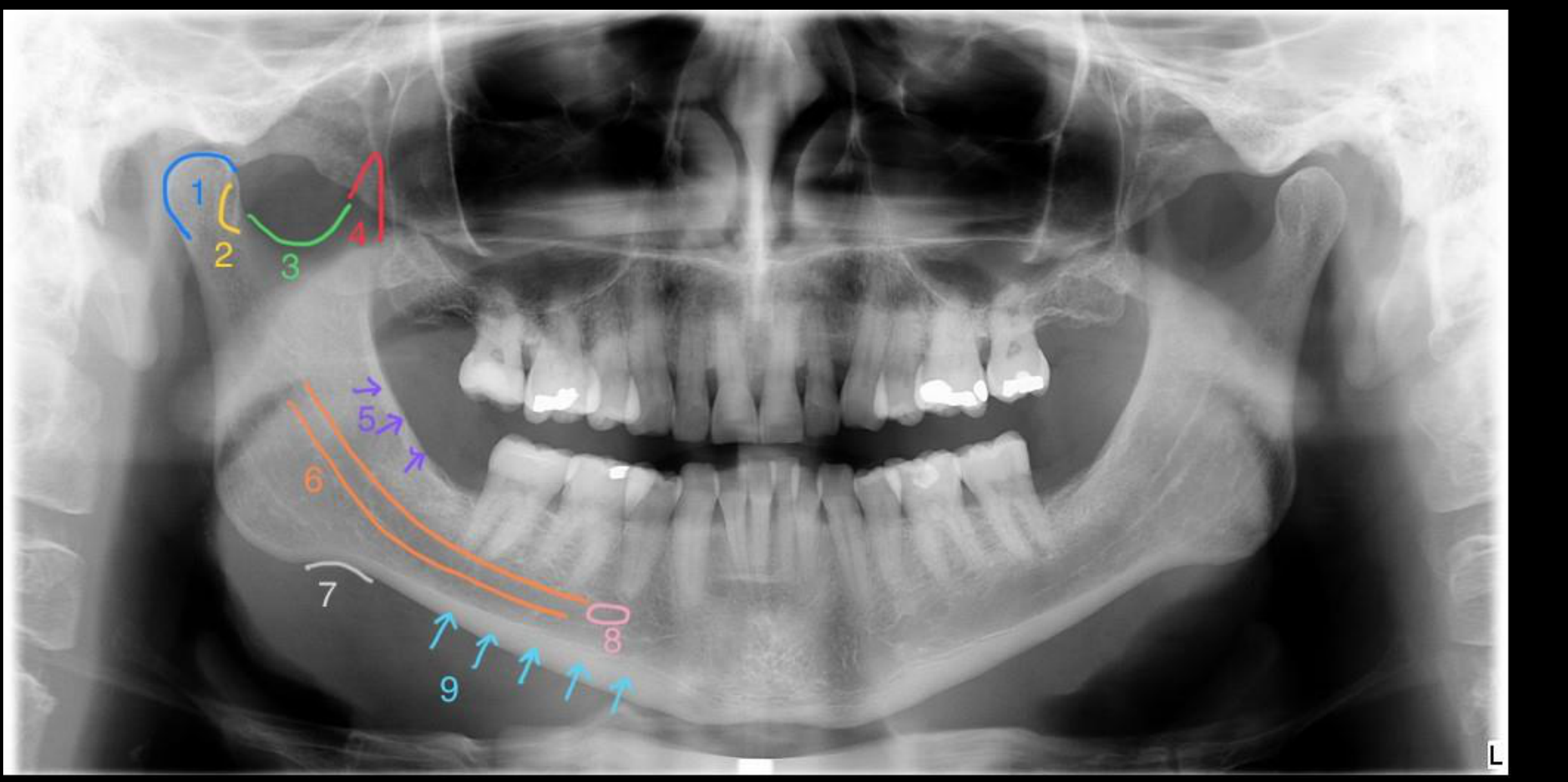
Comprehensive Radiographic Landmark Review13
Dental panoramic X-ray with radiographic landmarks labeled with numbers
Limitations of OPG
While OPGs are excellent for screening, specific issues such as TMJ pathology, fine sinus details, or root resorption often require 3D volumetric imaging (Cone Beam CT or Multi-slice CT) for definitive assessment.
Footnotes
-
Original PDF page 1: L9 (OPG excerices) Panoramic Radiographic Anatomy - Interactive Lecture, p.1 ↩
-
Original PDF page 2: L9 (OPG excerices) Panoramic Radiographic Anatomy - Interactive Lecture, p.2 ↩
-
Original PDF page 3: L9 (OPG excerices) Panoramic Radiographic Anatomy - Interactive Lecture, p.3 ↩
-
Original PDF page 4: L9 (OPG excerices) Panoramic Radiographic Anatomy - Interactive Lecture, p.4 ↩
-
Original PDF page 5: L9 (OPG excerices) Panoramic Radiographic Anatomy - Interactive Lecture, p.5 ↩
-
Original PDF page 6: L9 (OPG excerices) Panoramic Radiographic Anatomy - Interactive Lecture, p.6 ↩
-
Original PDF page 7: L9 (OPG excerices) Panoramic Radiographic Anatomy - Interactive Lecture, p.7 ↩
-
Original PDF page 8: L9 (OPG excerices) Panoramic Radiographic Anatomy - Interactive Lecture, p.8 ↩
-
Original PDF page 9: L9 (OPG excerices) Panoramic Radiographic Anatomy - Interactive Lecture, p.9 ↩
-
Original PDF page 10: L9 (OPG excerices) Panoramic Radiographic Anatomy - Interactive Lecture, p.10 ↩
-
Original PDF page 11: L9 (OPG excerices) Panoramic Radiographic Anatomy - Interactive Lecture, p.11 ↩
-
Original PDF page 12: L9 (OPG excerices) Panoramic Radiographic Anatomy - Interactive Lecture, p.12 ↩
-
Original PDF page 13: L9 (OPG excerices) Panoramic Radiographic Anatomy - Interactive Lecture, p.13 ↩