Clinical Case Discussion Of Asymptomatic Oral Lesion
Patient Presentation and History1
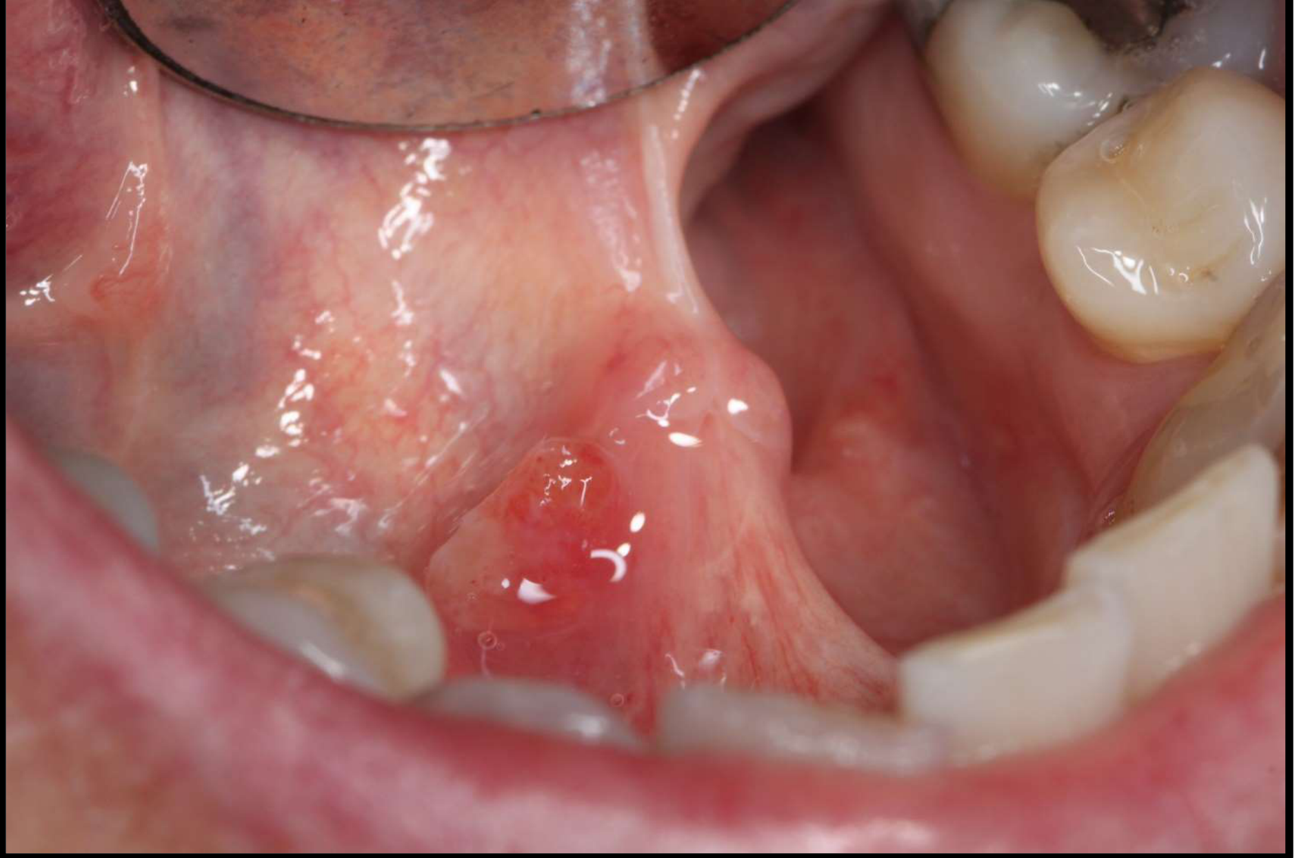
A 48-year-old female presented with an asymptomatic oral lesion. The patient has been aware of the lesion’s presence for approximately two months.
Key Patient Factors:
- Medical History: Non-contributory.
- Social History: Non-smoker; does not consume alcohol.


Clinical Assessment And Diagnostic Activity
Clinical Assessment Activity
1. Identify the pathosis and describe the clinical features.
- Pathosis: The lesion is a mucocele, specifically a mucous retention cyst.
- What can cause obstruction?
- infection, inflammation, scarring (from trauma) , sialolith, neoplasm
- Clinical Features:
- Appearance: Described as a fluid-filled lesion.
- Location: Associated with the submandibular gland duct.
- Symptoms: Asymptomatic; present for approximately two months.
- Palpation: Small mucoceles often feel “nice and soft” and perfectly round. Irregularity or tenderness may indicate a need for removal.
2. What is the differential diagnosis?
- Salivary gland neoplasm
- Malignant Salivary Gland Tumor: Considered because the lesion is in a “high-risk area” in a female non-smoker/non-drinker.
- Mucoepidermoid Carcinoma: Mentioned as a possibility where fluid retention is part of the tumor pathology.
- SCC - could be blocking the duct
- Though the lesion does not look like a typical SCC, it is noted as a theoretical consideration for a lesion in a high-risk area.
- Mucous Extravasation Phenomenon: Distinguished from retention by the lack of an epithelial lining and the presence of an inflammatory response.
3. What are the relevant clinical investigations?
- YOU DON’T NEED AN INVESTIGATION
- this looks like a mucocele and you don’t have to do imaging
- If you mucocele is small you can watch it, sometimes it can even resolve
- Most you should do is a sialogram or a CT sialogram
- Imaging Details:
- Occlusal Radiograph: Used to look for sialoliths (salivary stones). However, its effectiveness is limited by the degree of calcification of the stone.
- Sialogram / CT: A CT scan is advantageous to determine if there is a single stone or multiple stones along the duct.
- Imaging Details:
- Biopsy:
- Incisional Biopsy: Not recommended as it may cause further injury to the duct or simply drain the fluid without providing a definitive specimen.
- Excisional Biopsy: Preferred to confirm the diagnosis and ensure no other pathology is present.
- Note on Palpation: While not a formal “investigation” in a lab sense, palpation for irregularity or tenderness is a key clinical assessment step.
4. What is the diagnosis?
- mucocele in the submanddibular gland duct - the most common cause is a sialolith
- Specific Diagnosis: Mucous Retention Cyst.
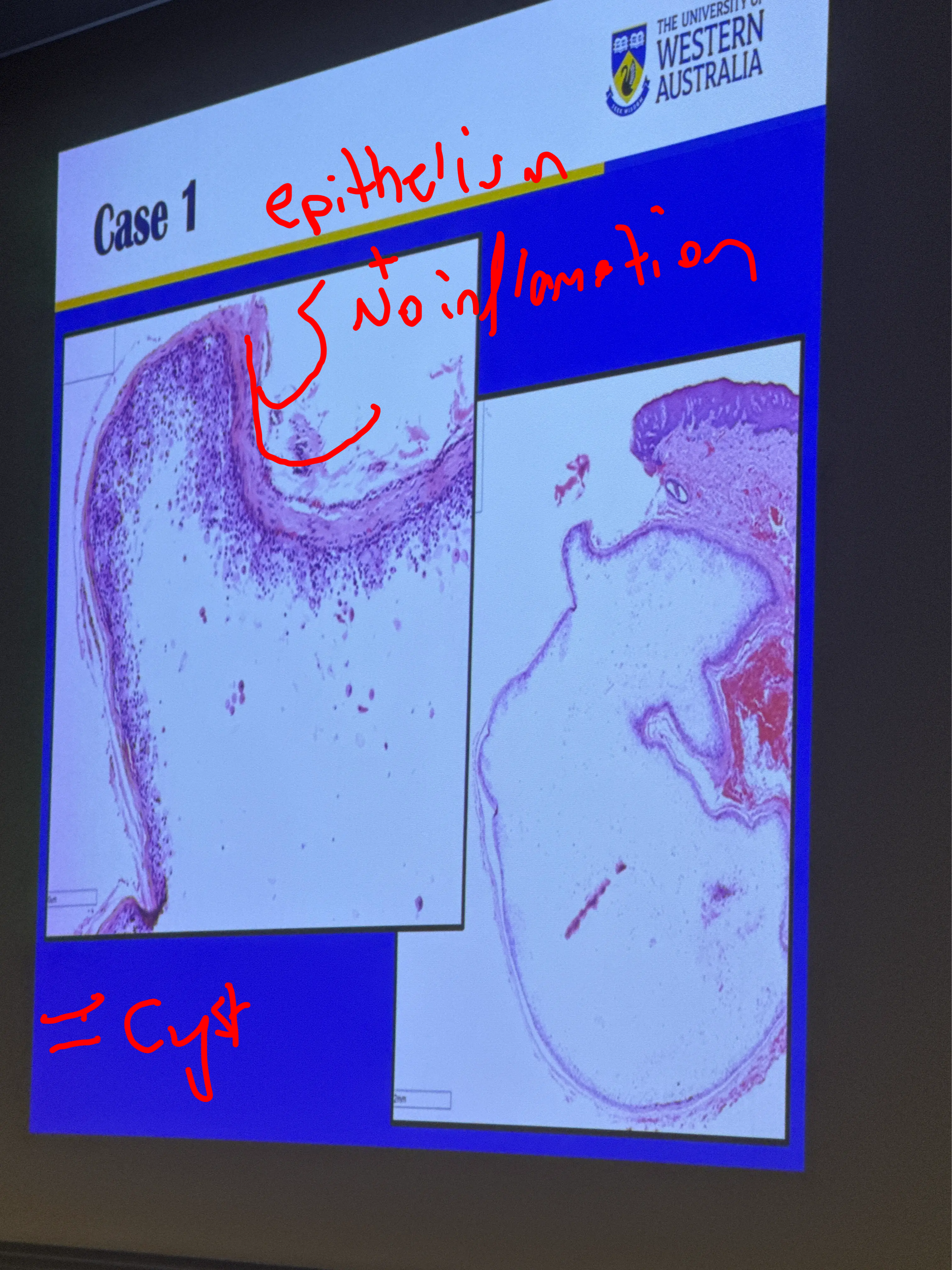
- Histopathology:
- Characterized by an epithelial-lined cavity.
- The mucus is contained within the extended duct; there is no spillage into the surrounding connective tissue.
- Absence of inflammation: Unlike extravasation, there are typically no chronic inflammatory cells or macrophages in the surrounding soft tissue because the saliva (an irritant) is contained.
- Etiology (Causes of Obstruction):
- Mucus plugs or Sialoliths (most common).
- Trauma leading to scarring or strictures.
- Infection (e.g., Sialadenitis).
- Compression by an adjacent neoplasm.
5. How would you manage this patient?
- these lesions should actually be removed to make sure that there isn’t anythign else there
- Observation: If the mucocele is very small, soft, and asymptomatic, it can be monitored as some may resolve spontaneously.
- Surgical Excision:
- Recommended if the lesion has persisted (e.g., over two months) or if it feels irregular/tender.
- Risks: Patients must be warned about the risk of scarring due to ductal injury and a high recurrence rate (approximately 10%).
- Recurrence Factors: High recurrence is often due to surgical scarring or the patient’s saliva having a higher mineral content.
Extra:
- risk of recurrence is 10 percent
- due to scarring from excision
- Comparison of Mucocele Types:
- Extravasation: Caused by severance of the duct; lacks an epithelial lining (pseudocyst); triggers an inflammatory response (macrophages) because saliva enters the connective tissue.
- Retention: Caused by obstruction; possesses an epithelial lining; no inflammatory response in the surrounding tissue.
Footnotes
-
Original PDF page 1: 13. Salivary gland disease case 1, p.1 ↩