Clinical Discussion Case
Patient Profile and History1
- Patient: 58-year-old female
- Medical History: Hypertension
- Social History: Non-smoker; social alcohol consumption
- Dental History: Wears a removable partial denture
- Chief Complaint: Troublesome gingival lesion in the 21 region
- Previous Diagnosis: 2-year history of oral lichen planus
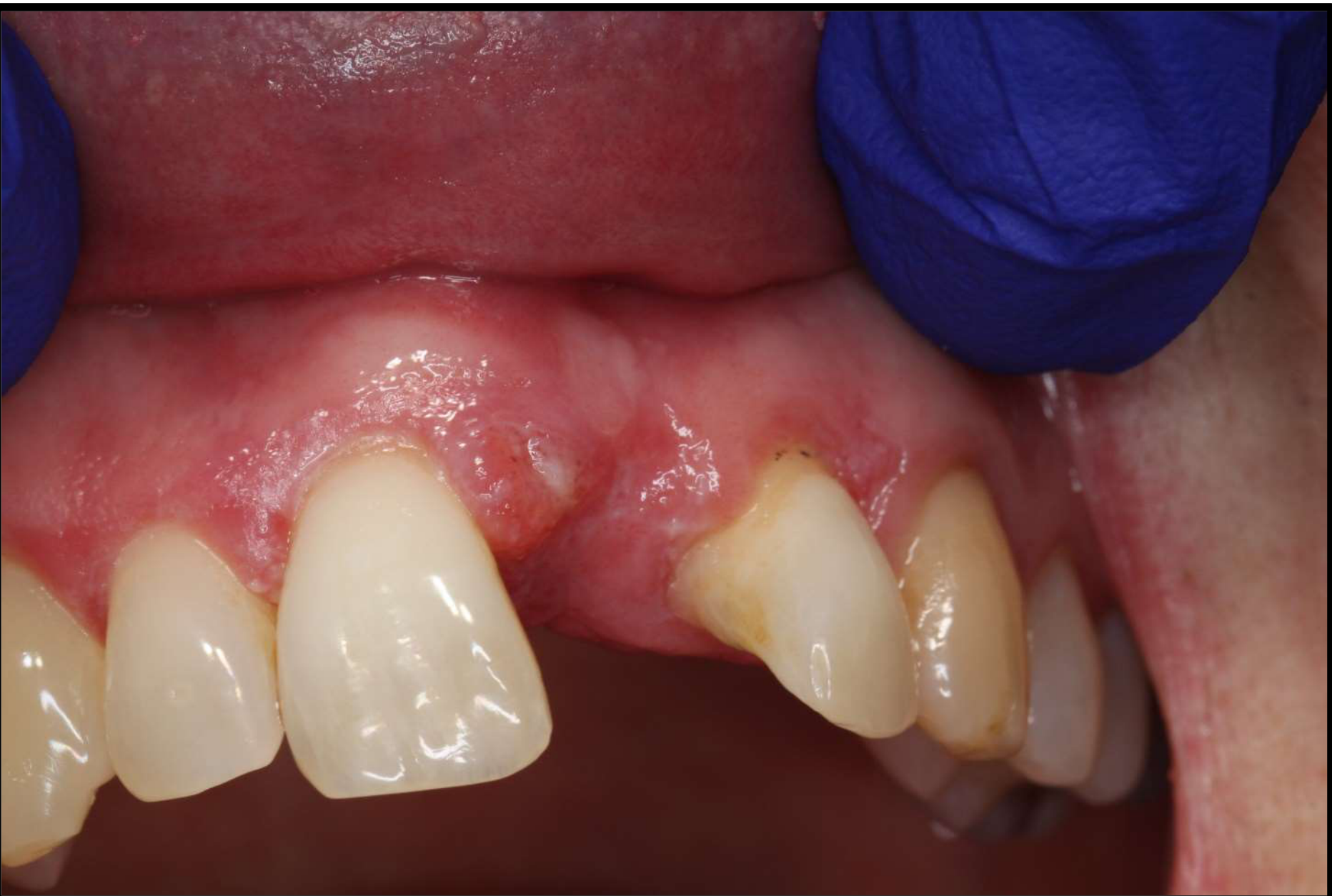
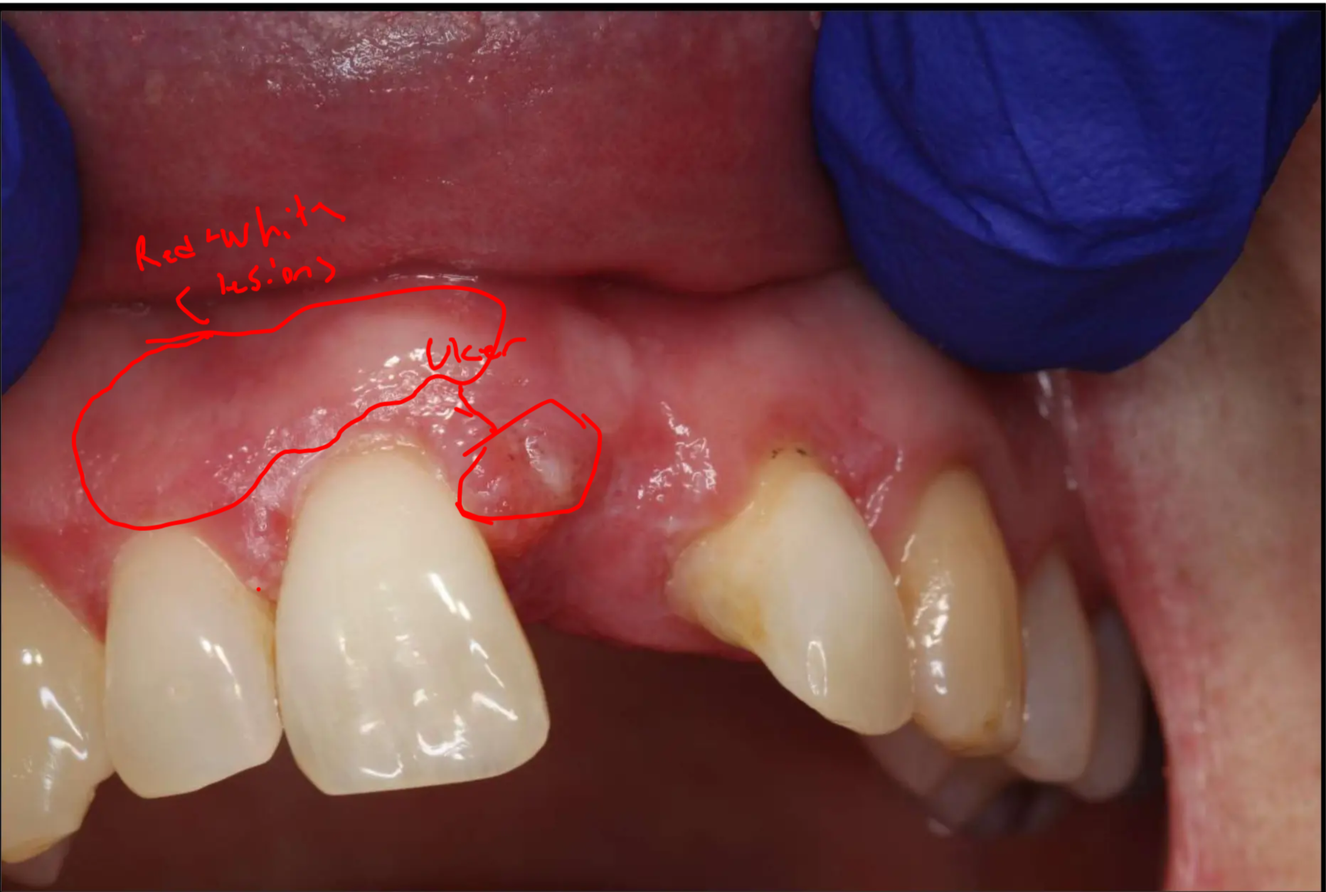
Identify the pathosis and describe the clinical features.
- Red and white patches on gingiva
- Transcript notes: Red and white lesions, including a white patch with a “sakila white” appearance.
- Areas are described as red and pink compared to the surrounding tissue.
- ulcer
- The ulcerated area is located on the gingiva (predominantly maxillary).
- Swelling within the lesions (Note: Lichen planus is typically flat, so swelling is a significant clinical finding).
- Clinical History Details:
- The patient has a two-year history of biopsy-proven lichen planus.
- She experiences periodic soreness that usually settles within a few days with corticosteroids.
- The current area has been sore for six weeks and is not responding to topical steroid ointment (betamethasone).
- The soreness is interfering with the patient’s ability to wear her denture.
What is the differential diagnosis?
- SCC
- A primary concern due to the non-healing nature of the ulcer and the known (small) risk of malignancy associated with lichen planus.
- Lichenoid drug reaction
- due to medications for hypertension
- The patient takes antihypertensive medication, which can be associated with these reactions.
- Lichen Planus
- The patient has a known history; however, the lack of response to steroids in this specific area is a concern.
- Traumatic ulcer
- Denture Trauma: Irritation from an ill-fitting denture causing gingival swelling and ulceration.
- Erythroleukoplakia: Considered if the lesion is isolated and inflammatory causes are ruled out.
- Desquamative Gingivitis: Discussed as a descriptive clinical term rather than a final diagnosis; it encompasses conditions like Lichen planus, Linear IgA disease, or other autoimmune/inflammatory diseases.
- Pyogenic Granuloma: Mentioned as a possibility for the clinical appearance.
- Secondary Infection: Specifically candidal infection (secondary to steroid use or denture wear).
What are the relevant clinical investigations?
Make sure to look at the denture
that its seating well and not irritating the gingiva
- Ask the paitent how she manages the lichen planus:
- she uses topical corticosteroids and bethamethasone ointment to settle flare ups
- her flareups settle within a few days and she stops the ointment, but this area she has been treating it daily for 6 weeks
- she uses topical corticosteroids and bethamethasone ointment to settle flare ups
Treatment of Traumatic vs Inflammatory Ulcers
- You shouldn’t treat traumatic ulcers with topical corticosteroids, but you can treat inflammtory ones with them
Blood Tests
- Hematomics: Full blood count.
Glucose tolerance
- we don’t know if the patient is diabetic
- Fasting Glucose: To check for underlying systemic issues like diabetes.
Dentures
- adjustment or asking patient to stop earing them
- Clinical Examination: Check if the denture is well-fitting.
- Adjustment: Adjust the denture to see if the lesion resolves once the source of irritation is removed.
Biopsy
- she is at 1% risk of malignancy due to having OLP
- We are worried about swelling because lichen planus is flat so there shouldn’t be any swelling
- Timing: Can be performed on the first visit if there is high clinical suspicion of malignancy, or after a two-week trial of conservative management (denture adjustment/antiseptics).
- Procedure: Multiple biopsies can be performed if other areas are also not responding to treatment, though the primary focus is the non-healing ulcerated area.
Histology
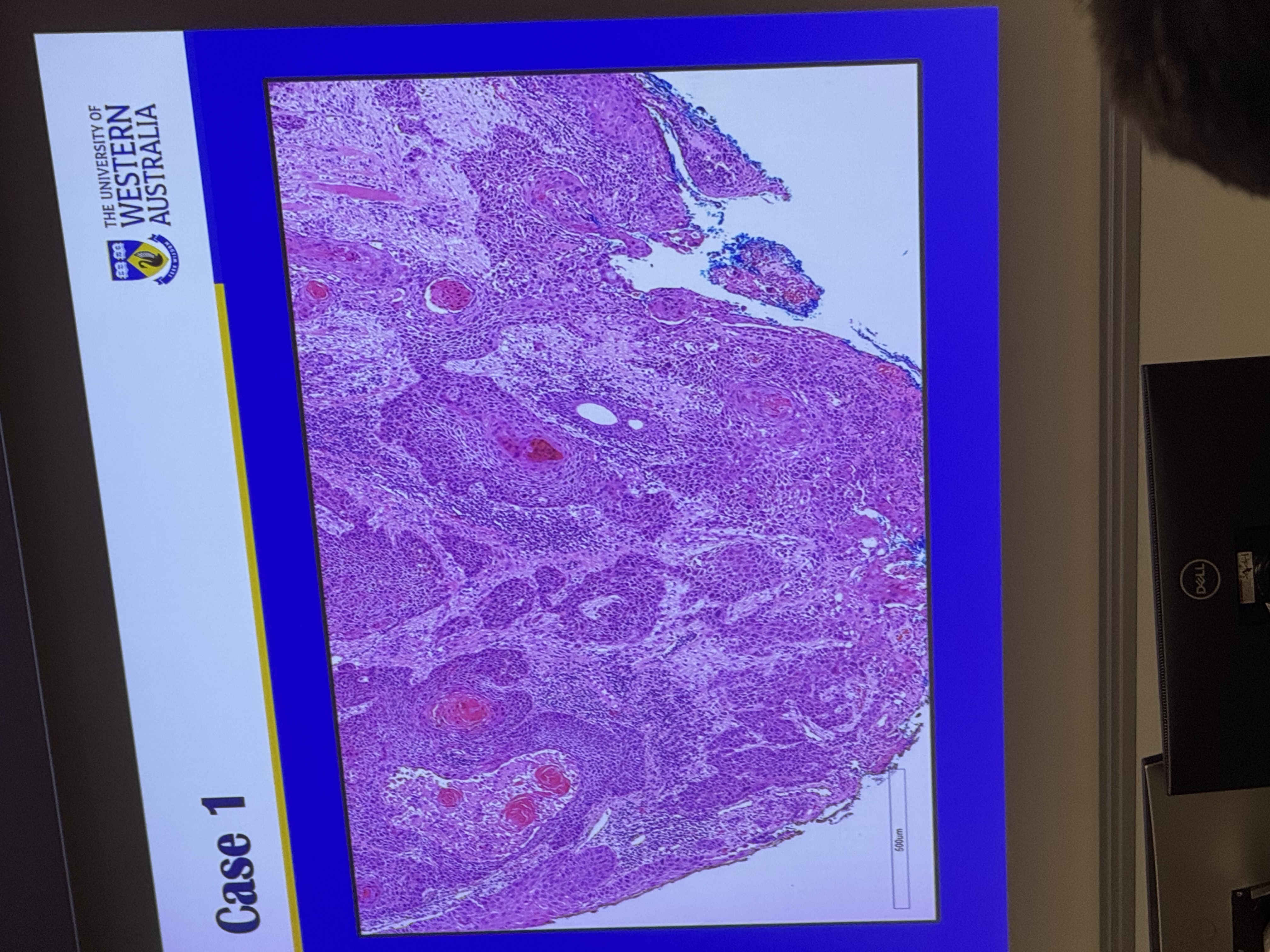
- Nuclear pleomorphism
- Variation in the shape and size of epithelial cells and nuclei.
- Lymphocytic infiltrate
- Hyperchromatism: Dark-staining lymphocytic infiltrate (seen as small blue dots).
- Mitotic figures
- Presence of abnormal mitotic figures (nuclear material arranged in lines).
- The histology is classic for a sqaumous cell carcinoma
- Invasion: Epithelial cells invading the underlying connective tissue; small islands of keratin-producing cells seen within the tissue.
- Differentiation: Presence of “keratin pearls” or well-formed keratin indicates a well-differentiated malignancy.
What is the diagnosis?
- Early Squamous cell carcinoma that has developed
- Developed within a pre-existing area of lichen planus.
How would you manage this patient?
- should still make the patient wait 2 weeks without hte dneture, as those two weeks wont make a difference , but two months will
- Conservative Trial (Initial Phase):
- Adjust the denture and instruct the patient to leave the denture out.
- Prescribe antiseptic mouthwash (e.g., Chlorhexidine) instead of steroids if trauma is suspected.
- Review in two weeks: This is the critical window; if the lesion does not improve, a biopsy is mandatory.
- Communication:
- Be honest but careful. Inform the patient that while the denture might be the cause, lichen planus carries a small risk of mouth cancer, and a biopsy is necessary if it doesn’t heal.
- Referral:
- Once a biopsy confirms SCC, refer the patient to a specialist head and neck surgeon.
- The patient will then be managed through a multidisciplinary team (MDT) process.
Footnotes
-
Original PDF page 1: 9. epithelial Pathosis II, p.1 ↩