Joint crepitus: A grinding sound indicating structural issues within the joint.
[!example] Differential Diagnosis
Muscle Pain: If symptoms appear muscular, they may be managed through standard clinical protocols first.
Degenerative Changes: Often described as “wear and tear” (similar to dental attrition). These changes do not always correlate directly with the severity of symptoms; a patient may have radiographic degeneration without significant pain or limitation.
Internal Derangement: This occurs when the disc is displaced out of the joint. This is often associated with degenerative changes and requires further investigation.
Radiographic Evaluation[^13]
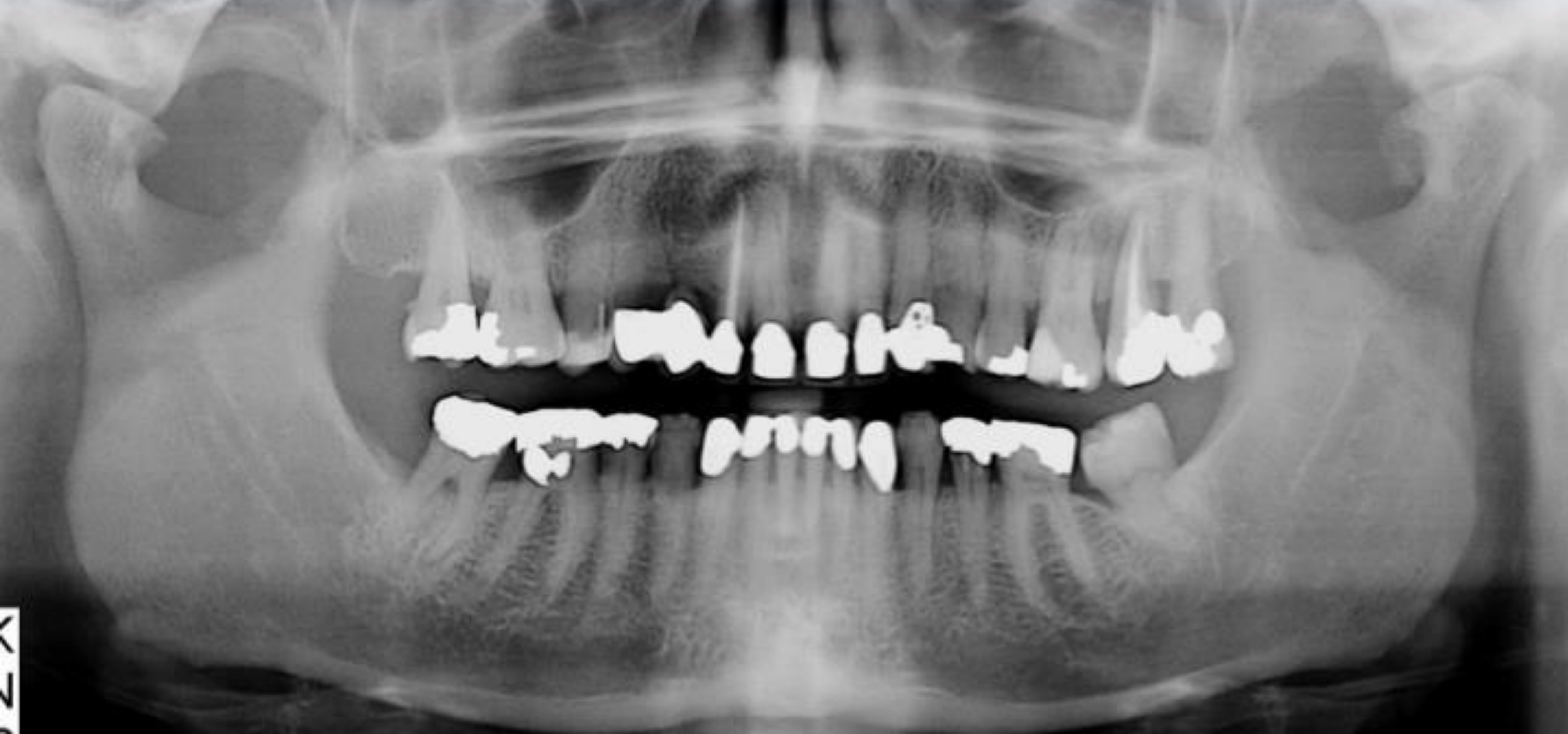
Describe the appearance and condition of the TMJs.
asymmetric condyles
Sclerotic bone towards articular surface of the left condyle
shows mostly signs of degenerative changes
Observed Degenerative Morphologies
Flattening: Loss of convexity in the articular surface.
Osteophytic Changes: Often appears as “peaking” or Anterior Osteophytic Lipping (prominent bone growth on the anterior aspect of the condyle).
Subchondral Sclerosis: Increased bone density (opacity) close to the articular surface.
General Opacity: Increased radiopacity anterior to the condyle.
Utility of the Orthopantomogram (OPG) in TMJ study
Indications: Frequently used to assess TMJs, though it has limitations. It is justified primarily in trauma cases to rule out fractures.
Limitations: An OPG cannot visualize the disc or internal derangement. It primarily shows bony changes.
Incidental Findings (Carotid Artery)
Carotid Atheromatous Calcification: May be seen on OPGs, typically in elderly or middle-aged male patients.
Management: Correlate with medical history (e.g., high cholesterol, use of statins). If the patient is unaware, refer them to a GP for a cardiovascular workup or ultrasound/Doppler study.
Advanced Imaging
Would you consider further imaging? Yes, specifically if symptoms persist or crepitus is present.
Specify which modality should be used and provide the clinical reasoning.
MRI (Magnetic Resonance Imaging): This is the Gold Standard and the definitive imaging modality for all TMJ diseases and internal derangements.
Clinical Reasoning: Unlike OPG, MRI allows for the visualization of soft tissues, specifically the position and condition of the articular disc.
Specialist Referral
It is recommended to refer patients to Oral Medicine Specialists for diagnosis.
Benefits of Referral: Specialists can provide a definitive diagnosis and refer for MRIs under Medicare benefits, significantly reducing the cost for the patient.