Case 12
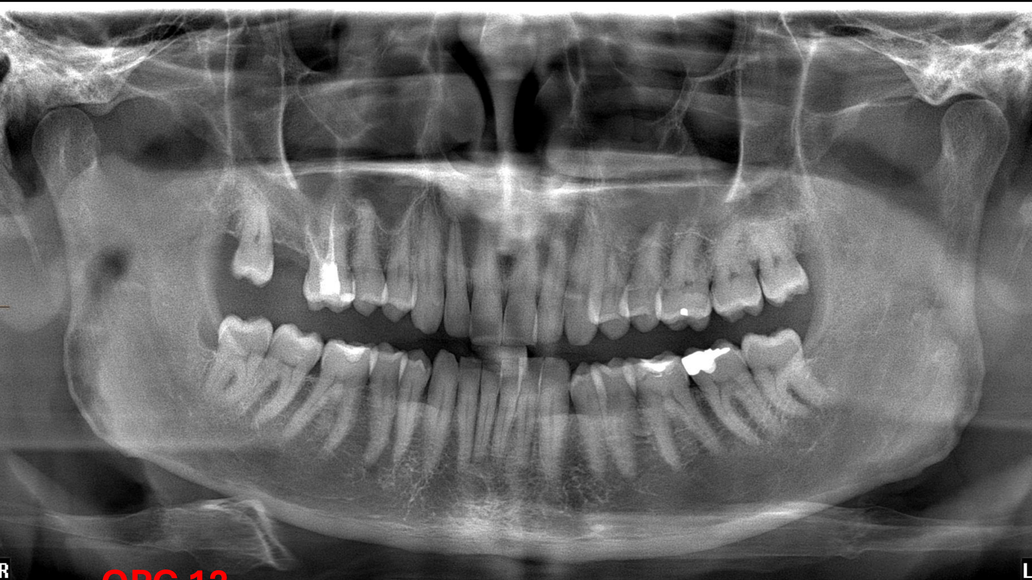
Radiographic Findings and Analysis
- Mandibular Third Molar Region (48):
- Suspected conditions include an odontogenic cyst, advanced periodontal issues, or pericoronitis.
- Pericoronitis is considered the most likely diagnosis for this area.
- There is a noted “big gap” between the 18 and the adjacent tooth.
- Angle of the Mandible:
- There is a lucency visible on the right-hand side at the angle of the mandible.
- A similar, though less prominent, lucency is visible on the left side.
- Interpretation: Because it is bilateral and has the same shape, it is identified as an anatomical airspace (specifically the oropharynx).
- Maxillary Anterior Region (12):
- A prominent lucency is noted around the 12 region.
- Differential Diagnosis:
- Radicular cyst.
- Lateral fossa (incisive fossa).
- It was noted that the lucency does not appear continuous with the nasal palate.
- Maxillary Sinuses:
- Evidence of mucosal thickening.
- Presence of an opacity, which is suspected to be a bone island.
Investigations
- Imaging for the 12 Region Lucency:
- First step: Take a Periapical (PA) radiograph.
- Second step: If the PA is inconclusive, proceed to a Cone Beam CT (CBCT).
Diagnosis
- Primary Clinical Suspicion: Pericoronitis (associated with the 48).
- Incidental Findings: Oropharyngeal airspace, mucosal thickening in the sinus, and a suspected bone island.