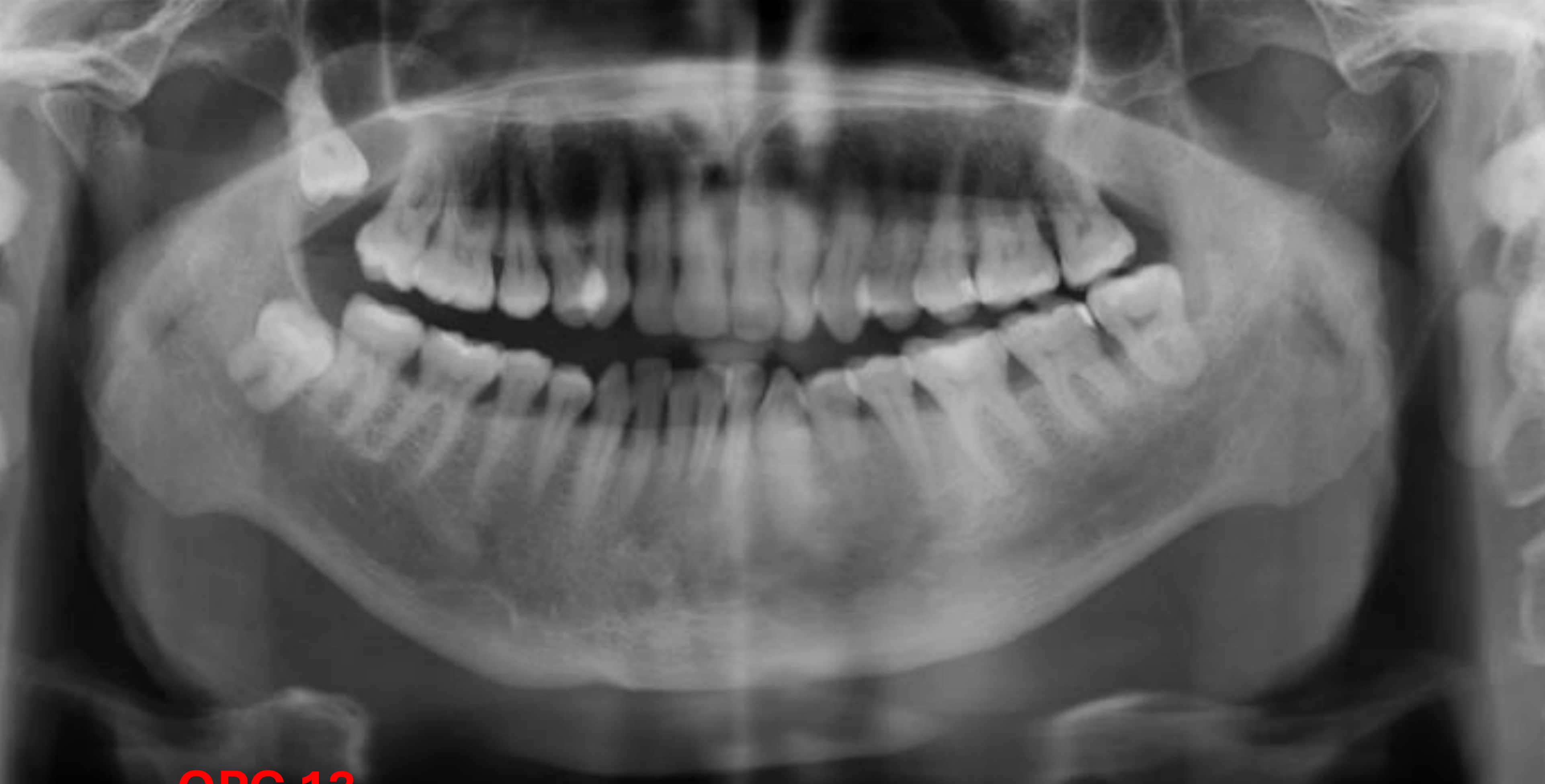
A round, soft tissue density superimposed over the sinus.
The lesion has elevated and displaced the sinus floor.
Location: Mesial to the 18, extending to the periapical region of the 17 and 16.
There is possible communication with the oral cavity.
Mandibular Observations:
Condyles: Both condyles are very small (hypoplastic) with minimal condylar neck. The left side is flattened, while the right side remains somewhat round.
Mandible: Hypoplastic mandible (both hemi-mandibles are very small).
Occlusion: Potential anterior open bite with posterior contact only, which is associated with the mandibular hypoplasia.
Sinus Observations:
The left maxillary sinus is very small (hypoplastic), sitting at or slightly above the level of the nasal floor/hard palate.
This is noted as a developmental finding rather than a pathologic one.
# Differential Diagnosis
Dentigerous Cyst:
The primary differential.
Large dentigerous cysts can “fold over” the tooth.
Communication with the oral cavity may disrupt its natural characteristics.