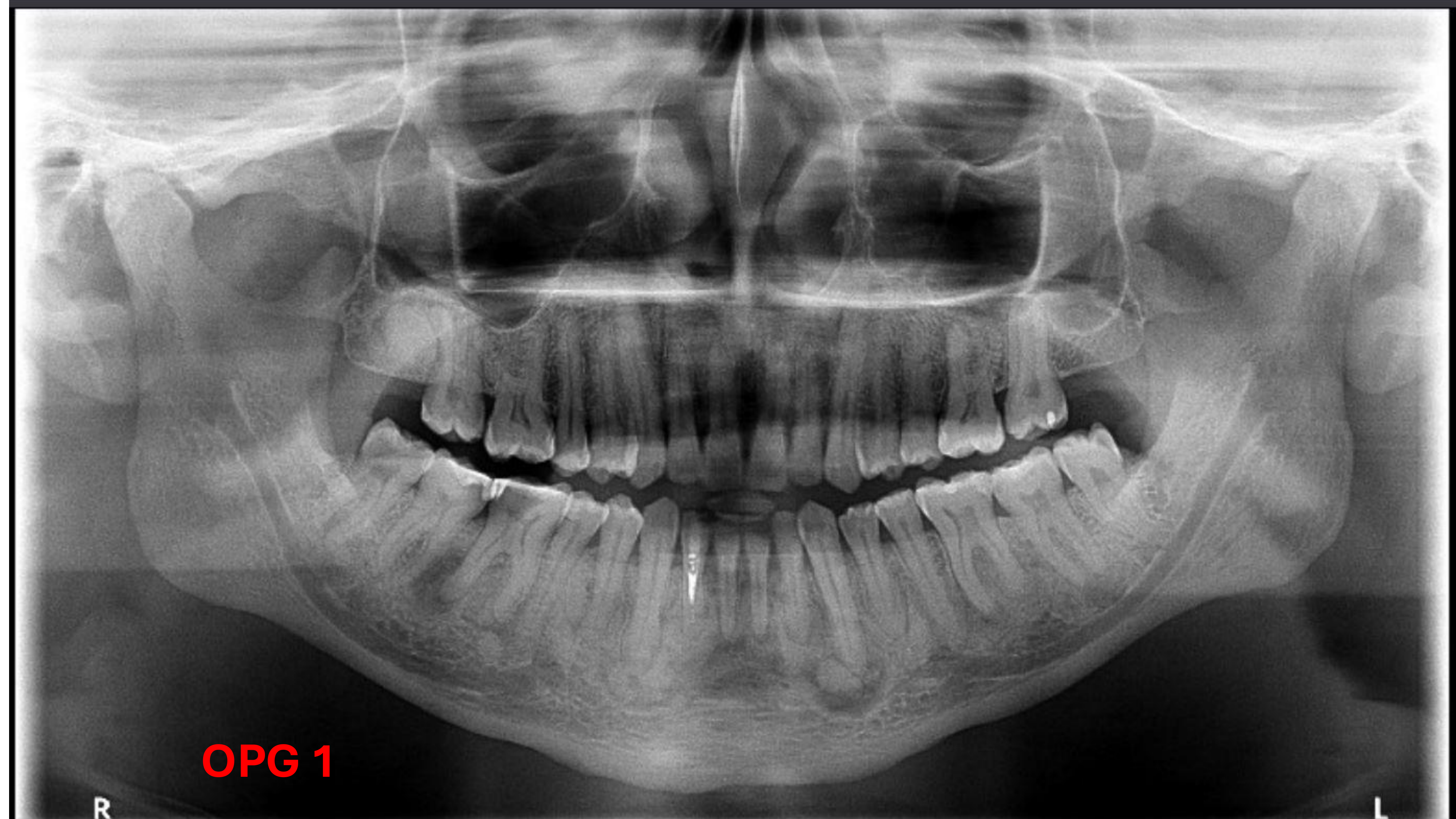
The patient presents with multiple mixed density lesions in the mandible, suggesting florid cementosseous dysplasia of varying maturity.
1.8 Region: The opacity in this region raises the possibility of a highly matured cementosseous dysplasia.
Anterior Mandible: These lesions typically favor the anterior region of the mandible but can also occur in the maxilla and near third molars.
Fibro-osseous Lesions:
This is a family of lesions including fibrous dysplasia, ossifying fibroma, and cementosseous dysplasia.
These lesions consist of both fibrous tissue (represented by radiolucency) and bony tissue (represented by opacity).
4.6 Region Lucency:
Periapical Inflammatory Lesion: Due to the presence of caries on the 4.6, a periapical inflammatory lesion is the primary differential.
Infected Cementosseous Dysplasia: Because the patient has multiple focal or florid CODs, the possibility that a COD has become infected cannot be excluded.
Other Considerations:
Root remnants or hypercementosis of root remnants.
Analysis
Lesion Appearance and Maturation:
Early-stage lesions appear as round, periapical radiolucencies, resembling inflammatory lesions.
As they mature, they form internal or central calcifications.
As the calcification grows, the outer radiolucent border narrows, and the lesion becomes more opaque.
Clinical Presentation:
The lesions are generally round in shape.
They exhibit different patterns of internal calcification depending on the stage of maturation (medium maturation vs. advanced/mature).
Management
Infection Control:
The 4.6 region requires treatment regardless of whether the cause is a standard periapical lesion or an infected COD.
Treatment options include endodontic therapy or extraction.
Clinical Considerations:
If the diagnosis is an infected COD, management is noted to be slightly more difficult than a standard periapical inflammatory lesion.