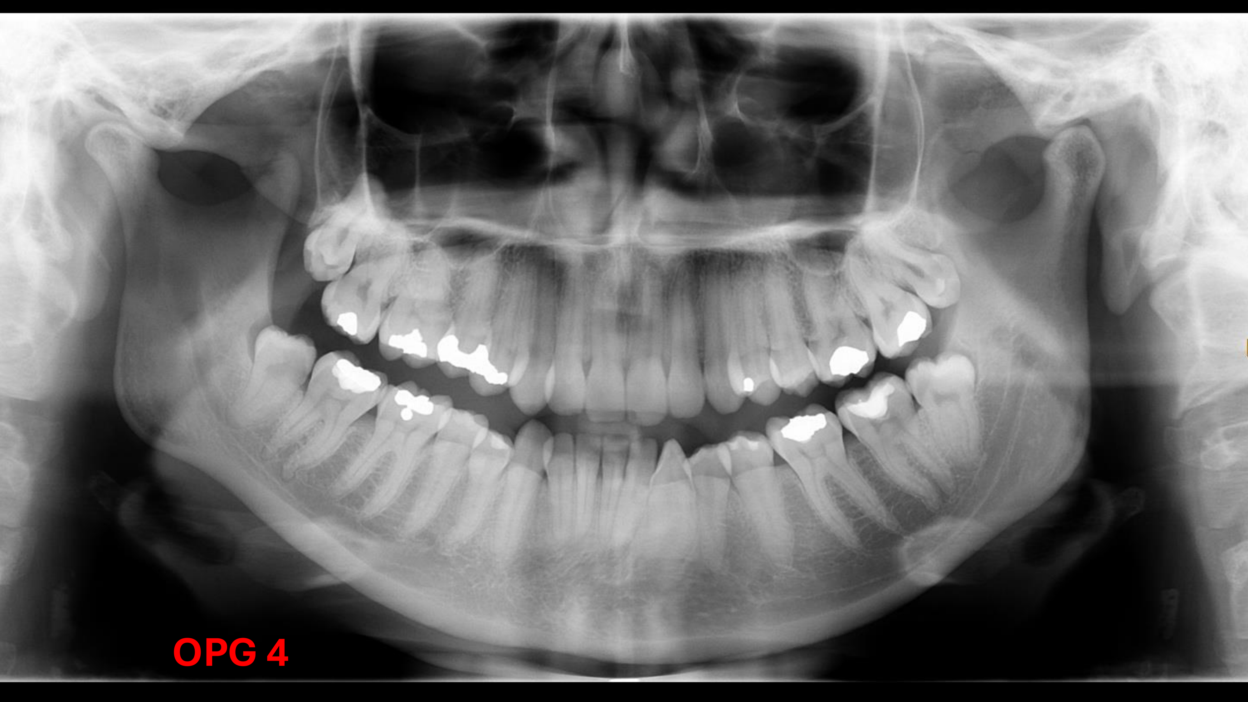
Joker Smile: Present due to the anchor plane being oriented downwards.
Airspace: No airspace visible because the tongue was not positioned on the roof of the mouth.
Ghost Images: A ghost image of the hyoid bone is visible on the left hand side of the mandible.
Dental Analysis
Tooth Count: All 32 teeth are present.
Impacted/Ectopic Teeth:
3, 8 and 4, 8 are vertically impacted and associated with pericoronitis.
Dental Anomalies:
3, 3 is rotated and infra-occluded.
4, 4 and 1, 8 show buccal rotation.
Dental Pathology:
3, 7 has a restoration extending into the pulp floor.
No bone loss is noted.
Occlusion: Occlusal asymmetry is present, with an asymmetrical deviation of mandibular teeth on the right hand side.
Bony Structures and Pathology
General: No abnormal radiolucencies, radio-opacities, or fractures are present. The trabecular pattern is unaltered.
Maxillary Sinus: Bilateral sinuses are in close proximity to the maxillary molars.
Zygomatic Arch: The zygomaticotemporal suture is visible bilaterally; it is a normal anatomical feature and not a fracture, though it appears distinct in this patient.
Mandibular Asymmetry and TMD
Morphology:
Left Side: The mandible is larger. The angle is very obtuse and rounded, with a loss of the antigonial notch (a normal anatomical feature). This is suggestive of left hemi-mandibular hyperplasia.
Right Side: The mandible is smaller. The condyle is small and tilted backwards rather than flattened. This is suggestive of right condylar hypoplasia.
Condylar Comparison: The left condyle appears normal and rounded at the superior point. The right condyle is smaller and tilted.
Symmetry: The left side is more rounded, while the right side is flattened on the condylar range and shorter than the left.
Diagnosis and Management
Diagnosis: Asymmetric mandible (congenital rather than traumatic) characterized by left hemi-mandibular hyperplasia and right condylar hypoplasia.
Referral Path:
Orthodontist: Initial referral should be to an orthodontist to assist in diagnosis and planning, as correction involves orthodontic treatment.
Maxillofacial Surgeon: For surgical correction of the bony asymmetry (orthognathic surgery).
Further Investigations:
Cone Beam CT (CBCT) or standard CT scan to better visualize the bony abnormalities and asymmetry.