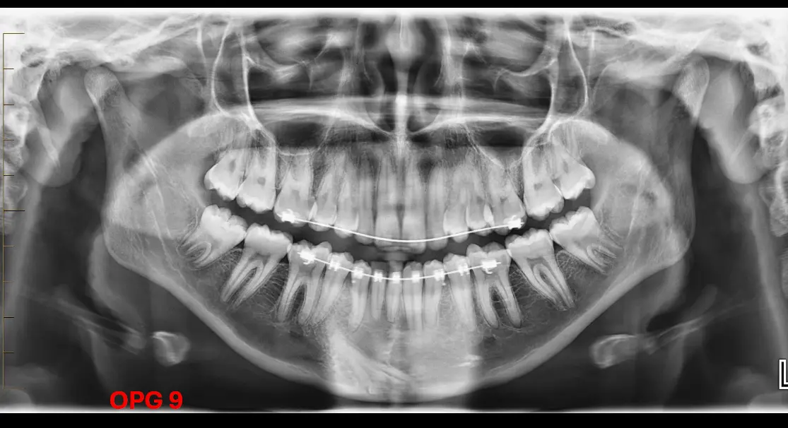
Missing Teeth: Auxiliary canines appear to be missing.
Quadrant Three: There appear to be almost three premolars in the third quadrant, described as “twin sizes.”
Quadrant Four:
A well-defined radio-opacity is located mesial to the 4/5 root apices.
This opacity extends down to the anterior border of the mandible and has a mixed appearance.
The structure is identified as an inverted tooth.
Analysis of the Impacted Tooth
Morphology:
The tooth has a conical shaped crown, similar to a canine.
The narrowest part is identified as the apex, leading to the root and the neck of the tooth.
A pulp canal and pulp chamber are visible as lucent structures within the tooth.
Crown Resorption:
There is significant lucency within the crown, which is identified as resorption rather than caries.
Only bits of enamel remain on the crown.
Supporting Structures:
The follicular space is lost.
The PDL space is not visible, suggesting tooth-to-bone contact.
The tooth is likely ankylosed; because the PDL/follicular space is gone, the tooth undergoes normal bone turnover.
Investigations
Counting Teeth: To identify the structure, count the teeth in quadrant four to determine what is missing.
3D Imaging: It is noted that OPGs are difficult for identification; 3D imaging (CBCT) would be required to see the structure clearly and confirm the diagnosis.