Unit A - Part 1 : Malocclusion: what is it and why should we treat it?
Describe ideal occlusion regarding tooth relationship to the line of occlusion.
Angle’s Description of “line of occlusion” Lecture, Contemporary Orthodontics (sixth edition) TOC
- Angle used specific suffixes to identify tooth positions:
- Bucco-version
- Linguo-version
- Labio-version
- Torsio-version (rotation)
- “The line of occlusion is a smooth (catenary) curve passing through the central fossa of each upper molar and across the cingulum of the upper canine and incisor teeth. The same line runs along the buccal cusps and incisal edges of the lower teeth, thus specifying the occlusal as well as interarch relationships once the molar position is established” (“Contemporary Orthodontics (sixth edition)”, p. 13) (pdf)

- “Note that the Angle classification has four classes: normal occlusion, Class I malocclusion, Class II malocclusion, and Class III malocclusion. Normal occlusion and Class I malocclusion share the same molar relationship but differ in the arrangement of the teeth relative to the line of occlusion. The line of occlusion may or may not be correct in Class II and Class III malocclusion.” (“Contemporary Orthodontics (sixth edition)”, p. 12) (pdf)
Describe ideal occlusion in transverse, antero-posterior, and vertical planes.
Ideal Occlusion in the A-P Plane

- Posterior Occlusion in the anterio-posterior plan is defined by the molar relationship.
- Divided into 4 classes (Angle’s):
- Class I (normal occlusion): Mesiobuccal cusp of maxillary fist molar occludes in the buccal groove of the lower first molar
- Class I Malocclusion: Molar relationships remain the same but differ in the arrangement of teeth relative to the line of occlusion.
- Class II Malocclusion: Lower molar distally positioned relative to upper molar, line of occlusion not specified
- Class II Division 1: Proclined maxillary incisors (increased overjet); associated with convex profile and lip incompetence
- Class II Division 2: Retroclined maxillary incisors + proclined lateral incisors; associated with a deep bite
- Class III: Lower molar mesially positioned relative to upper molar, line of occlusion not specified
- Divided into 4 classes (Angle’s):
- Anterior occlusion in the anterio-posterior plane is defined by overjet
- Overjet: the horizontal overlap of the incisors
- Normally the incisors are in contact, with the upper incisors ahead of the lower by only the thickness of their incisal edges (i.e., overjet of 2 to 3 mm is the normal relationship).
- If the lower incisors are in front of the upper incisors, the condition is called reverse overjet or anterior crossbite.” (“Contemporary Orthodontics (sixth edition)”, p. 16) (pdf)

- Overjet: the horizontal overlap of the incisors
Ideal Occlusion in the Transverse Plane
- Occlusion in the transverse plane is dictated by the location of the posterior teeth.
- Normal transverse occlusion is: Upper teeth buccal to lower teeth (i.e. BULL Rule )
- The most common major deviation from the transverse dental relationship is lingual posterior crossbite
- Lingual posterior cross bite: upper teeth lingual to the lower teeth
Ideal Occlusion in the Vertical Plane
- Ideal Occlusion in the vertical plane is measure by overbite
- Overbite is defined as the vertical overlap of the incisors
- Normally the lower incisal edges contact the lingual surface of the upper incisors at or above the cingulum
- there is normally a 1 - 2 mm overbite
- In open bite, there is no vertical overlap and the vertical separation of the incisors is measured to quantify its severity
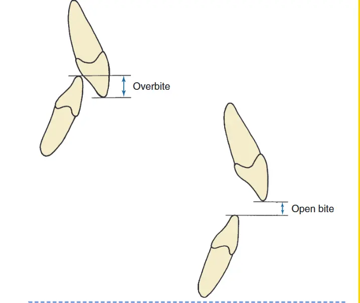
- Normally the lower incisal edges contact the lingual surface of the upper incisors at or above the cingulum
List dental characteristics for the Angle classification and discuss its advantages.
See above
Describe functional, health, and psychosocial reasons for treatment.
Primary Objectives for Orthodontics
- Reduce psychosocial handicap
- Improve oral function
- Adjunct to disease control (not to treat disease )
Describe realistic goals for orthodontic treatment and their evolution.
The modern objectives for orthodontic treatments fall under the “Soft Tissue Paradigm”
- Treatment goals of the soft tissue Paradigm
- Normal soft tissue proportions and adaptations
- Functional occlusion is a secondary goal
Historically the angle paradigm for ideal dental occlusion was to have an ideal dental occlusion and skeletal relationships. This goal has shifted to the soft tissue paradigm in the modern era because a patients appearance is often much more of concern to them and functional occlusion can still occur even if the scheme is not ideal.
| Feature | Angle Paradigm | Soft Tissue Paradigm |
|---|---|---|
| Primary goal of treatment | Ideal dental occlusion | Ideal soft tissue proportions and adaptation |
| Secondary goal of treatment | Jaw relationships | Functional occlusion |
| Hard vs soft tissue relationship | Ideal skeletal/dental produces ideal soft tissue | Ideal soft tissue defines ideal skeletal/dental |
| Diagnostic emphasis | Dental casts, cephalometric x-rays | Clinical examination of soft tissues |
| Treatment approach | Obtain ideal dental and skeletal relationships, and the soft tissues will be OK | Determine ideal soft tissue relationships, and then place the jaws and teeth as needed to obtain them |
Modern Patient Perception
Patients often identify themselves by their profile in the mirror rather than molar relationships. Modern standards favor fuller profiles (e.g., the “Angelina Jolie era”) over the straight or concave profiles favored in the past.
Discuss treatment need versus demand in the current environment.
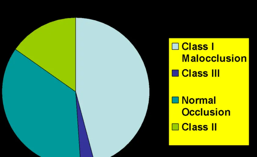
Treatment Need 01 - Malocclusion Definition and Prevalence The key points to remember from modern population studies of malocclusion:
-
About half the population have well-aligned teeth or slight irregularity, while about 15% have irregularity severe enough that either major expansion of the dental arches or extraction of a permanent tooth in each quadrant of the arch is likely to be needed if the teeth are to be aligned.
-
With increasing age, incisor irregularity gets worse and the number of people with ideal alignment decreases.
-
15% of the US population are Class II, and about half of these individuals have overjet severe enough to predispose them to problems in social interactions.
-
Open bite is much more prevalent and deep bite less prevalent in African-Americans.
-
Class III and reverse overjet are much more prevalent in those of Asian descent.
-
Other racial / ethnic differences are modest and unimportant.
Who seeks Orthodontic treatment
- 65% of the population have malocclusion
- Dentists feel that 55% need treatment
- Patients feel that 35% need treatment
 **Treatment Acceptance Rates **
- 55% need treatment
- 50% accept treatment at no cost
- 5% wouldn’t accept treatment even if it was free
Increasing Treatment Demand
Older patients are more likely to want orthodontic treatment now. Some say its because its more socially acceptable although no one can say why
Unit A - Part 2 The etiology of Malocclusion: Definition and rational
Categorize known causes of malocclusion and their relative prevalence.
02 - Known Causes of Malocclusion There are four major factors in the etiology of malocclusion :
- Hereditary factors
- Interference with normal development
- Trauma
- Disturbance in normal function
Hereditary Factors
Recent Development
Framgents of prehistoric jaws show mostly good development, so malocclusion may be a modern problem
- Evolutionary tred towards a decrease in teeth and jaw size
- Interracial mixing DOES NOT produce an increase in malocclusion
- Familial patterns of malocclusion like the Hapsburg jaw is possible
- 50% may be the maximum contribution to malocclusion by hereditary factors , the environmental contribution is likely to be significantly higher than the genetic one
Interference with normal development
Interferences with Pre-Natal Development
- Genetic and environmental insults in utero
- Fetal alcohol syndrome
- Teratogens like thalidomide
- Zika Virus
- Treacher collins syndrome
- Even birth trauma to the mandible is possible but more rare
- Intra-uterine molding
- Pre-Natal interferences with the Dentiion
- hyper and hypo dontia
Anything affecting things like neural crest cell migration or neural plate closure
Interference with Post-Natal Development
- look at it with the soft tissue theory of growth
- scarring from an injury is more likely to affect development than anyh hard tissue injury
- Fracture of the condylar process has 75% chance of normal growth and 25% chance that it causes assymmetry
- that 25% chance is normally due to soft tissue scarring around the TM joint
Effects of Trauma on the dentition
- Trauma can effect the occlusal balancing forces
- for example :
- a young child crawling around on the floor bites down on an electrical cord, the result is likely to be a severe burn at the corner of the mouth, as in the girl shown in image 1. As it heals and scars down, pressure against the teeth creates a marked asymmetry in the mandibular dental arch, and makes normal arch form almost impossible to maintain.
- for example :
The reverse effect is seen when cheek tissues are lost (image 2). Then unopposed pressure by the tongue tips the teeth facially.
 Image 1: Scarring as this burn heals will distort dental arch form Image 1: Scarring as this burn heals will distort dental arch form |  Image 2: Loss of cheek tissue to a tropical infection has led to buccal displacement of the teeth Image 2: Loss of cheek tissue to a tropical infection has led to buccal displacement of the teeth |
Direct Trauma to the teeth
- This can also lead to malocclusion
- If a tooth is lost the adjacent teeth will tend to drift into its space and normal alignment and position of the teeth will be lost unless **space is maintined **
Disturbences in function
- See thumb sucking and thrusting
Identify malocclusions linked to inherited jaw proportions and provide supporting evidence.
Twin studies for skeletal and dental overject show that up to 50% of maloccluions have origins in hereditary factors
- Certain types of malocclusion do run in families
- Haris and Johnson experiments shows that heritability of skeletal chracteristics is high but dental characteristics is actually low
- Brazilian studies show that heritability of mandibular prognatishm is high with second highest being the long-face pattern of deformity to run in families
Explain how mandibular trauma affects future growth.
- mostly if soft tissue scaring occurs (loook at 75/25 distribution in the condyles )
Identify the magnitude and duration of force required for tooth movement (e.g., thumb sucking habits).
- “Sucking habits during the primary dentition years have little if any long-term effect.” (“Contemporary Orthodontics (sixth edition)”, p. 136) (pdf)
- The classic presentation of a habitual thumb sucker is :
- protruding upper incisors
- anterior open bite
- maxillary constriction causing the child to shift into a unilateral posterior crossbite
- the negative pressure of sucking actually isn’t responsible for this, its because the tongue is depressed ! so it shifts the balance of the upper teeth as usually the tongue is supposed to be pushing back on them
- A little more forward growth of the maxilla is also possible
- lingually positioned lower incisors
“There can be considerable variation in which teeth are affected and how much.” (“Contemporary Orthodontics (sixth edition)”, p. 137) (pdf)
Duration of force
- this can be found in unit 1 but essential related back to Equilibrium theory
- how much the teeth are displaced correlates with the number of hours per day of sucking than with the magnitude of pressure
- p. 139:

Describe the maturation of oral function and swallowing patterns from infancy to adulthood.
-
For most patients the position of the tongue probably is more an adaptation to the position of the the teeth than the cause of their displacement
-
“Tongue thrust swallowing simply has too short a duration to have an impact on tooth position. Pressure by the tongue against the teeth during a typical swallow lasts for approximately 1 second” (“Contemporary Orthodontics (sixth edition)”, p. 141) (pdf)
-
the mature or adult swallow pattern appears in some normal children as early as age 3 but is not present in the majority until about age 6 and is never achieved in 10% to 15% of a typical population. Tongue thrust swallowing in older patients superficially resembles the infantile swallow (described in Chapter 3), and sometimes children or adults who place the tongue between the anterior teeth are spoken of as having a retained infantile swallow. This is clearly incorrect. Only braindamaged children retain a truly infantile swallow in which the posterior part of the tongue has little or no role.” (“Contemporary Orthodontics (sixth edition)”, p. 140)
-
“During the transition from an infantile to a mature swallow, a child can be expected to pass through a stage in which the swallow is characterized by muscular activity to bring the lips together, separation of the posterior teeth, and forward protrusion of the tongue between the teeth. This is also a description of the classic tongue thrust swallow. A delay in the normal swallow transition can be expected when a child has a sucking habit. When there is an anterior open bite and/or upper incisor protrusion, as often occurs from sucking habits, it is more difficult to seal off the front of the mouth during swallowing to prevent food or liquids from escaping. Bringing the lips together and placing the tongue between the separated anterior teeth is a successful maneuver to close off the front of the mouth and form an anterior seal.” (“Contemporary Orthodontics (sixth edition)”, p. 140)
-
The modern viewpoint is that tongue thrust swallowing is seen in two circumstances:
- younger children with reasonably normal occlusion in which its a transitional stage in normal maturation
- individuals of any age with displaced incisors
-
The presence of a large overjet often and anterior overbite (nearly always) conditions a child or ault to place the tongue between the anterior teeth
Discuss myofunctional therapy for tongue thrusting and its validity for anterior open bite.
- no validity, the tongue thrusting is most likely an adaptation to the anterior open bite, not the cause of it!
“As Fig. 5.38 shows, at every age above 6, the number of children reported to have a tongue thrust swallow is about 10 times greater than the number reported to have an anterior open bite.” (“Contemporary Orthodontics (sixth edition)”, p. 141) (pdf)
Describe the role of nasal obstruction in malocclusion etiology.
“Because total nasal obstruction in humans is so rare, the important clinical question is whether partial nasal obstruction, of the type that occurs occasionally for a short time in everyone and chronically in some children, can lead to malocclusion; more precisely, how close to total obstruction does partial obstruction have to come before it is clinically significant? The question is difficult to answer, primarily because it is difficult to know what the pattern of respiration really is at any given time in humans. Observers tend to equate lip separation at rest with mouth breathing (see Fig. 5.39), but this is simply not correct. It is perfectly possible for an individual to breathe through the nose while the lips are apart” (“Contemporary Orthodontics (sixth edition)”, p. 142)
“The best experimental data for the relationship between malocclusion and mouth breathing were derived from studies of the nasal/oral ratio in normal versus long-face children.40 The relationship is not nearly as clear-cut as theory might predict. It is useful to represent the data as in Fig. 5.42, which shows that both normal and long-face children are likely to be predominantly nasal breathers under laboratory conditions. A minority of the long-face children had less than 40% nasal breathing, but none of the normal children had such low nasal percentages. When adult long-face patients are examined, the findings are similar: the number with evidence of nasal obstruction is increased in comparison to a normal population, but the majority are not mouth breathers in the sense of predominantly oral respiration.” (“Contemporary Orthodontics (sixth edition)”, p. 143)