Clinical Overview of Natal and Neonatal Teeth
1. Definitions and Classification
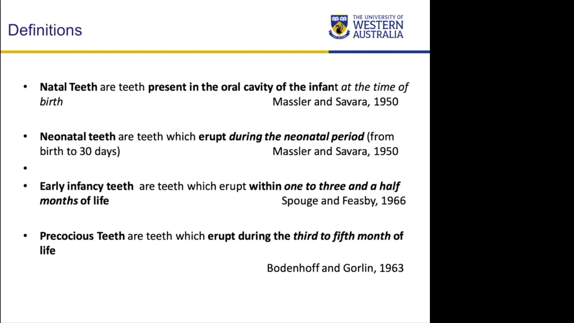
Natal and neonatal teeth represent rare dental eruption anomalies observed in newborns. According to established diagnostic criteria (e.g., Massler and Savara):
- Natal Teeth: Teeth present in the oral cavity at birth.
- Neonatal Teeth: Teeth that erupt within the first 30 days of life.
Structural Classifications
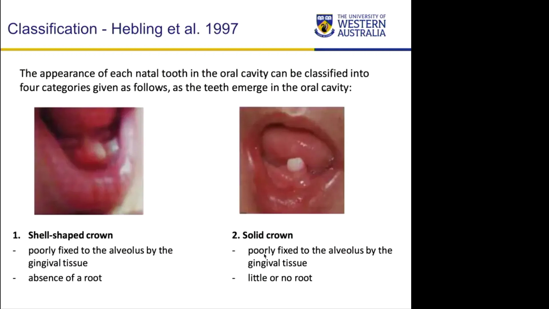
While academic classifications categorize these teeth by maturity (e.g., mature vs. immature) or morphological presentation, clinically they generally present in one of four forms:
- A fully formed crown attached loosely to the gingiva by soft tissue (lacking root structure).
- A solid crown loosely attached to the alveolar ridge.
- An incisal edge barely erupted through the mucosal tissue.
- A mucosal swelling containing an unerupted, palpable tooth shell.
2. Epidemiology and Etiology
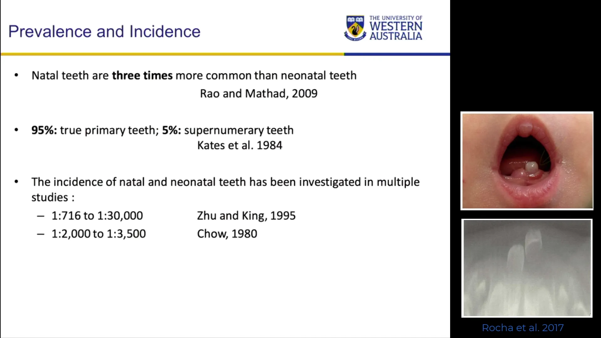
- Incidence Ratio: Natal teeth are approximately three times more common than neonatal teeth.
- Tooth Type: Over 95% of natal and neonatal teeth are true primary teeth, rather than supernumerary teeth. Premature loss of these teeth typically results in an edentulous space until the permanent teeth erupt.
- Anatomic Location: Most frequently observed in the mandibular primary central incisor region (teeth 71 and 81 / O and P), corresponding to the normal sequence of primary tooth eruption.
* *Clinical Red Flag:* Eruption of posterior teeth (e.g., primary molars) at birth without anterior eruption indicates severe underlying systemic or developmental pathology.
- Gender: No statistically significant gender predilection has been established.
Etiology
- Primary Etiology: The most widely accepted cause is an abnormally superficial position of the tooth germ, leading to accelerated eruption.
- Associated Factors: While maternal malnutrition or systemic health issues have been correlated in observational studies, direct causation remains unproven. Increased prevalence is noted in infants with cleft lip and palate or complex congenital syndromes.
3. Cultural and Social Considerations
In several cultures worldwide (e.g., regions within East Africa and West Africa), natal teeth carry deep-rooted superstitions and cultural stigmas. They may be associated with bad omens, evil spirits, or severe social marginalization for both the child and parents. Clinicians should approach these cases with cultural sensitivity, providing empathetic communication and clear scientific explanation alongside clinical management.
4. Clinical Presentation and Histological Features
Clinical Presentation
- Morphology: Usually resemble normal primary incisors in size and shape, though frequently smaller, discolored, or malformed.
- Development: Highly immature. Crowns may present as soft enamel shells or dysplastic structures attached to a floating mucosal base.
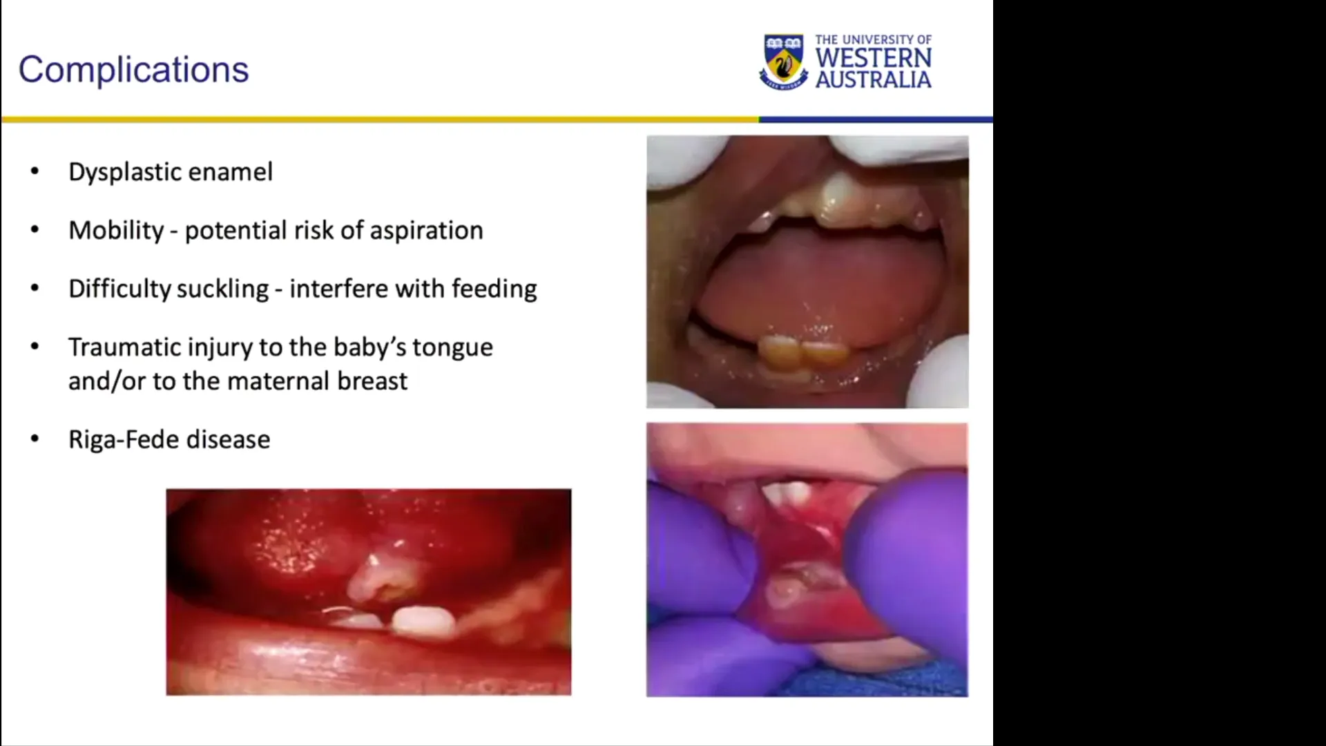
- Mobility: Typically exhibit severe mobility due to incomplete or absent root formation and lack of alveolar bone support.
Histological Characteristics
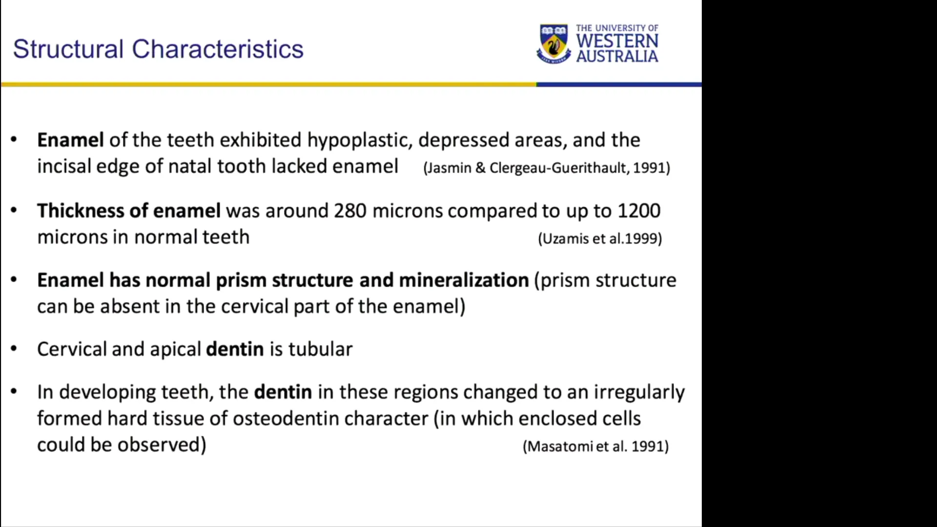
- Enamel: Hypoplastic, hypomineralized, and substantially thinner than normal primary enamel.
- Dentin: Irregular and poorly organized dentinal tubule structure.
- Cementum: Absent or underdeveloped due to premature eruption prior to root formation.
- Pulp: Hypervascularized with an enlarged pulp chamber, increasing the risk of pulp exposure, breakdown, and periapical infections/abscesses.
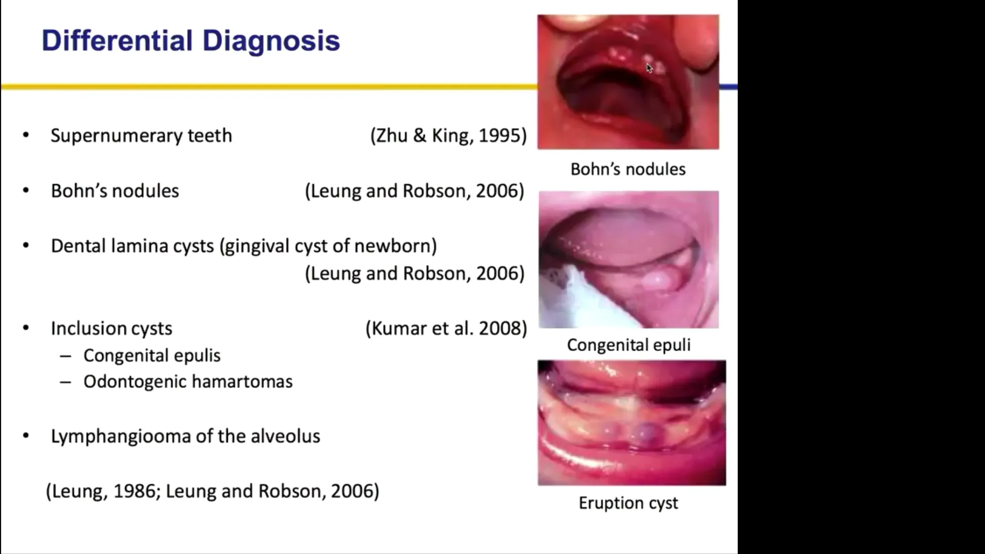
5. Differential Diagnosis of Neonatal Oral Lesions
When evaluating a infant presenting with oral structures or swellings, the following non-dental conditions must be differentiated from natal teeth:
| Condition | Clinical Characteristics | Location | Management |
|---|---|---|---|
| Natal / Neonatal Teeth | Mineralized structures; primary tooth morphology or enamel shell. | Alveolar ridge (predominantly mandibular anterior). | Conservative monitoring or extraction if indicated. |
| Bohn’s Nodules | Keratin-filled cysts derived from dental lamina remnants; high prevalence (~60%). | Buccal and lingual aspects of the alveolar ridge. | Spontaneous exfoliation; no treatment required. |
| Epstein Pearls | Keratin-filled palatal cysts entrapped during palatal shelf fusion. | Midpalatal raphe / palatal suture line. | Spontaneous resolution; no treatment required. |
| Congenital Epulis | Soft tissue benign mesenchymal tumor present at birth. | Anterior alveolar ridge. | Surgical excision if feeding/airway is compromised. |
| Eruption Cysts | Fluid-filled tissue sac overlying an erupting tooth structure. | Directly over the alveolar ridge. | Monitoring; usually resolves with tooth eruption. |
Other rare entities include lymphangiomas and melanotic neuroectodermal tumors of infancy. Ambiguous lesions require immediate specialist referral.
6. Clinical Complications
- Feeding Interference and Maternal Discomfort: Sharp or mobile natal teeth can disrupt nursing and cause discomfort to the mother.
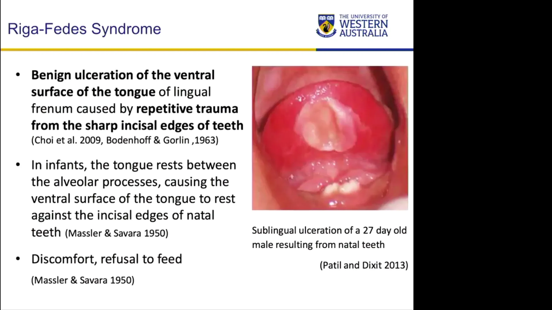
- Riga-Fede Disease: Ulceration on the ventral surface of the infant’s tongue caused by repetitive trauma against the sharp incisal edges of lower natal teeth during feeding.
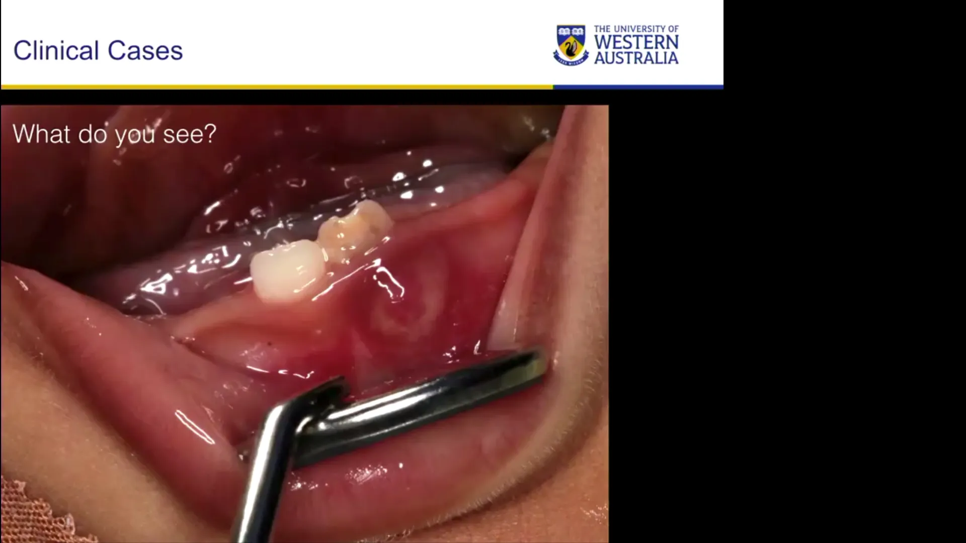
- Rapid Caries and Pulpal Abscess: Due to enamel dysplasia, thin dentin, and large vascular pulps, these teeth break down rapidly, predisposing the infant to pulpal exposure and abscess formation.
- Risk of Ingestion or Aspiration: Frequently cited in literature as a rationale for extraction due to mobility. However, true documented cases of aspiration are virtually non-existent, as these teeth typically maintain strong gingival mucosal attachments even when highly mobile.
7. Diagnostic Evaluation
- Clinical Examination: Physical assessment is the primary diagnostic tool. Gently palpate the tooth, evaluate mobility, inspect for tongue ulcerations, and examine the alveolar ridge for signs of infection.
- Radiographic Examination:
- Routine radiographs are generally discouraged in neonates in accordance with ALARA
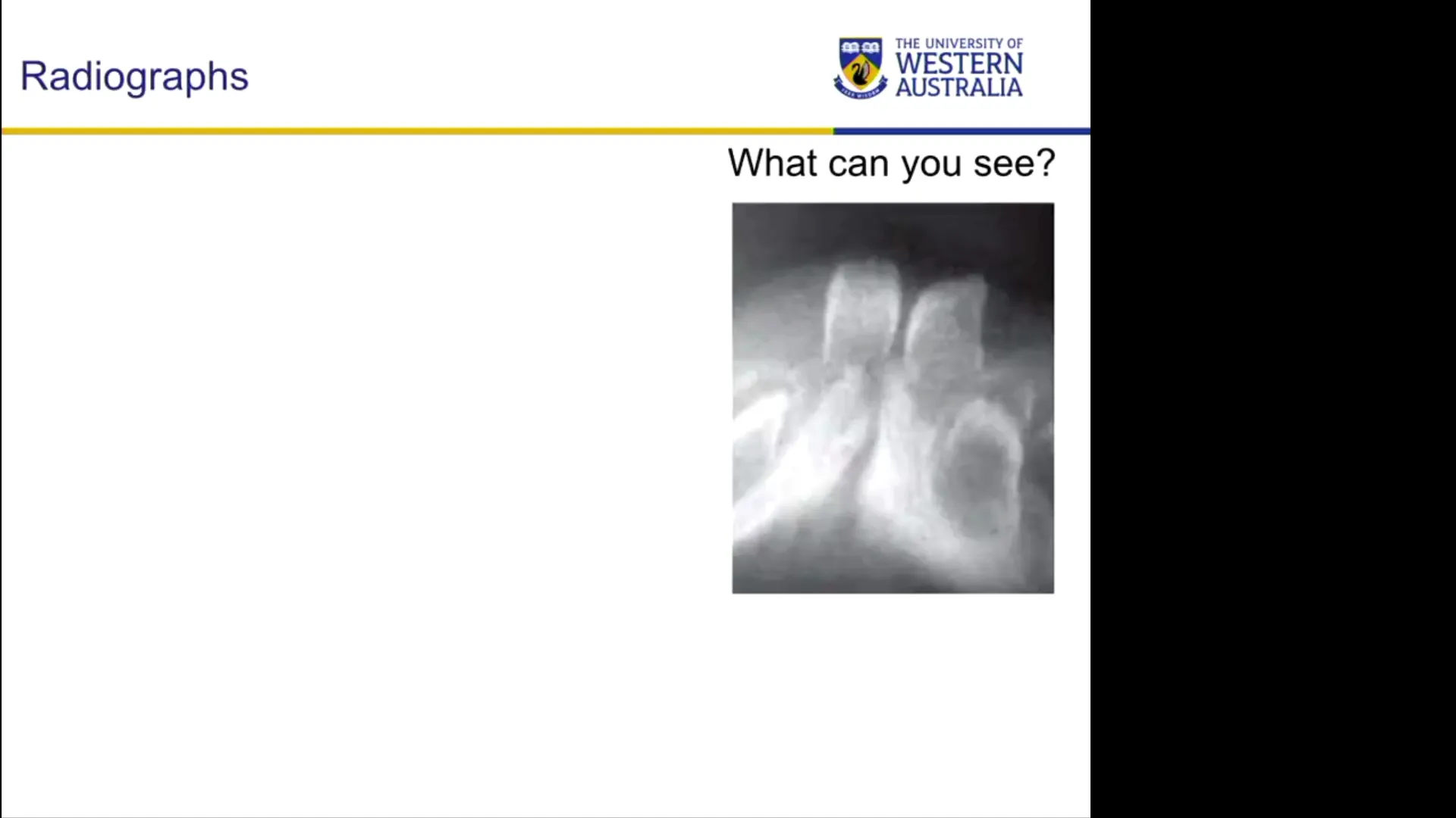 (As Low As Reasonably Achievable) principles.
* Calcification of primary incisors begins in utero, but resolution on neonatal radiographs is often poor, providing minimal diagnostic benefit.
* Radiographs should only be considered if foreign body impaction, structural pathology, or complex underlying anomalies are suspected.
(As Low As Reasonably Achievable) principles.
* Calcification of primary incisors begins in utero, but resolution on neonatal radiographs is often poor, providing minimal diagnostic benefit.
* Radiographs should only be considered if foreign body impaction, structural pathology, or complex underlying anomalies are suspected.
8. Management Strategies
Management should prioritize conservative care wherever possible. Surgical extraction is reserved for specific clinical indications and should generally be referred to a pediatric dental specialist.
                  [ Neonatal Patient with Tooth Present ]
                                     |
                -------------------------------------------
               |                                           |
    [ Asymptomatic & Stable ]                   [ Symptomatic / Severe ]
               |                                           |
    - Reassurance & Education                  ---------------------------------
    - Monitor Root Formation                  |                                 |
    - Monitor Hygiene                     [ Feeding Injury / ]           [ Abscess / Severe Decay / ]
                                          [ Ventral Ulceration ]         [ Extreme Mobility Hazard ]
                                                  |                                 |
                                        -------------------                 - Vitamin K Verification
                                       |                   |                - Controlled Local Anesthetic
                                 (Conservative)        (Surgical)           - Extraction & Socket Curettage
                                       |                   |                - Post-Op Hemostasis Check
                                 - Smooth Edges        Extraction
                                 - GIC / Composite
                                 - Stomahesive Wafer
A. Conservative Non-Surgical Management (First-Line)
If the child is feeding well, asymptomatic, and free of severe ulcerations or infection:
- Reassurance: Educate parents regarding the benign nature of true primary natal teeth and the likelihood of mobility decreasing as root development progresses.
- Smoothing Sharp Edges: Cautiously smooth sharp incisal edges to prevent soft tissue trauma.
- Protective Coverings: Apply a small amount of Glass Ionomer Cement (GIC) over sharp edges to cushion the tongue.
- Stomahesive Wafers: Use biocompatible adhesive wafers over the teeth
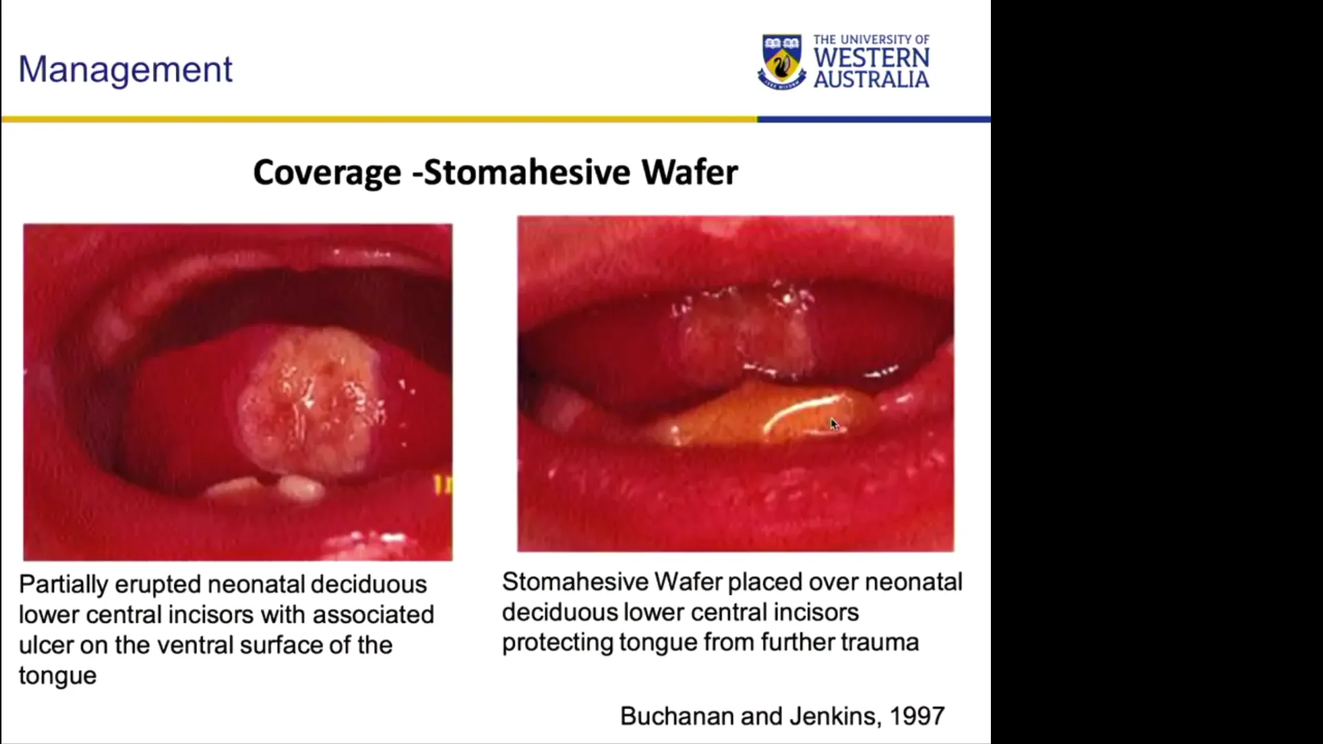 to act as a barrier, allowing ventral tongue ulcers to heal without interfering with feeding.
- Note on Topical Medications: Topical corticosteroid/anesthetic pastes (e.g., Kenalog) are contraindicated in neonates due to systemic absorption and toxicity risks.
B. Surgical Extraction Protocol
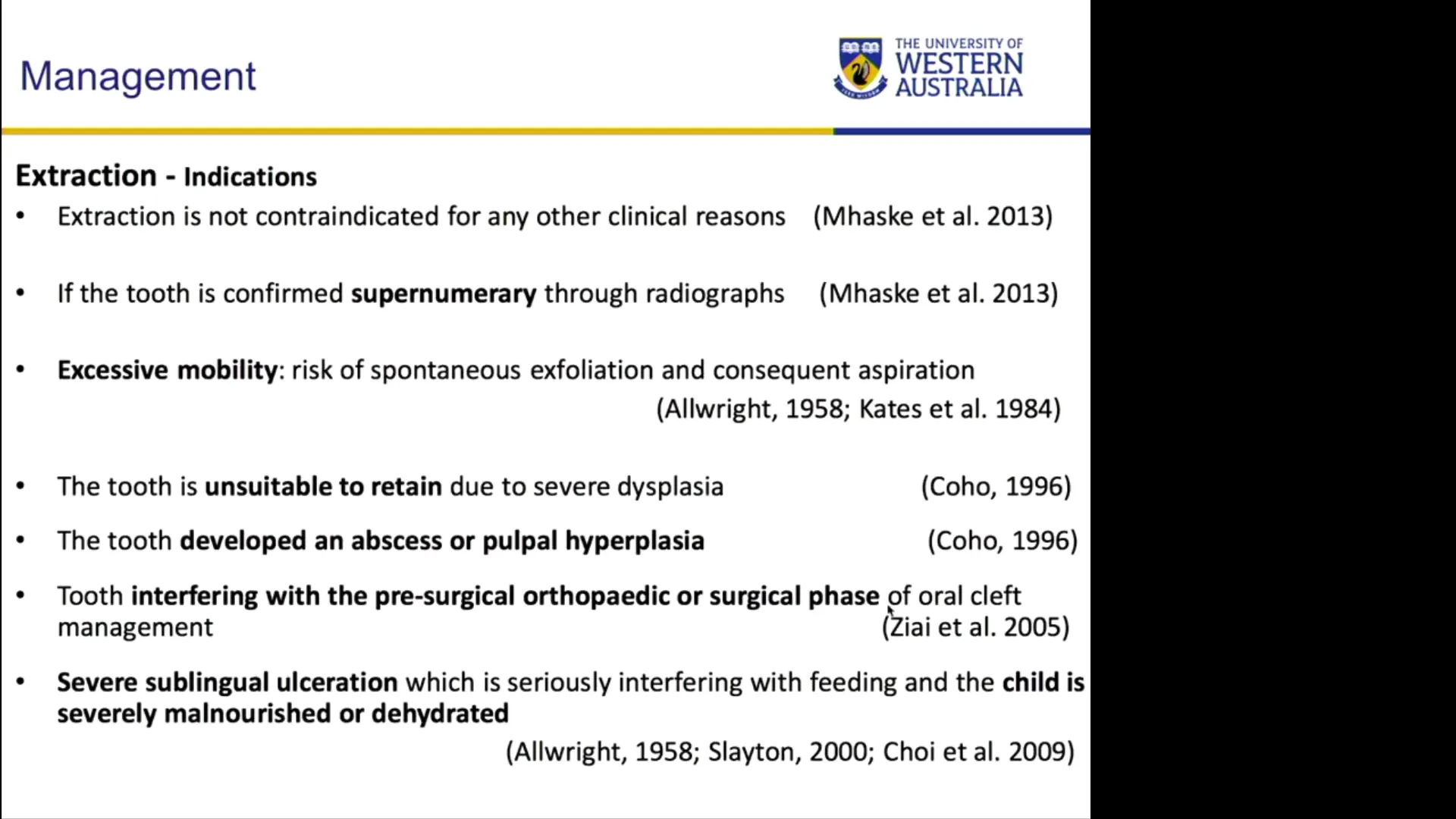
Extraction is indicated only under the following conditions:
- Severe Riga-Fede ulceration unresponsive to conservative measures.
- Structural breakdown, advanced caries, or pulpal infection/abscess.
- Interference with surgical obturator management in patients with cleft lip/palate.
Critical Surgical Considerations:
- Systemic Verification: Ensure the neonate has received their post-birth Vitamin K injection to prevent severe neonatal hemorrhage.
- Local Anesthetics Dosing: Neonates (2–3 kg) are highly sensitive to local anesthetic toxicity. Exact volumetric dosing must be strictly calculated and administered (e.g., utilizing an intraligamentary syringe such as a Lignaject at 0.2 mL increments).
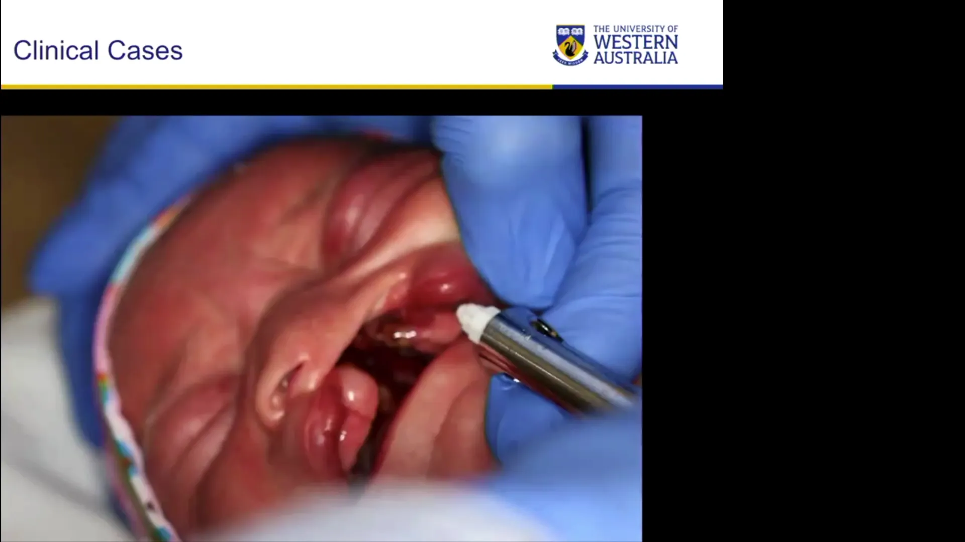
- Socket Curettage: Upon extraction, the follicle, Hertwig’s Epithelial Root Sheath (HERS), and residual dental papilla cells must be thoroughly curetted. Failure to do so can result in the continued development of residual root fragments in the jaw.
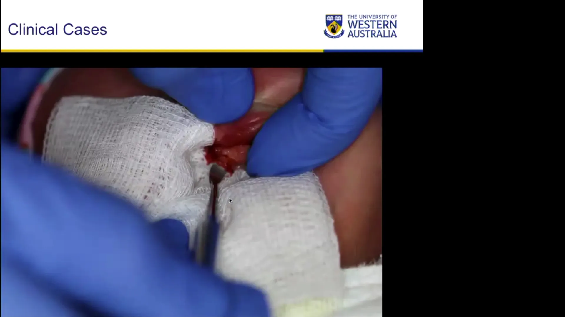
- Aspiration Risk Control: Secure physical control of the tooth during extraction using adequate gauze protection to prevent displacement into the pharynx or nasal cavity (especially in cleft palate cases).
C. Space Maintenance Implications
Premature extraction of anterior primary teeth generally does not require a space maintainer. While mild initial space loss may occur in the anterior segment, arch expansion during subsequent facial growth normally restores adequate space prior to the eruption of permanent successors.
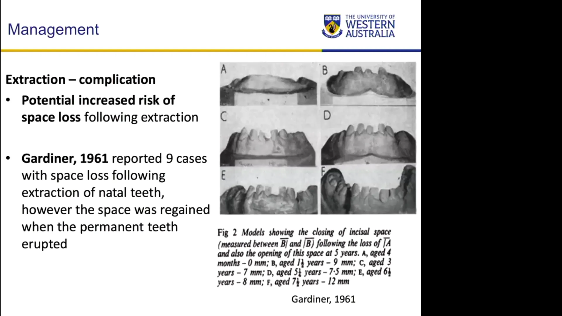 (This contrasts with primary molar extractions, which always cause significant space loss).
9. Summary Recommendations for Dental Practice
- Confirm Tooth Identity: Recognize that natal teeth are typically normal primary teeth, not extra/supernumerary teeth.
- Prioritize Conservative Care: Retain natal teeth whenever possible through monitoring, smoothing, or applying protective materials.
- Refer Complex / Surgical Cases: Due to local anesthetic toxicity risks, severe bleeding potential, and small anatomic limits, extractions in neonates should generally be referred to pediatric dental specialists or hospital settings.
Clinical Cases
Case: Natal tooth in a preterm newborn
Question
Scenario: A preterm newborn is evaluated for a tooth present in the oral cavity at birth. What’s shown: An intraoral photograph of an erupted tooth and a radiograph of the same area. Consider: Identify the erupted tooth and determine its relationship to the underlying dentition based on the radiographic findings.
Answer
Observations:
- The intraoral image shows an erupted tooth in the mandibular incisor region (tooth 71).
- The radiograph confirms the erupted tooth is the primary 71, not a supernumerary tooth.
- The underlying primary teeth 31 and 41 are visible, along with tooth 81 which is close to erupting. Reasoning: The radiograph verifies that the natal tooth is a true primary tooth. This aligns with the fact that 95% of natal teeth are actual primary teeth rather than supernumerary teeth. Takeaway: Radiographs can confirm whether a natal tooth is a primary or supernumerary tooth, though the vast majority are primary teeth.
Case: Natal tooth extraction in a child with unilateral cleft lip and palate
Question
Scenario: A child with a unilateral cleft lip and palate presents with a natal tooth causing complications. What’s shown: Intraoral mirror views showing the cleft, nose, vomer, the natal tooth, and a feeding ulcer on the tongue. Images of the extraction process using an intraligamentary anesthetic system and the extracted tooth with its follicle. Consider: Identify the anatomical structures, the complication present, and the specific considerations for extracting the tooth in this patient.
Answer
Observations:
- The intraoral view shows the cleft, nose, vomer, the natal tooth, and a feeding ulcer on the ventral surface of the tongue.
- The extraction utilizes an intraligamentary anesthetic system delivering 0.2 ml per click to carefully control the local anesthetic dose.
- The extracted tooth is shown along with the entire surrounding follicle. Reasoning: The natal tooth is causing a traumatic feeding ulcer on the tongue, indicating the need for extraction. In a cleft lip and palate patient, there is a risk of the tooth or instruments displacing into the nasal cavity. Additionally, the entire follicle and Hertwig’s epithelial root sheath must be curetted to prevent future root formation. Precise anesthetic dosing is critical to avoid toxicity in a small infant. Takeaway: Extracting a natal tooth in a cleft lip and palate patient requires careful anesthetic dosing, thorough curettage of the follicle, and awareness of the risk of displacement into the nasal cavity.
Case: Mobile natal tooth with hidden abscess and enamel defects
Question
Scenario: A fit and healthy newborn presents with a highly mobile natal tooth that appears to be floating on tissue. What’s shown: Images of the alveolus with the floating tooth, macro/magnified views of the area with the lip lifted, and the extracted tooth with its follicle. Consider: Identify the hidden pathology revealed upon careful examination and the structural defects of the extracted tooth.
Answer
Observations:
- The tooth is highly mobile and floating on its follicular tissue.
- Lifting the lip reveals a hidden abscess associated with the yellow, defective tooth.
- The extracted tooth shows qualitative (yellow) and quantitative (broken down) enamel defects, including a deep groove running through the lingual surface. Reasoning: The extreme mobility indicates the tooth is attached only to soft tissue, necessitating extraction. Careful examination by lifting the lip is crucial to avoid missing hidden pathology like an abscess, which might not present with obvious swelling. The structural defects are due to the superficial positioning of the tooth germ and immature development. Takeaway: Thorough clinical examination, including lifting the lip, is essential to identify hidden complications like abscesses in natal teeth, which often present with significant enamel defects and extreme mobility.