Management of Edentulism and the Terminal Dentition: Maxilla Fixed1
Clin A/Prof Glen Liddelow BDSc(WA), MScD(WA), DClinDent(Syd), MRACDS(Pros), FPFA, FICD, FRACDS
BRÅNEMARK CENTRE PERTH — Associated Brånemark Osseointegration Centre, Perth
Treatment Strategies for the Edentulous Jaw2
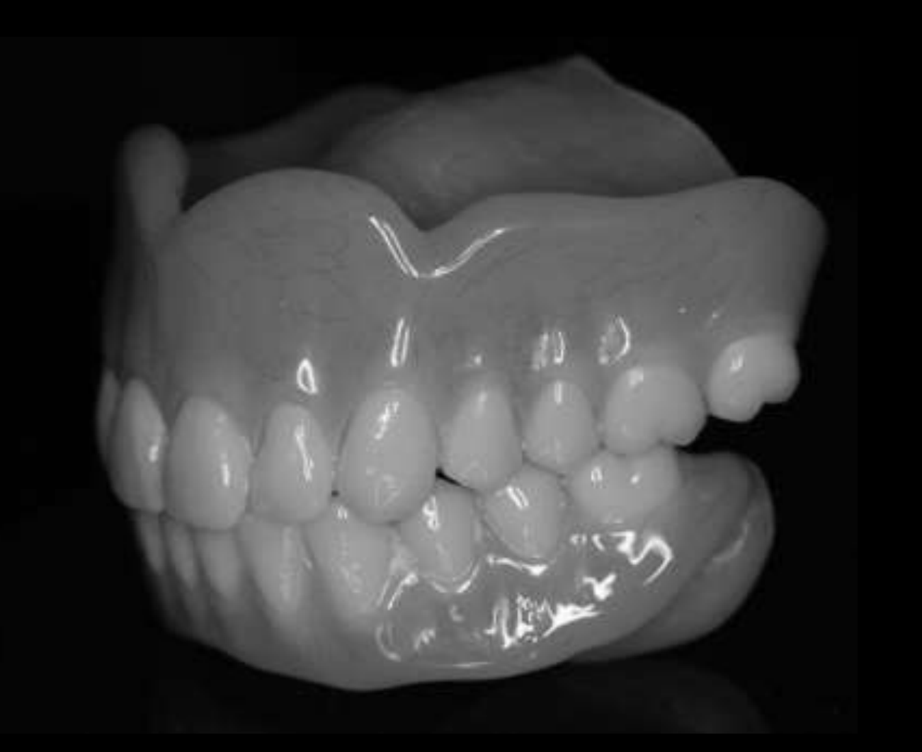 | 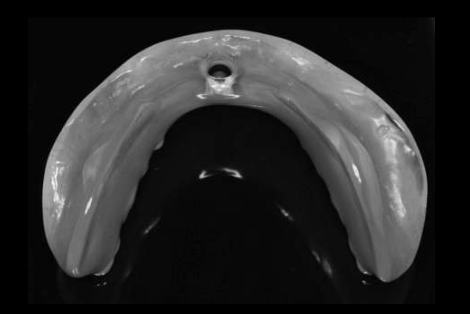 |
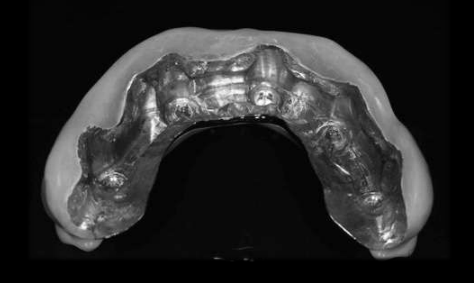 | 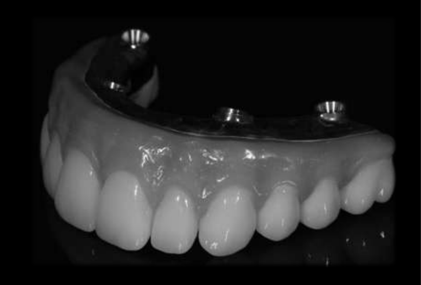 |
Lecture Introduction: Fixed Prosthesis for the Maxilla3456
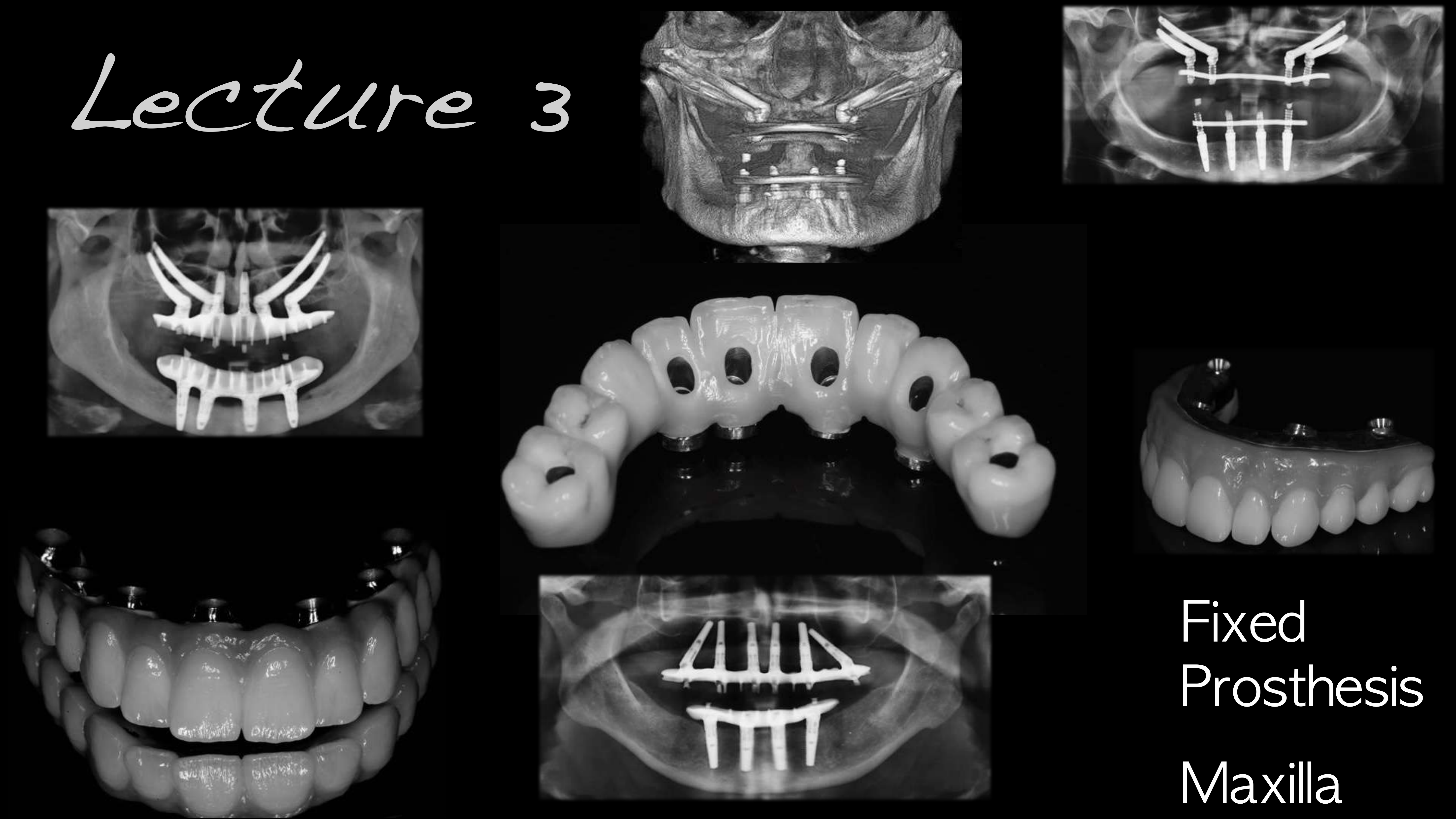
Lecture 3 — Fixed Prosthesis, Maxilla
- Maxillary complete denture prosthetics
- Mandibular prosthodontics
- Mn IOD
- Mn IFP (Least maintenance)
- Review at 12 months (75% satisfied)
Friberg B, Jemt T. Rehabilitation of edentulous mandibles by means of four TiUnite implants after one-stage surgery: a 1-year retrospective study of 75 patients. Clin Implant Dent Relat Res. 2010; 12 Suppl 1:56-62.
Friberg B, Jemt T. Rehabilitation of edentulous mandibles by means of five TiUnite implants after one-stage surgery: a 1-year retrospective study of 90 patients. Clin Implant Dent Relat Res. 2008; 10(1):47-54.
Lecturer — Maxillary Fixed Options
Patients increasingly seek a fixed maxillary solution because they want their own teeth back rather than a removable prosthesis.
- A maxillary overdenture remains removable, covers much of the palate, affects taste and speech, and allows the face to collapse when removed, producing a markedly sunken appearance.
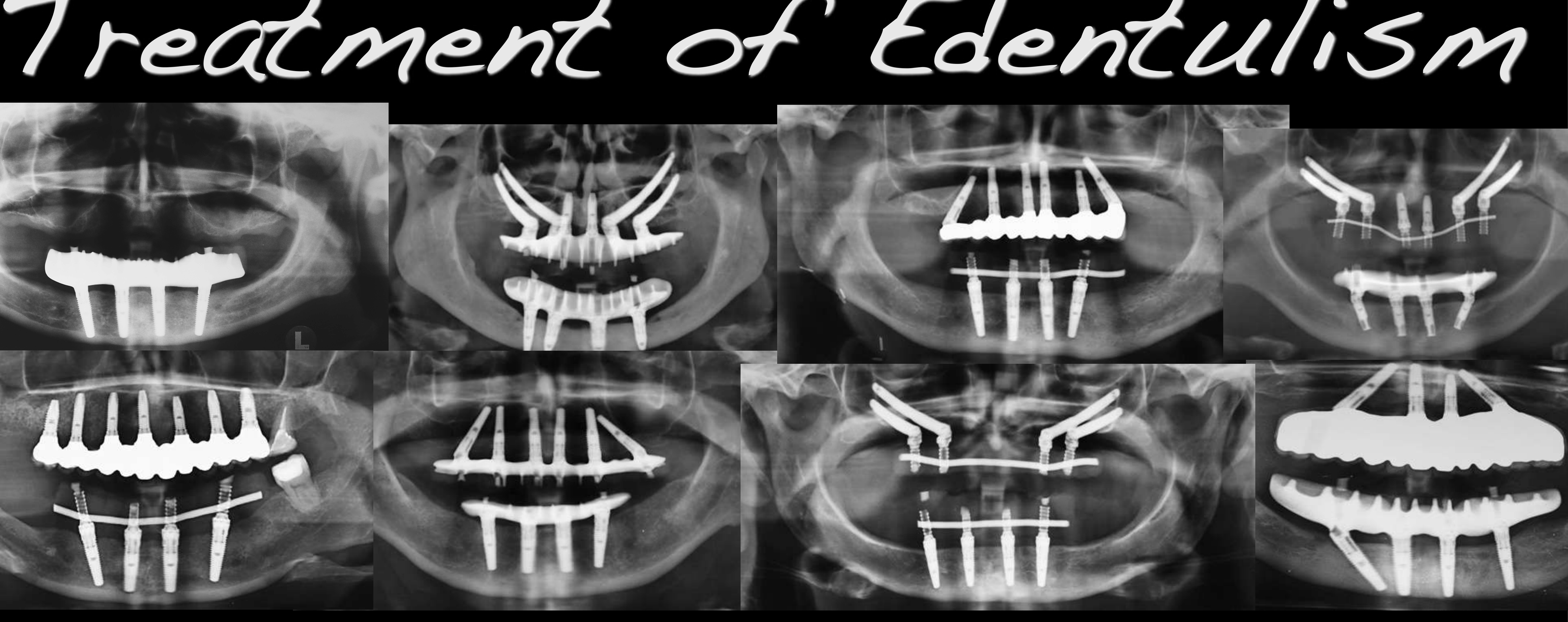
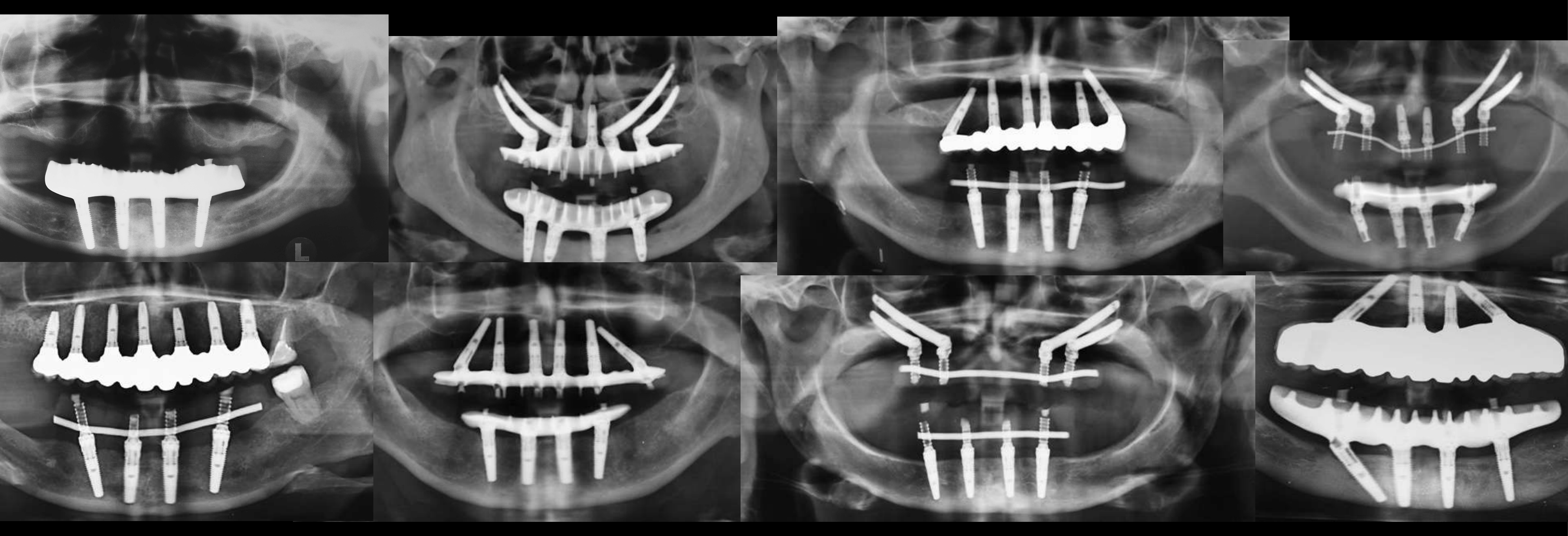
- Maxillary treatment may involve axial implants, tilted implants to avoid the sinus, grafted bone, zygomatic implants, or combinations of zygomatic and anterior implants.
- Four or six implants may be used; quad zygomatic treatment is an option when no usable maxillary bone remains.
- The prosthesis must provide facial-tissue support comparable to the support previously provided by the denture flange.
 |  |
 |
Evolution of Immediate Loading Protocols7
- 1991 — Single stage surgery and immediate loading — T-033C
- 1994 — Routine lower jaw on 4 implants, immediate loading
- 1998 — Branemark Novum — T-086
- 2000 — Immediate loading maxilla
- 2003 — Teeth in an hour (NobelGuide)
- 2008 — Immediate loading of Zygoma implants
Lecturer — Immediate Loading
Immediate loading is used to place patients into fixed teeth more quickly, but its feasibility depends on several clinical factors.
- Relevant factors include the quality of the native bone, implant stability, the amount of load placed on the implants, and the patient’s medical and behavioral risk factors.
- Native bone provides the option of loading when adequate stability is achieved, but healing and biological stability remain important even when immediate loading is selected.
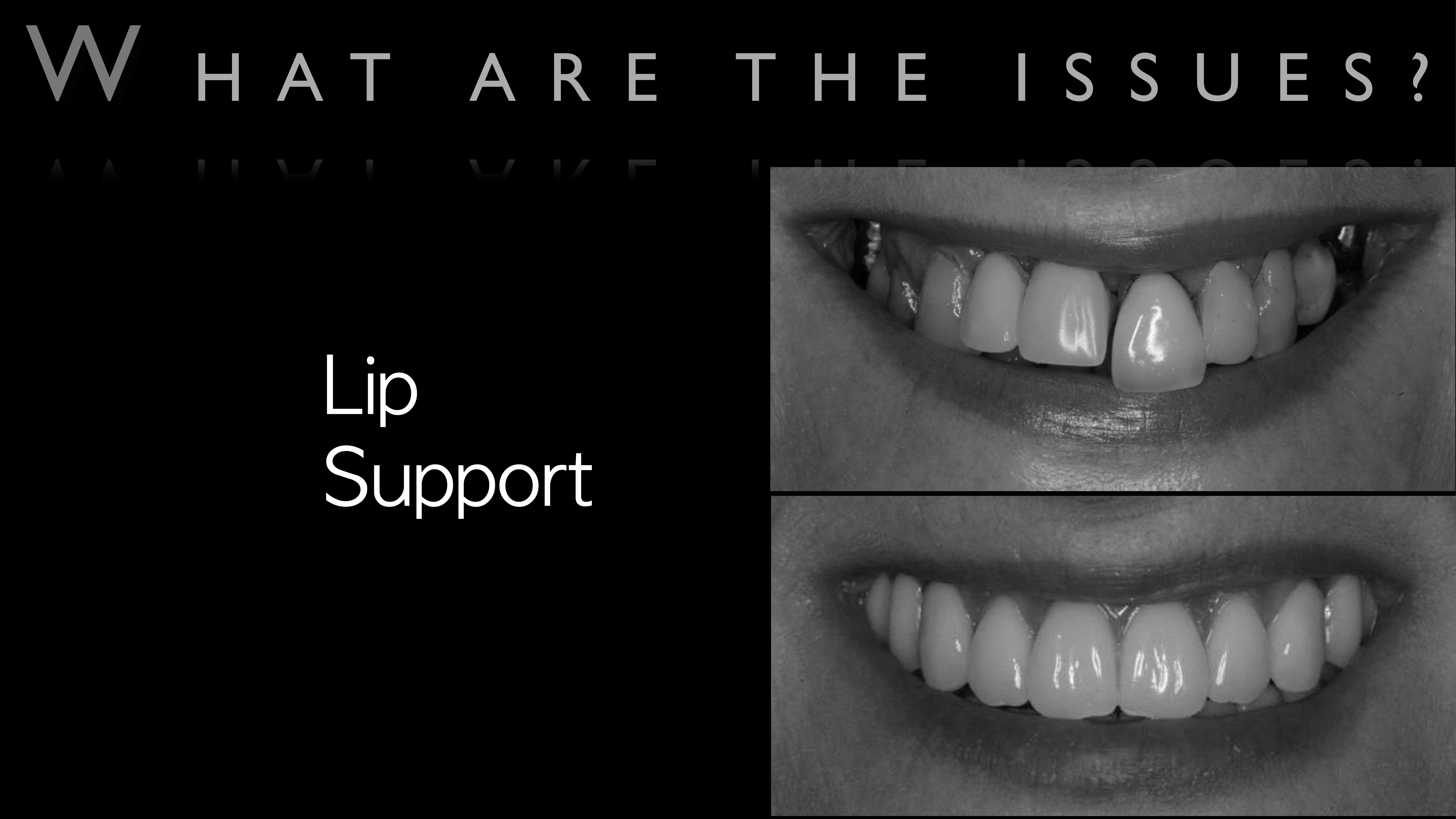
- Tooth loss is followed by substantial reduction of the alveolar bone, which contributes to loss of lip support.
- Assessment should include the previous prosthesis, the lip support it provided, facial aesthetics before treatment, tooth and gingival display during smiling, and individual upper-lip movement.
- Facial support also decreases with age as musculature and collagen change, contributing to wrinkles; the amount of support required differs between patients.
Lecturer — Facial Aesthetics
The goal is not merely to replace teeth but to maintain or improve the patient’s facial appearance.
- Before-and-after assessment should examine facial creases, vermilion support, changes in the lips and perioral tissues, and the appearance of the face with the proposed prosthesis.
- The prosthesis should be designed in relation to the patient’s existing facial form rather than according to a single idealized appearance.
Lecturer — Lip Support
A traditional denture flange supports the lips and perioral musculature, whereas an implant bridge cannot simply reproduce the flange because the tissues behind it must remain accessible for cleaning.
- The replacement must allow toothbrushing, flossing, use of a water jet, and use of interdental cleaning aids.
- Reducing the bulk of the flange-like support can cause the upper lip to collapse inward, particularly with a thin upper lip.
- Upper-lip mobility and individual orofacial musculature must be considered when designing the prosthesis.
Lecturer — Ridge Reduction
Ridge reduction may be necessary to create a prosthetic contour that provides adequate facial support while remaining cleansable.
- Reducing the ridge can allow the bridge contour to approach the support previously provided by a denture flange.
- Excessive reduction may improve prosthetic space and facial support but can also remove bone needed for implant placement.
- The amount of reduction must be planned in relation to the desired prosthetic contour and the available implant sites.
Lecturer — Tooth Position
Implant-supported teeth can be positioned more freely than denture teeth because the implant prosthesis is rigid, while denture teeth generally need to remain close to the ridge to avoid destabilizing the denture.
- Tooth position should be determined before implant placement and related to facial aesthetics, lip support, phonetics, the transition to natural gingiva, and final implant position and angulation.
- Digital smile design involves drawing on photographs and quantifying desired changes.
- Successful treatment requires close coordination between the dental laboratory and clinic during design and fabrication.
Lecturer — Replacement Classifications
FP1 replaces only the teeth, without replacing gingival or mucosal tissue, whereas FP2 includes some pink tissue replacement.
- FP1 is often presented as an ideal, but it may be limited in patients who have lost teeth and supporting bone.
- Even when tissue grafting creates papillae and root coverage, the result may still have long contours and small papillae, producing a band-like appearance if the ceramics are not carefully designed.
Patient Assessment and Factors to Consider
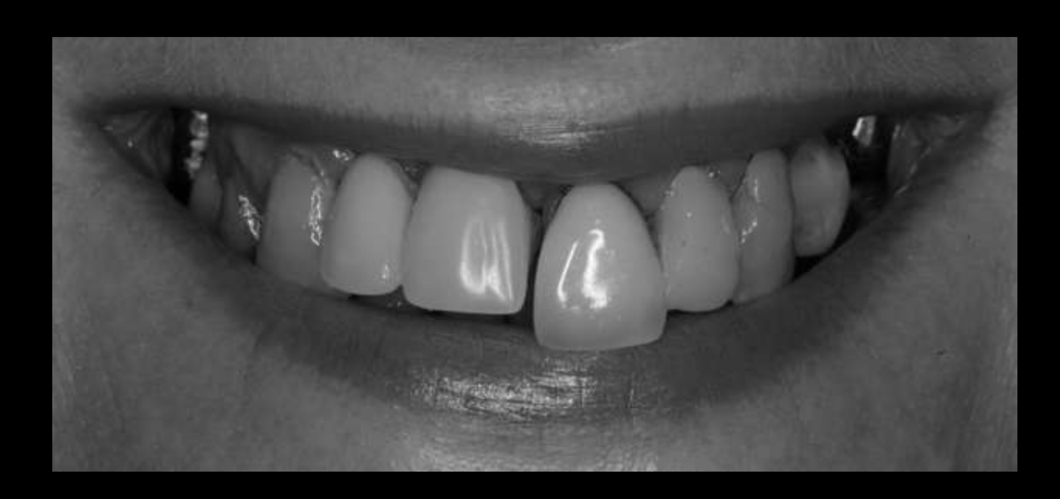
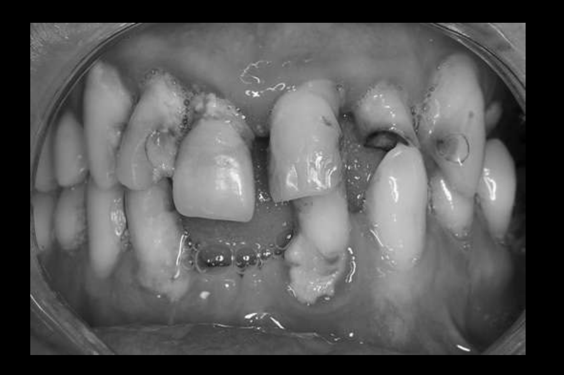
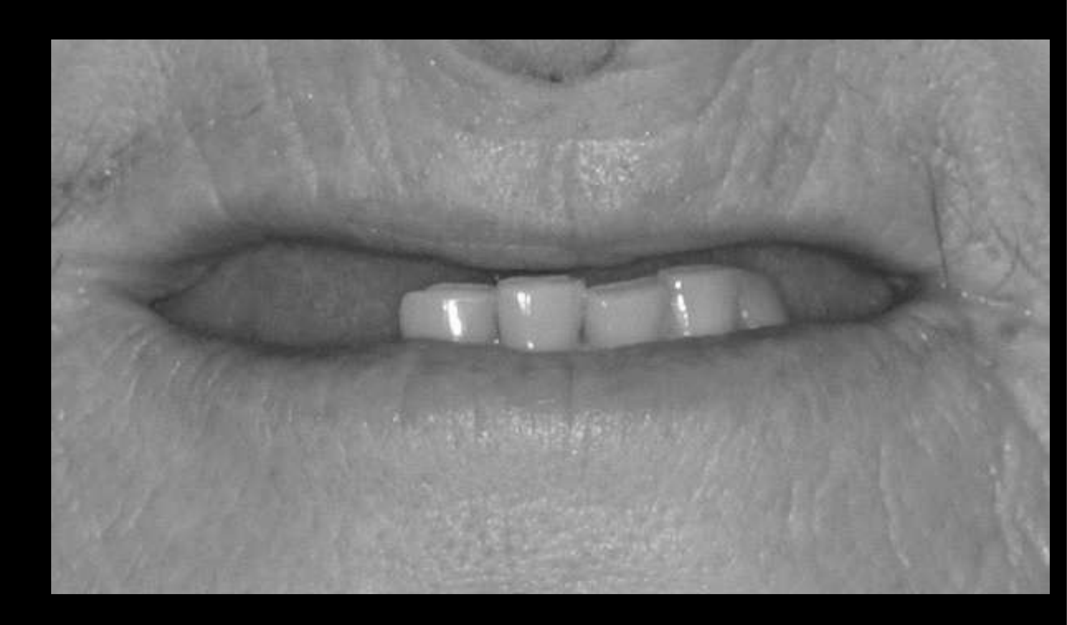
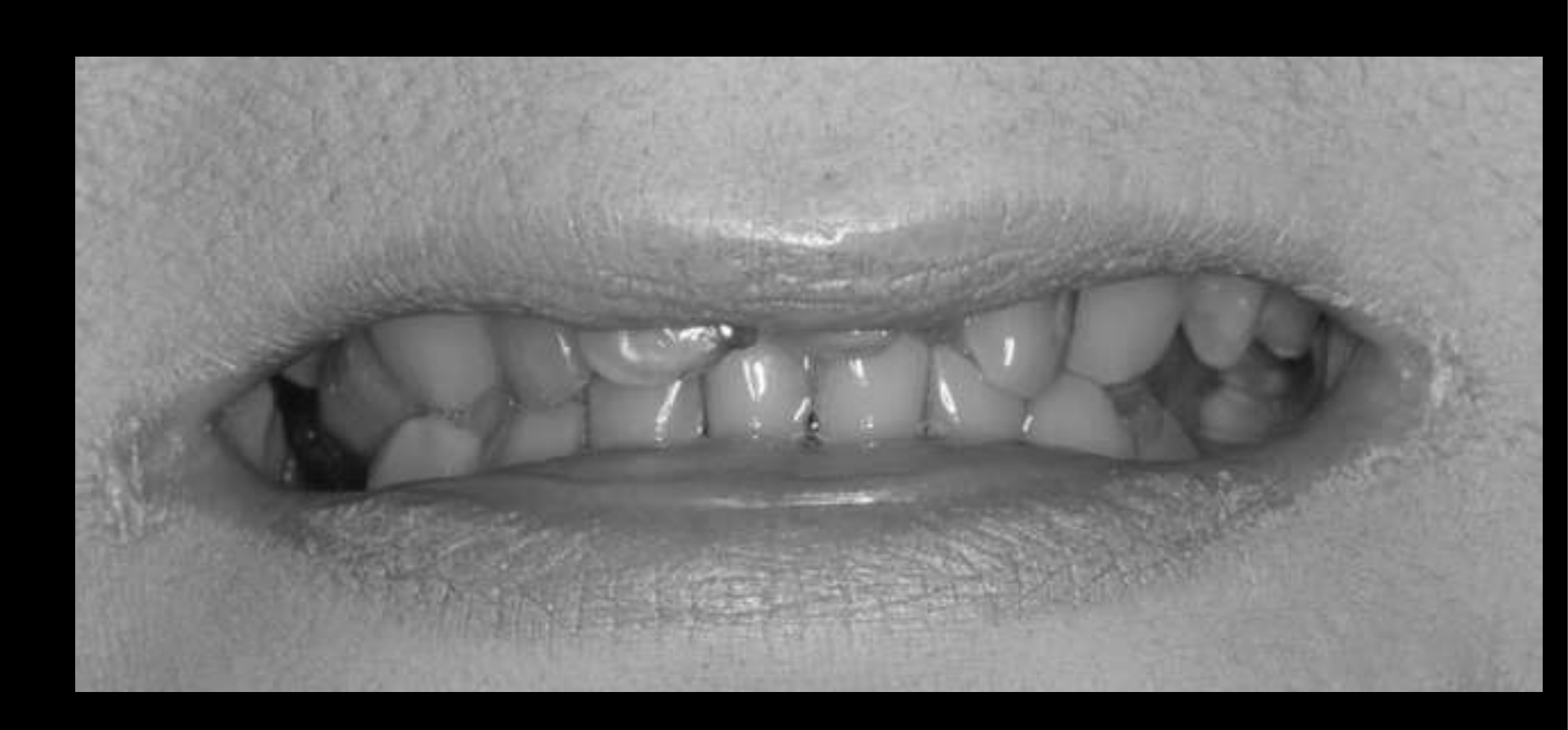
Sequelae of Tooth Loss89
Factors to consider
- Initial aesthetic expectation
- Current dentition, prostheses
 |  |
 |  |
Facial Aesthetics and Lip Support
What are the issues?
Facial Aesthetics10
What are the issues?
Lip Support11
Ridge Reduction and Facial Support12
Ridge Reduction13
Facial Support
Ridge Reduction — Facial Support14
Lecturer — Facial Support
Facial support was compared between a traditional denture and an implant-supported bridge.
- Anthropometric mask technology was used to evaluate changes in facial support between the pre-treatment prosthesis and proposed restoration.
- Three-dimensional simulations can help evaluate how prosthetic changes may affect facial support.
- The desired outcome is to maintain or improve facial support without creating a prosthesis that cannot be cleaned.
Tooth Position and Aesthetics Within the Face1516
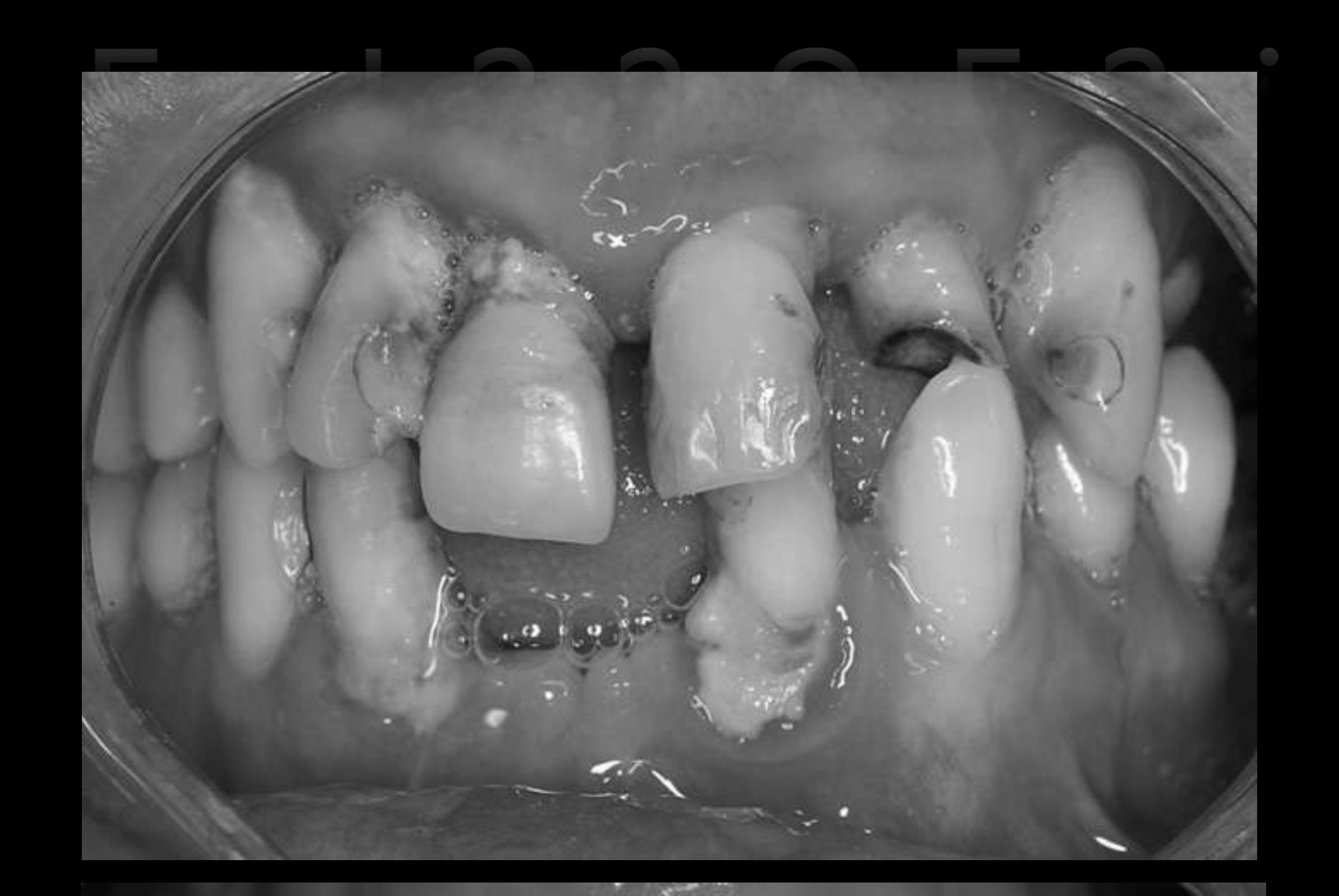
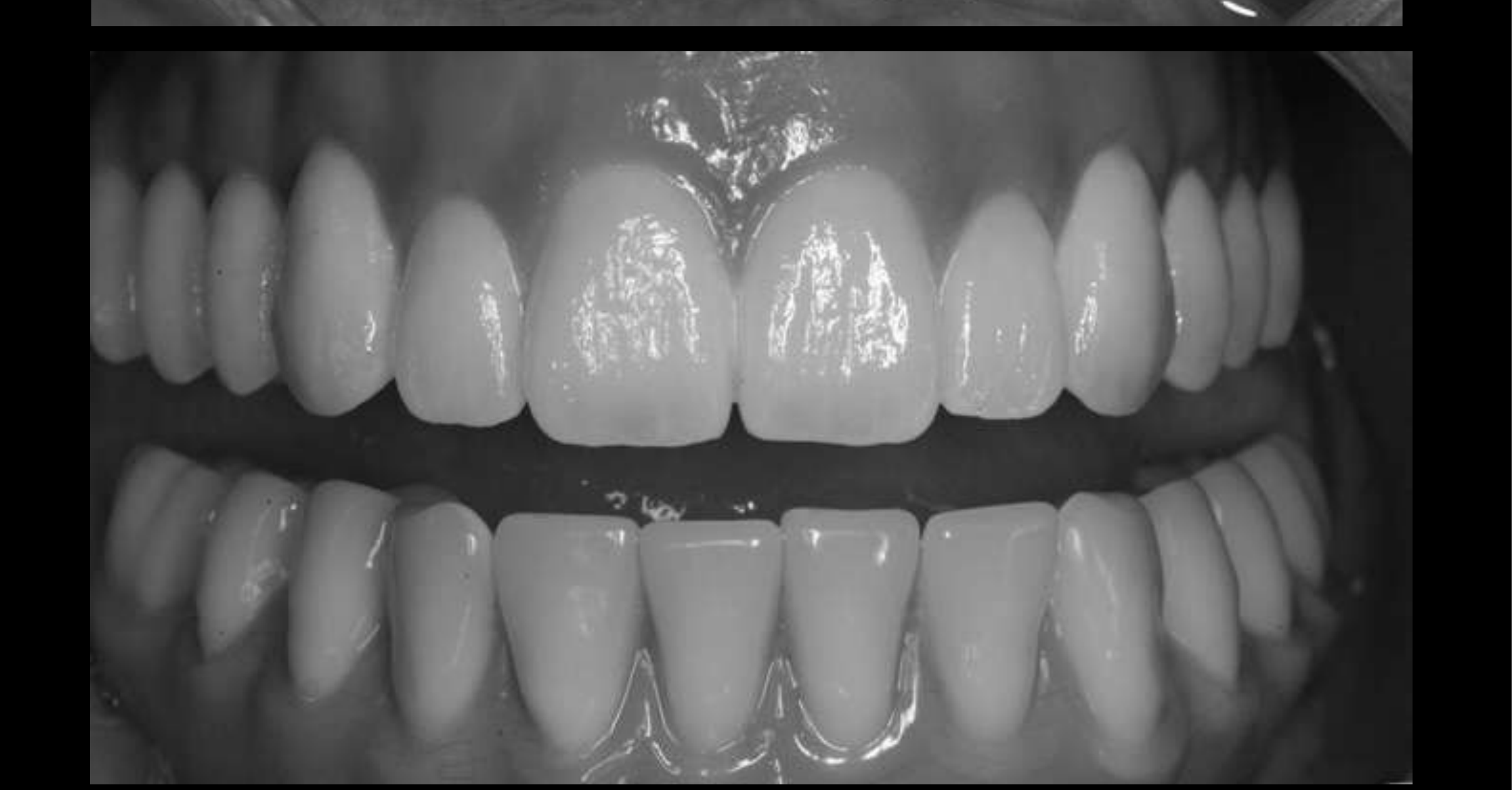
The Face — Determination of tooth position within the face
What are the issues?
Tooth aesthetics and function17
 |  |
Digital Planning and Work-Up
Digital Smile Design and Facebow Transfer1819202122232425
This page consists of figures only; the slide text serves solely as image labels.
Coachman C, Calamata M. Digital Smile Design: A Tool for Treatment Planning and Communication in Esthetic Dentistry. Quintessence Dental Technology 2012
Lecturer — Prosthetically Driven Planning
Radiographic work-up relates the available bone to the planned restoration.
- A CAT scan or cone-beam scan is used to evaluate the available bone.
- A trial prosthesis determines where implants should be placed in relation to the intended teeth.
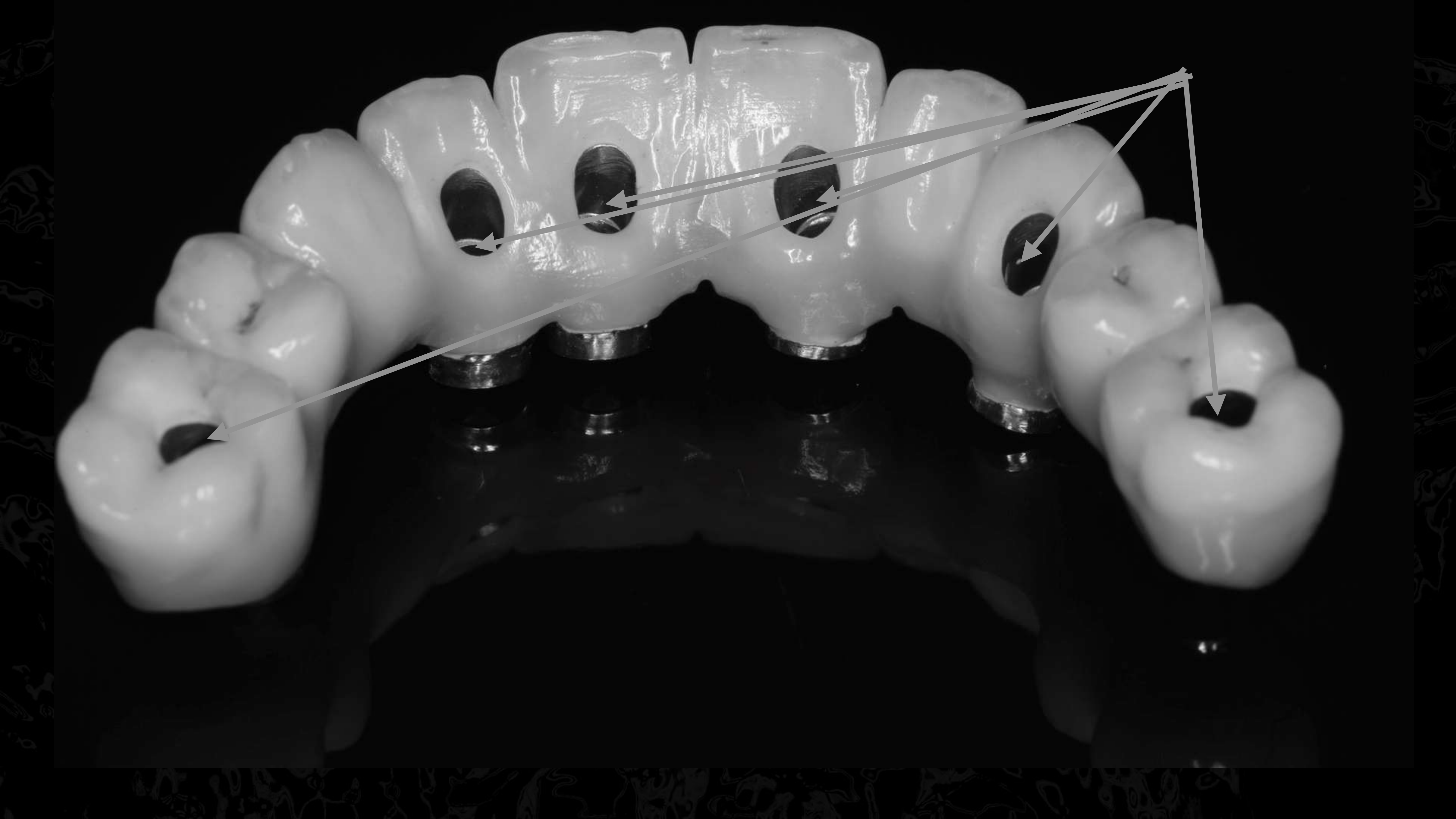
- The screw access should ideally emerge through an appropriate area of the tooth rather than through an unaesthetic or anatomically disruptive position.
- An implant placed at an unsuitable angle may require a large restorative bulge that looks non-anatomical, interferes with the tongue, and makes cleaning difficult.
- Implant positions may involve native bone, grafted bone, zygomatic bone, or other extra-alveolar positions.
- The key objective is for the implants to emerge through the basic position of the planned bridge and adapt properly to the prosthesis.
Lecturer — Digital Smile Design
Tooth position can be planned using analog denture principles or digital smile design.
- Digital smile design involves drawing on images of the patient, quantifying the changes to be made, and planning the appearance of the final teeth.
- The design process must be coordinated with the laboratory.
- Once tooth position and required lip support have been determined, implant positions can be planned in relation to the proposed teeth.
Digital Ruler
“Real Face Bow”
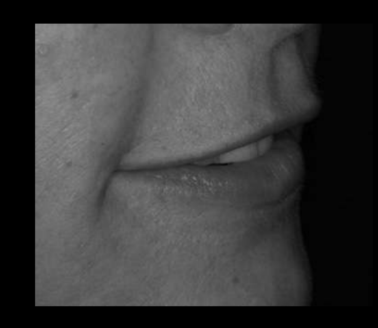
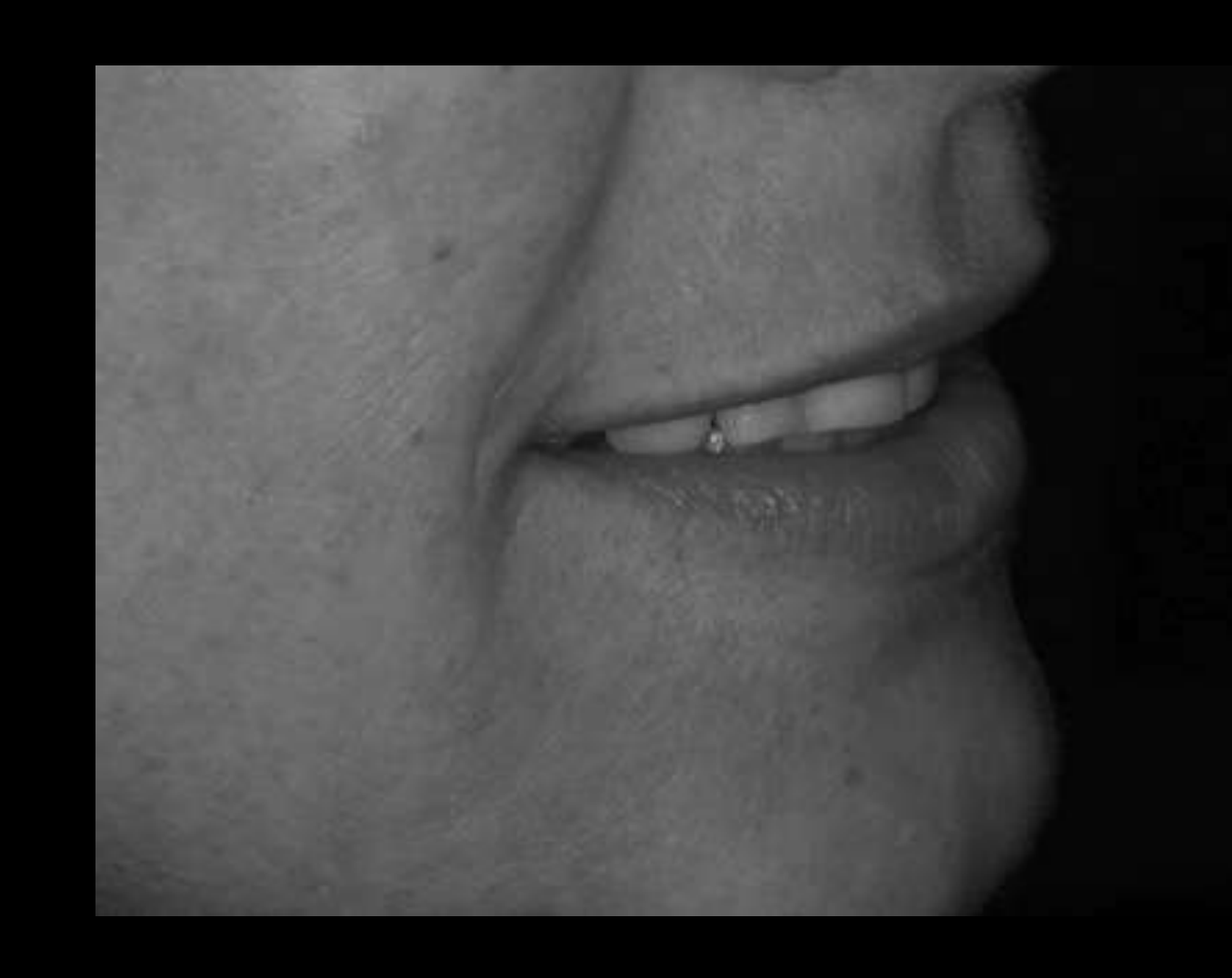
Junction between implant and prosthetic tooth determines lip support and phonetic surface
Image shows a collection of custom dental prosthetic devices (dentures or implant-retained suprastructures) against a black background.
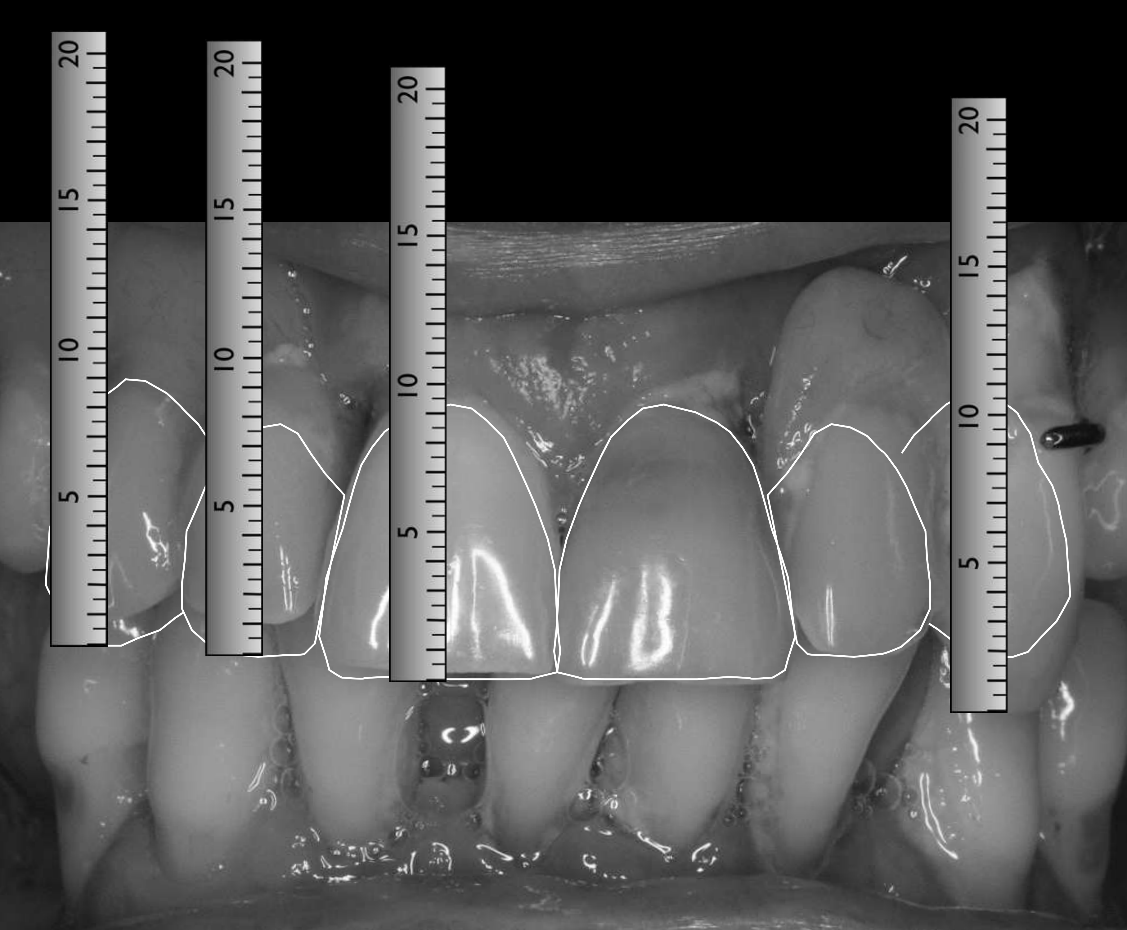
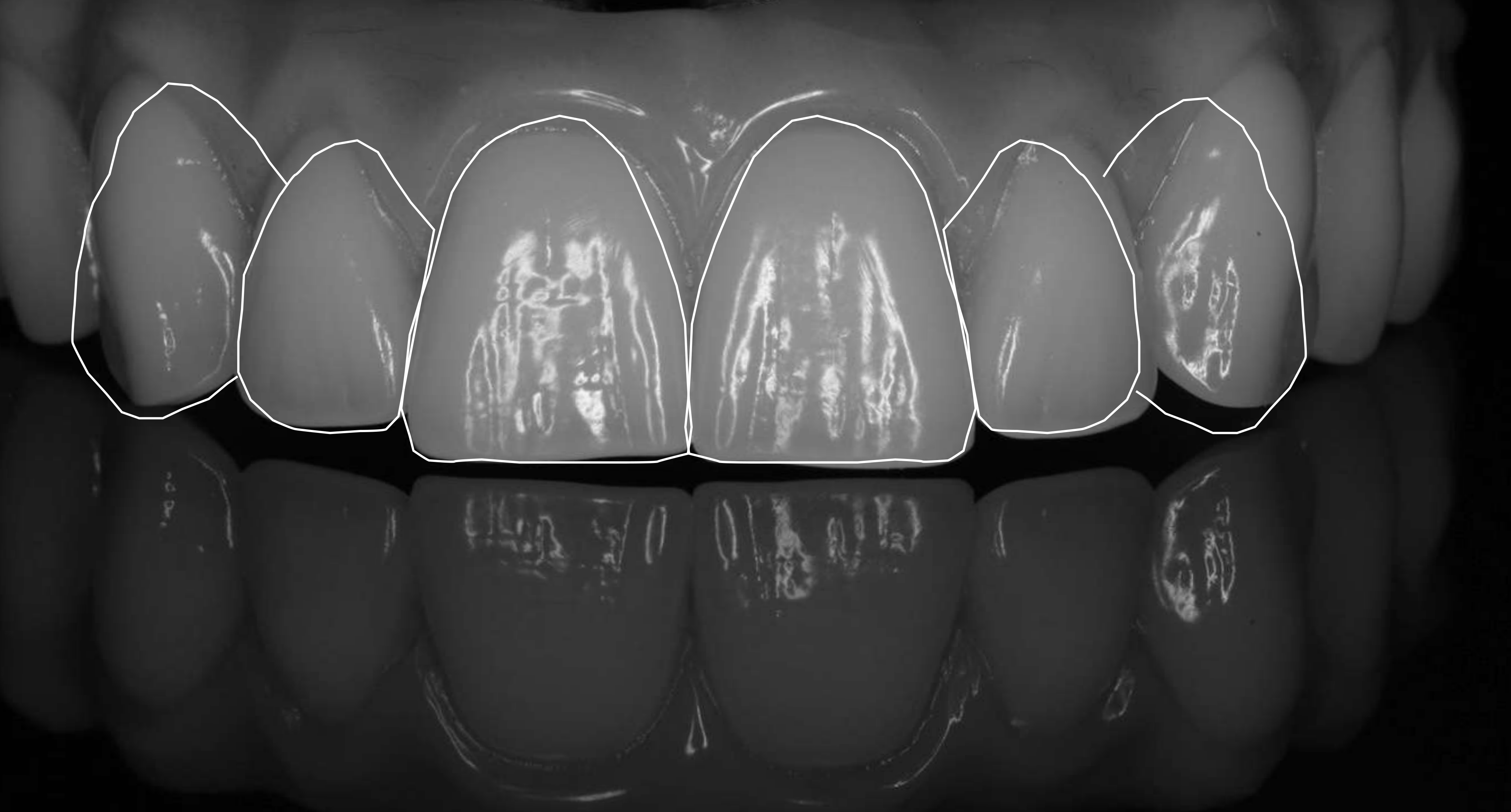
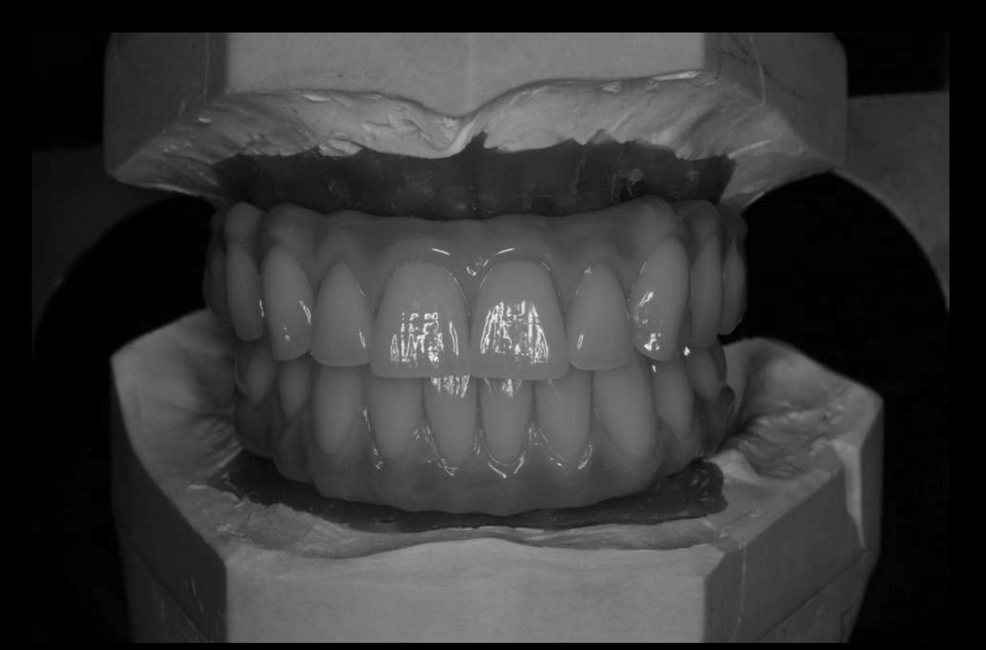
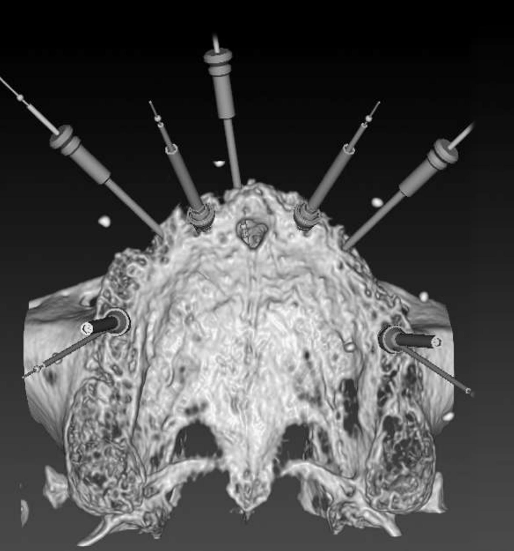
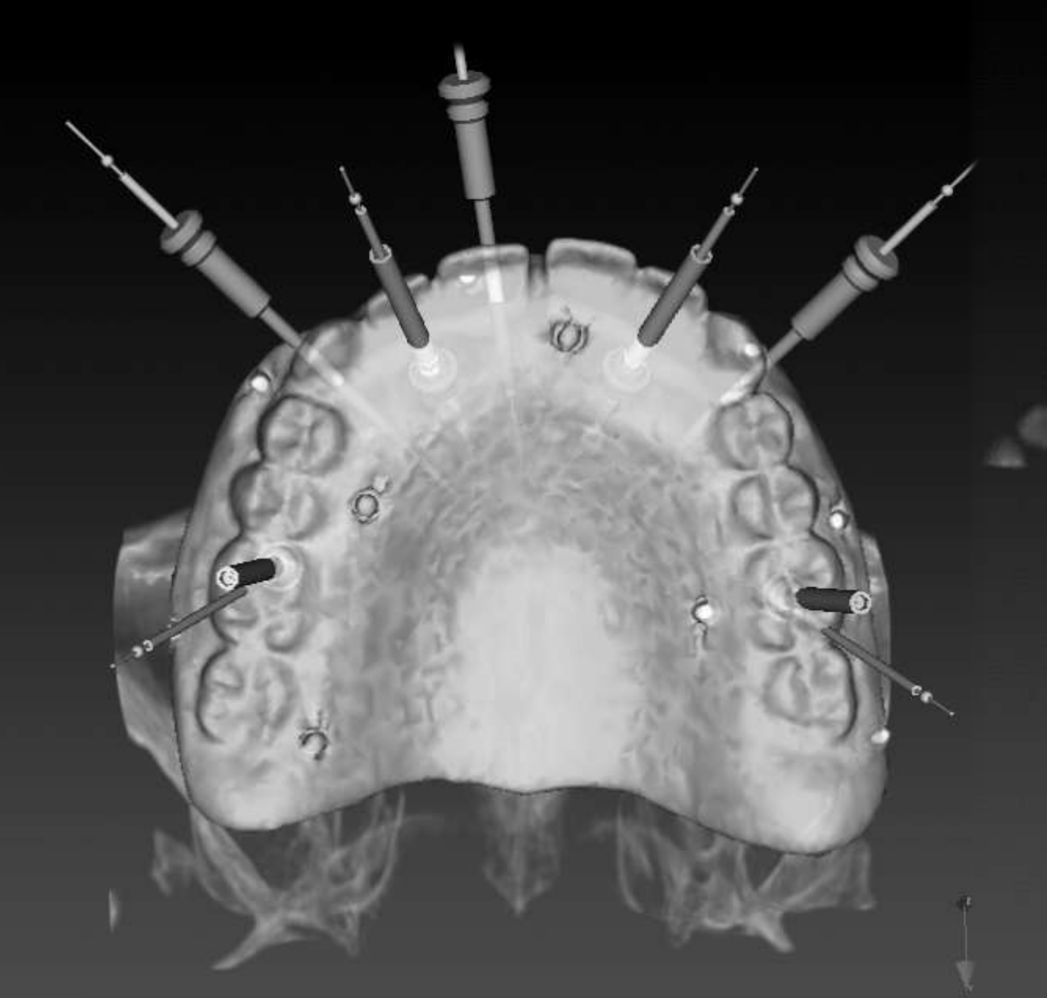
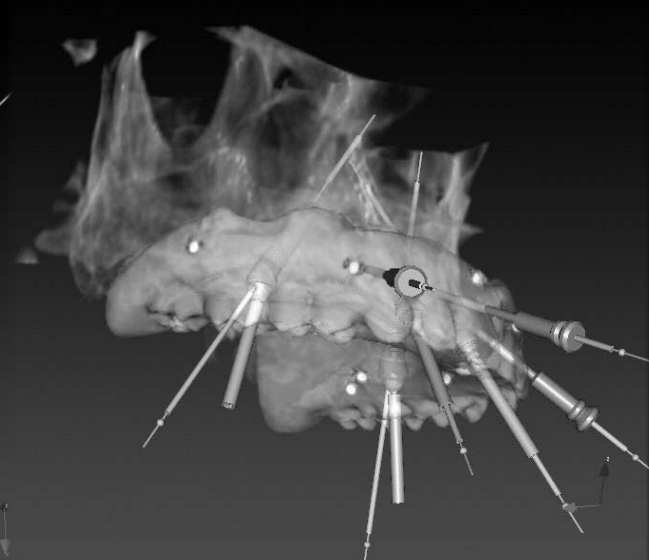
Radiographic Work-Up and Prosthetically Driven Planning26272829303132
The Face
Transfer to radiographic work up and NobelClinician software
Double Scan
Planning implant placement — prosthetically driven
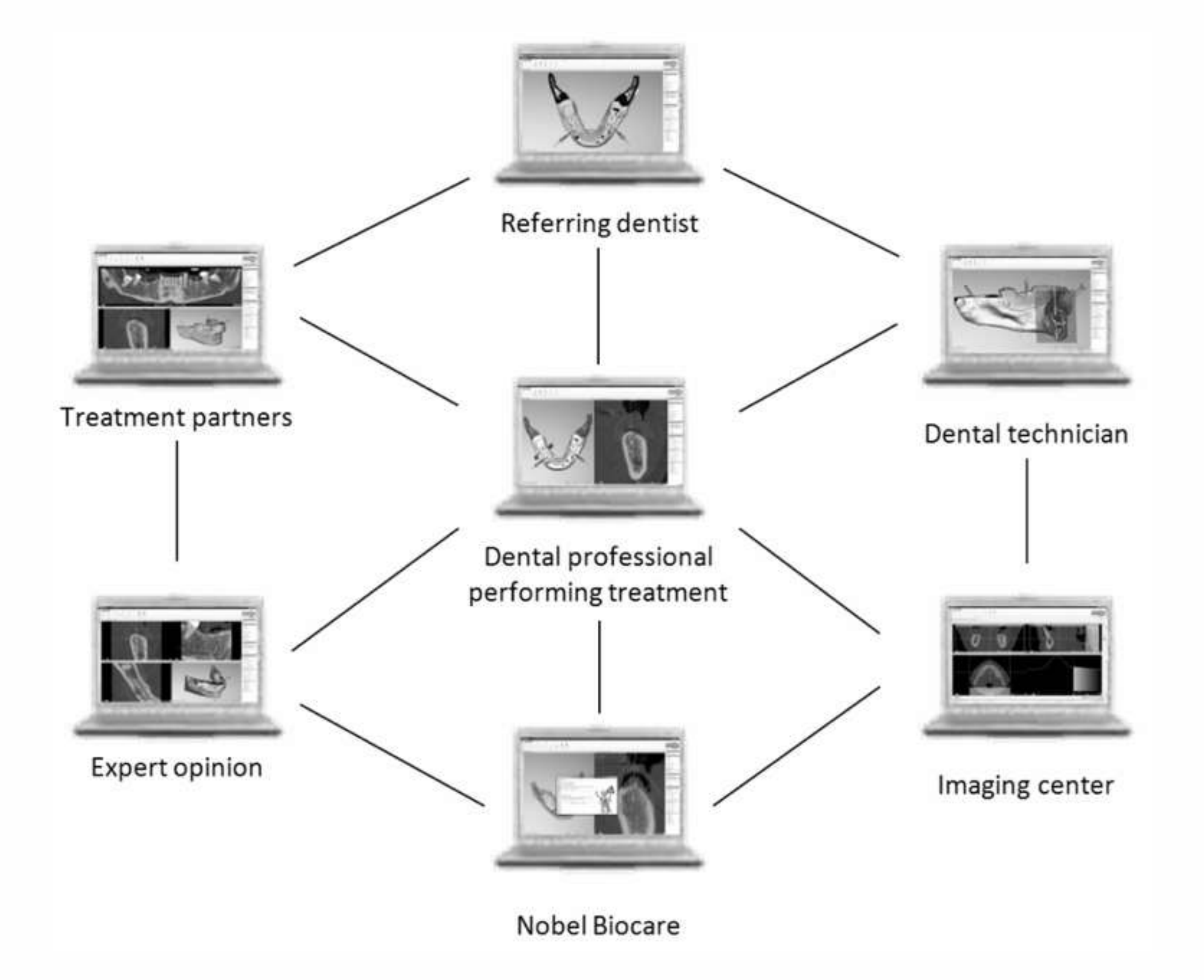
NobelConnect
- Critical for complex interdisciplinary treatment

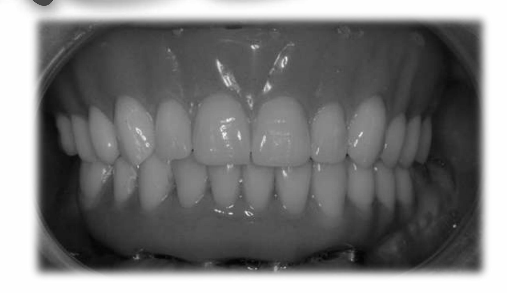
Implant Surgical Pathways33
Graftless
-
Axial
-
Tilted
-
Immediate loading
-
Implants engaging the zygoma Grafting
-
Sinus
-
Labial onlay
-
Ridge splitting
-
GBR
-
Interpositional
-
Distraction osteogenesis
-
LeFort 1
-
Free tissue transfer
Grafting material options:
- Autogenous
- Allograft
- Xenograft
- Synthetic
- BMPs
- Gene therapy
Lecturer — Surgical Planning
The surgical pathway depends on the available bone and the intended prosthetic result.
- The choice is influenced by the amount and location of available bone, the position of the sinus, the desired tooth position, the required facial support, and the patient’s risk profile.
- Straight axial implants are uncommon in abundant maxillary bone because many patients do not have sufficient bone.
- Implant placement should begin with the prosthetic plan, followed by identification of the bone that can support the implants.
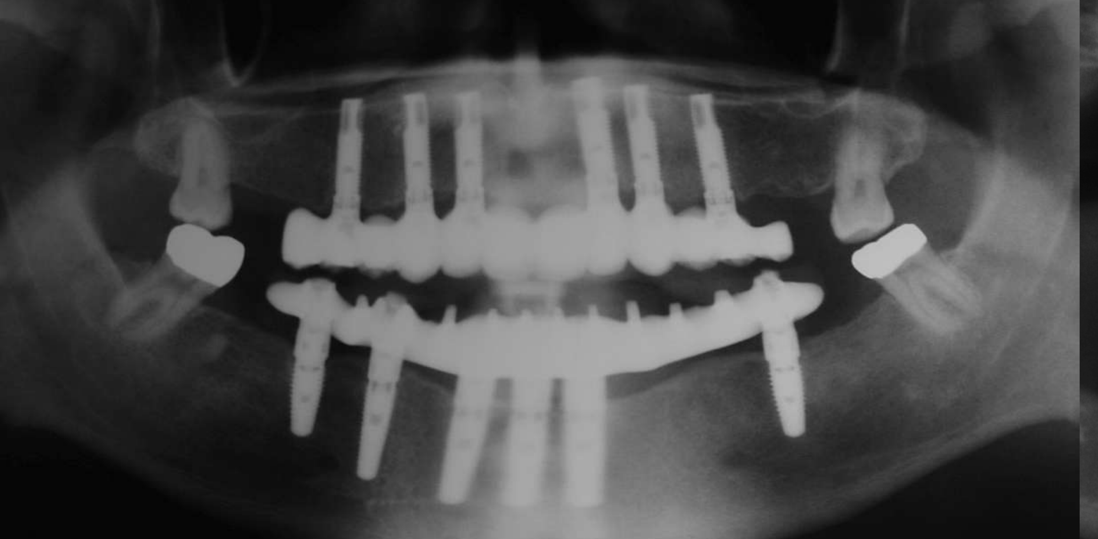 | 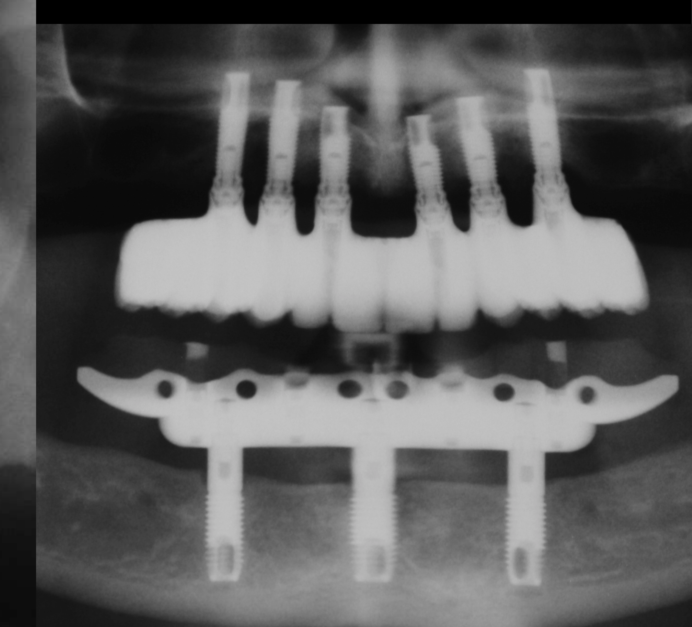 |
Clinical Studies: Axial Implants Without Grafting3435
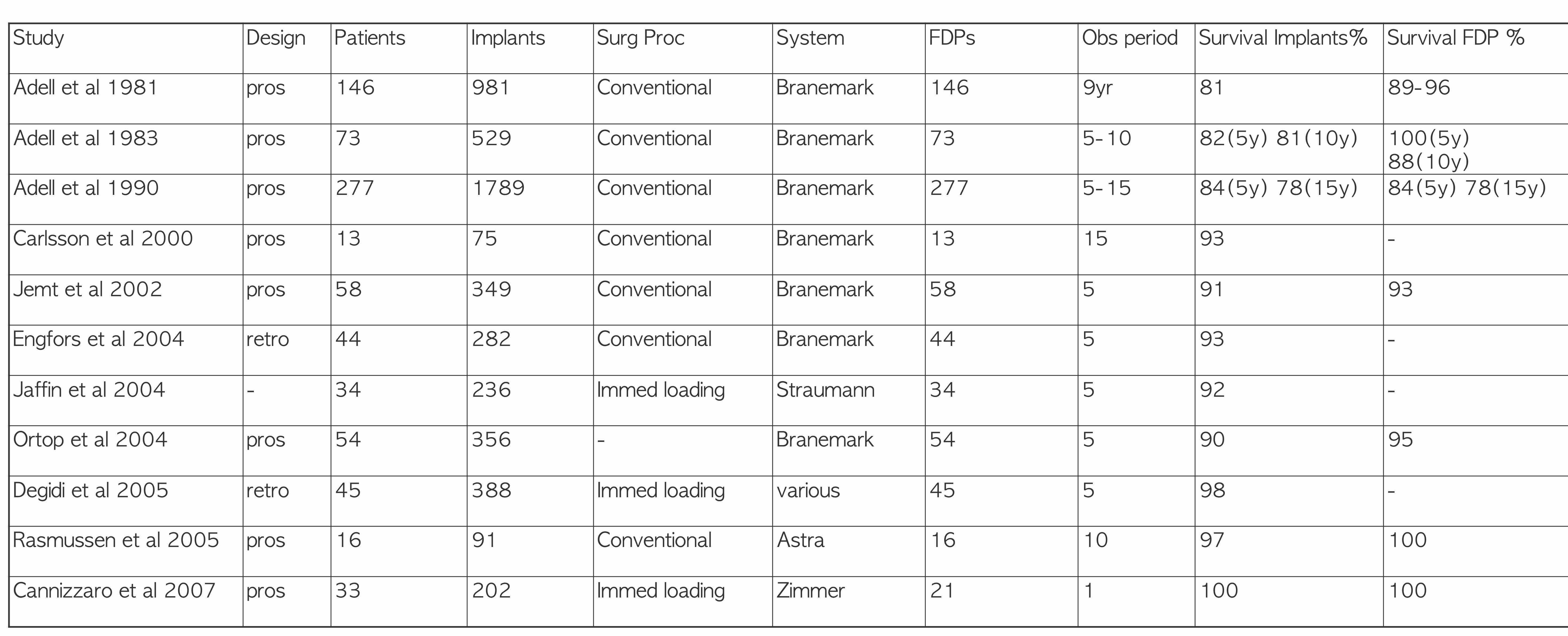
Studies restoring the edentulous maxilla with fixed prostheses — axial implants, no grafting:
| Study | Design | Patients | Implants | Surg Proc | System | FDPs | Obs period | Survival Implants % | Survival FDP % |
|---|---|---|---|---|---|---|---|---|---|
| Adell et al 1981 | pros | 146 | 981 | Conventional | Branemark | 146 | 9 yr | 81 | 89–96 |
| Adell et al 1983 | pros | 73 | 529 | Conventional | Branemark | 73 | 5?–10 | 82 (5y), 81 (10y) | 100 (5y), 88 (10y) |
| Adell et al 1990 | pros | 277 | 1789 | Conventional | Branemark | 277 | 5–15 | 84 (5y), 78 (15y) | 84 (5y), 78 (15y) |
| Carlsson et al 2000 | pros | 13 | 75 | Conventional | Branemark | 13 | 15 | 93 | – |
| Jemt et al 2002 | pros | 58 | 349 | Conventional | Branemark | 58 | 5 | 91 | 93 |
| Engfors et al 2004 | retro | 44 | 282 | Conventional | Branemark | 44 | 5 | 93 | – |
| Jaffin et al 2004 | – | 34 | 236 | Immed loading | Straumann | 34 | 5 | 92 | – |
| Ortop et al 2004 | pros | 54 | 356 | – | Branemark | 54 | 5 | 90 | 95 |
| Degidi et al 2005 | retro | 45 | 388 | Immed loading | various | 45 | 5 | 98 | – |
| Rasmussen et al 2005 | pros | 16 | 91 | Conventional | Astra | 16 | 10 | 97 | 100 |
| Cannizzarro et al 2007 | pros | 33 | 202 | Immed loading | Zimmerman | 21 | 1 | 100 | 100 |
Tilted implants, no grafting:
- Maxilla: 13-year follow-up
- 17-year follow-up
- Mandible: 24-year follow-up
Lecturer — Axial Implant Studies
Axial implants are placed in a relatively straight, tooth-axis position, and long-term studies extend back to the early 1980s. The studies discussed showed high success rates when sufficient bone is available, and native bone can provide enough stability for immediate loading.
 | 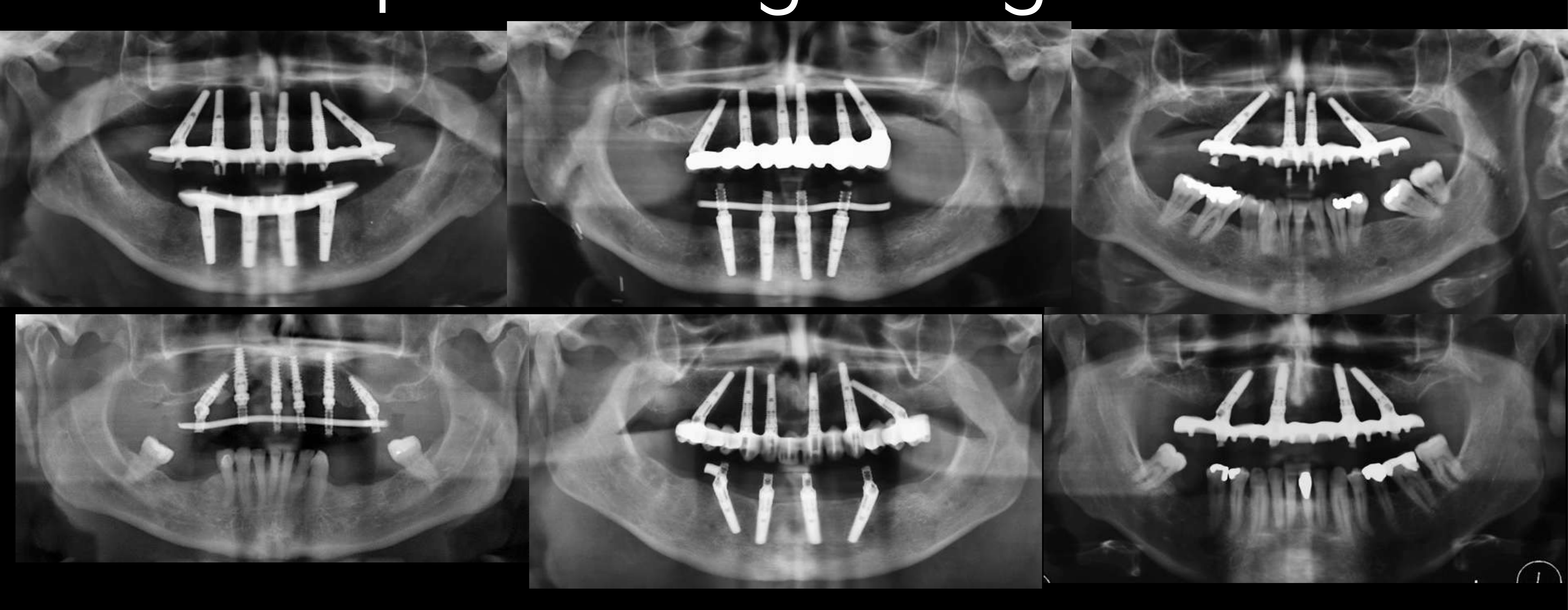 |
Clinical Studies: Tilted Implants Without Grafting363738
Superstructure cementation
Axial CT scan (C1) and osseous contour details
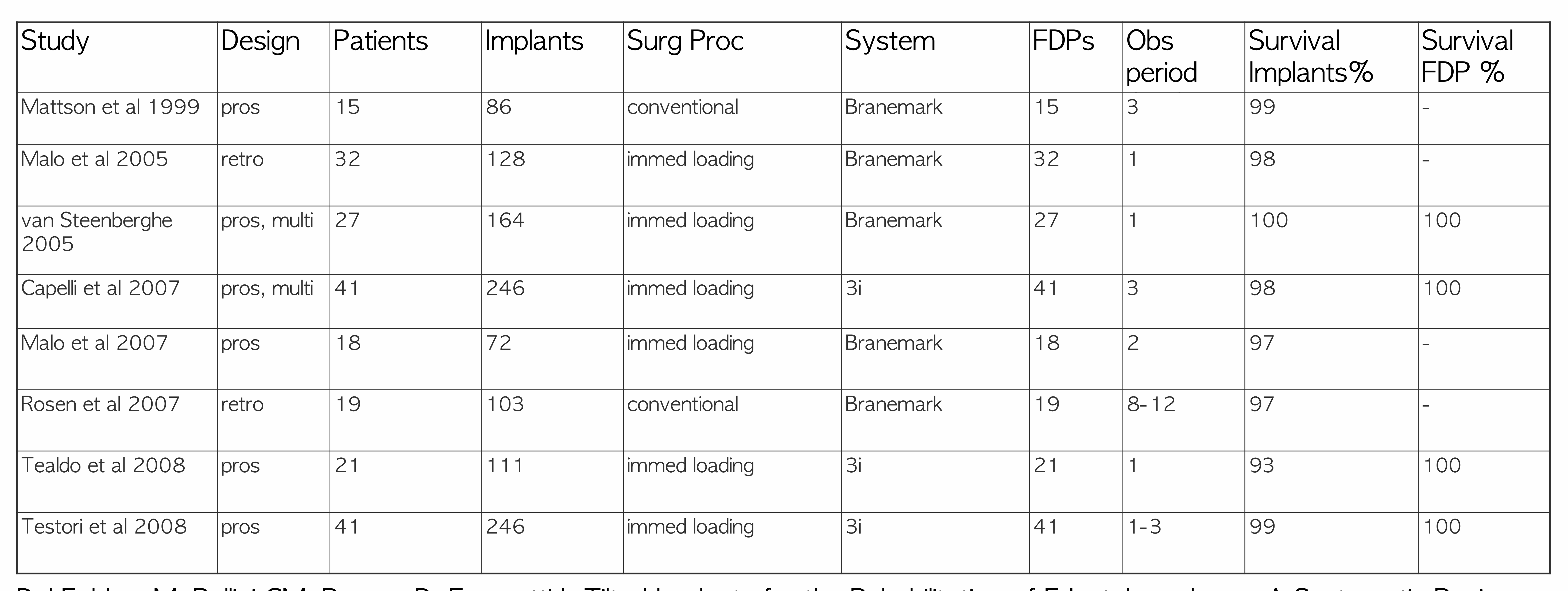
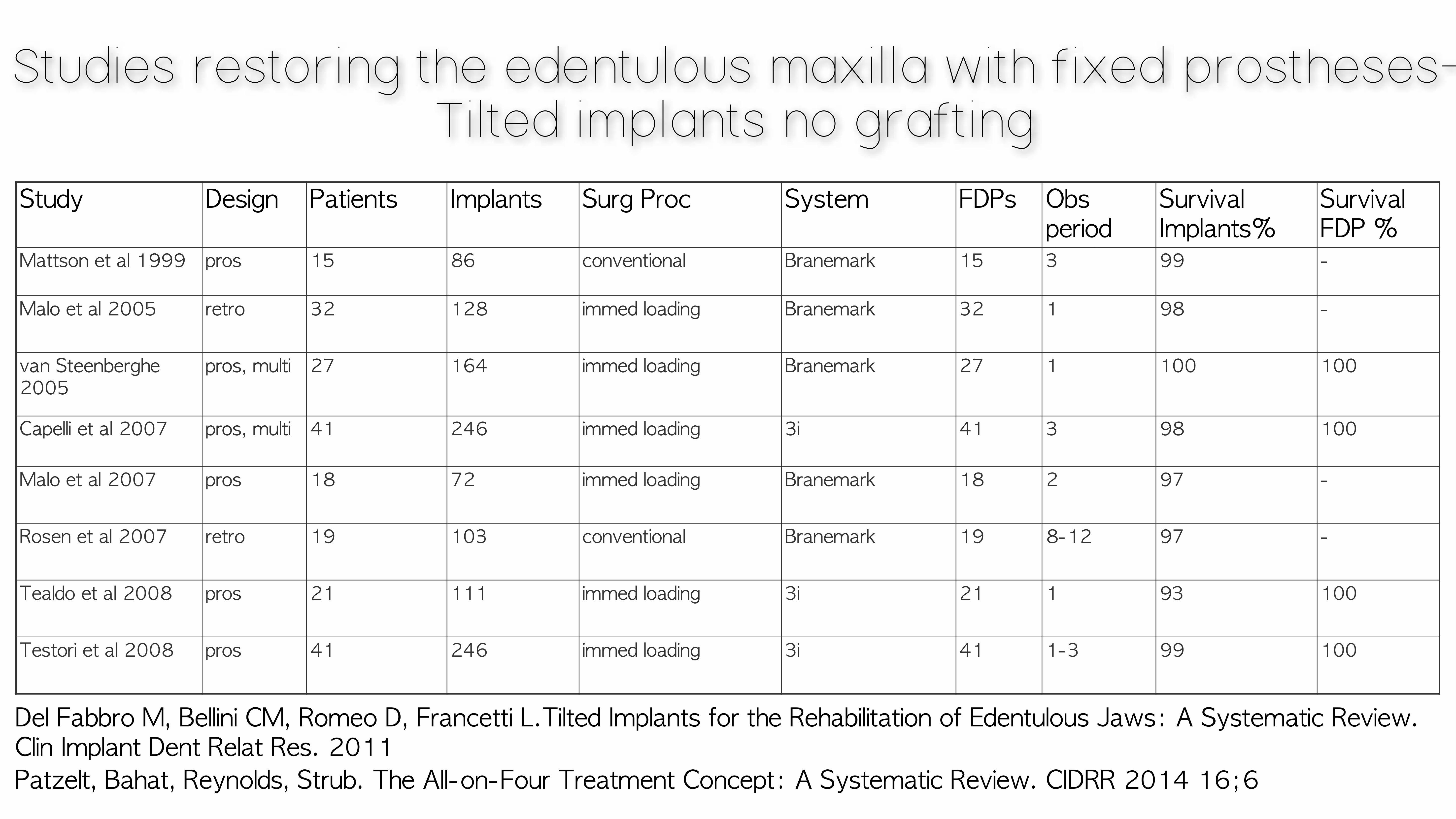
Studies restoring the edentulous maxilla with fixed prostheses — tilted implants, no grafting:
| Study | Design | Patients | Implants | Surg Proc | System | FDPs | Obs period | Survival Implants % | Survival FDP % |
|---|---|---|---|---|---|---|---|---|---|
| Mattson et al 1999 | pros | 15 | 86 | conventional | Branemark | 15 | 3 | 99 | – |
| Malo et al 2005 | retro | 32 | 128 | immed loading | Branemark | 32 | 1 | 98 | – |
| van Steenberghe 2005 | pros, multi | 27 | 164 | immed loading | Branemark | 27 | 1 | 100 | 100 |
| Capelli et al 2007 | pros, multi | 41 | 246 | immed loading | 3i | 41 | 3 | 98 | 100 |
| Malo et al 2007 | pros | 18 | 72 | immed loading | Branemark | 18 | 2 | 97 | – |
| Rosen et al 2007 | retro | 19 | 103 | conventional | Branemark | 19 | 8–12 | 97 | – |
| Tealdo et al 2008 | pros | 21 | 111 | immed loading | 3i | 21 | 1 | 93 | 100 |
| Testori et al 2008 | pros | 41 | 246 | immed loading | 3i | 41 | 1–3 | 99 | 100 |
Del Fabbro M, Bellini CM, Romeo D, Francetti L. Tilted Implants for the Rehabilitation of Edentulous Jaws: A Systematic Review. Clin Implant Dent Relat Res. 2011.
Patzelt, Bahat, Reynolds, Strub. The All-on-Four Treatment Concept: A Systematic Review. CIDRR 2014 16;6.
Studies restoring the edentulous maxilla with fixed prosthesis — tilted implants, no grafting:
No significant difference in failure rate or marginal bone levels between axial and tilted implants.
Menini M, Signori A, Tealdo T, Bevilacqua M, Pera F, Ravera G, Pera P. Tilted implants in the immediate loading rehabilitation of the maxilla: a systematic review. J Dent Res. 2012;91(9):821-7.
Del Fabbro M, Bellini CM, Romeo D, Francetti L. Tilted Implants for the Rehabilitation of Edentulous Jaws: A Systematic Review. Clin Implant Dent Relat Res. 2011.
Patzelt, Bahat, Reynolds, Strub. The All-on-Four Treatment Concept: A Systematic Review. CIDRR 2014 16;6.
Parel SM, Phillips WR. A risk assessment treatment planning protocol for the four implant immediately loaded maxilla: preliminary findings. J Prosthet Dent. 2011;106(6):359-66.
Maló P, Araújo Nobre MD, Lopes A, Rodrigues R. Double Full-Arch Versus Single Full-Arch, Four Implant-Supported Rehabilitations: A Retrospective, 5-Year Cohort Study. J Prosthodont. 2015;24(4):263-70.
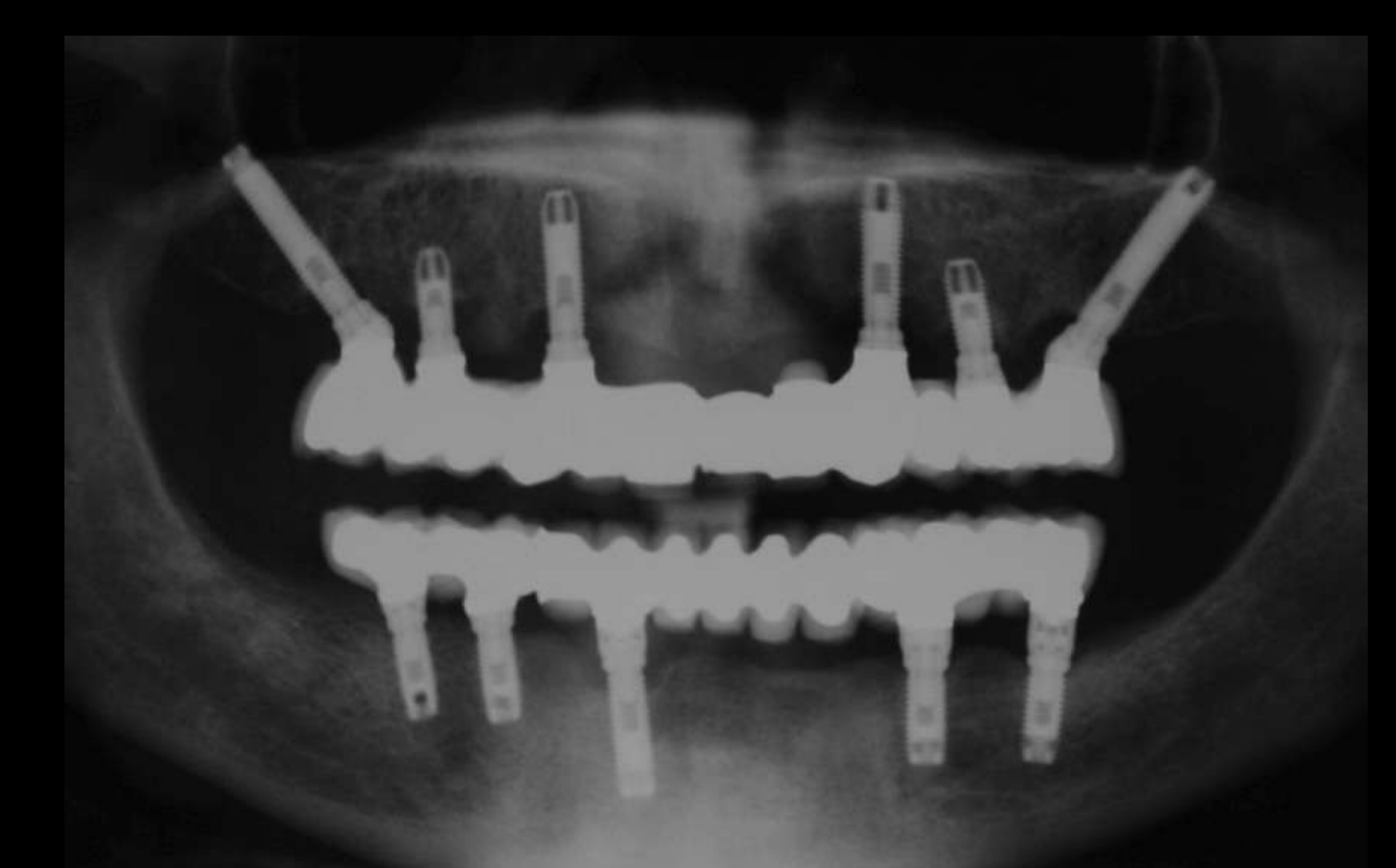 | 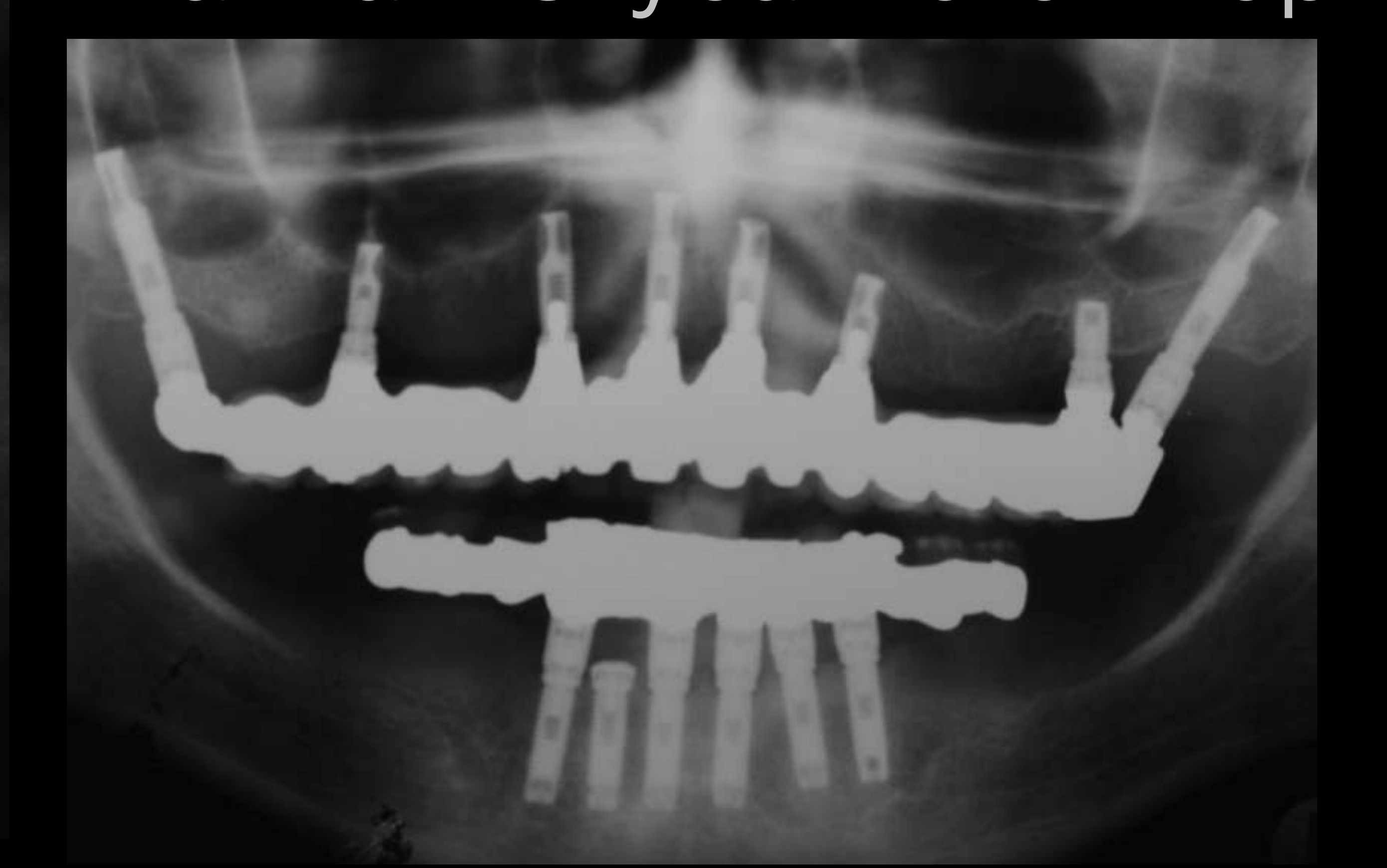 |
 |
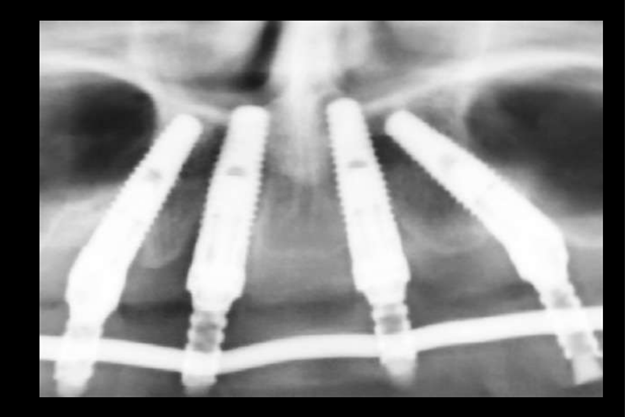
Advantages of Tilted Implants3940
-
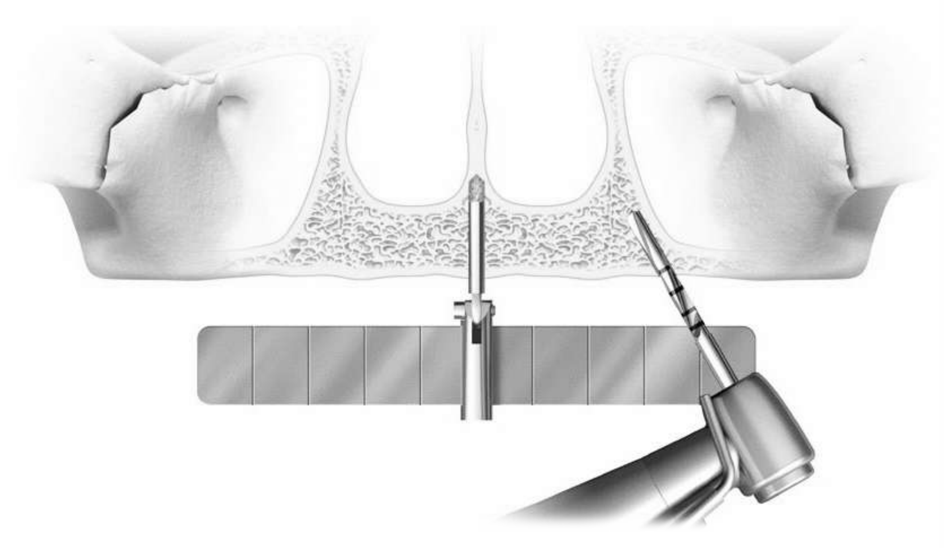
Allows for placement of longer implants — immediate loading
-
Allows for improved anchorage in dense bone
-
Allows for further distal extensions
-
Does not have a negative effect on load distribution
-
Relatively easier surgery compared to sinus lifting
-
Implants may be tilted anterior or posterior to the sinus to avoid it and increase implant distribution without grafting.
-
The further anterior the implant position, the better the bone was described as being.
Lecturer — Treatment Sequence
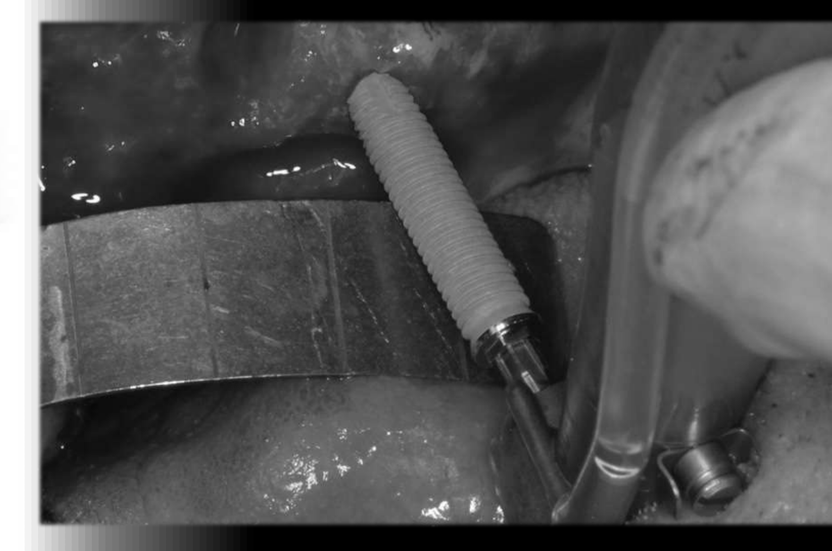
A tilted-implant treatment sequence may include several restorative and laboratory steps.
- Remove teeth requiring extraction and place the implants.
- Register the jaw using closed-tray impression copings and record the relationship between the implants and the planned teeth.
- Mount the records in an articulator, take a splinted implant impression, and create a model and provisional bridge.
- In one example, the provisional bridge remained in function for 15 years, illustrating the reported durability of this treatment.
 |  |
Graftless Full-Arch Outcomes: All-on-4 and All-on-6
All-on-4 Outcomes414243
CBCT showing dental implants with sinus lift
- 20 maxillae, 18–42 months, prospective single cohort, immediate loading
- Implant survival: 100%
- Marginal bone loss: 0.8 ± 0.4 mm axial, 0.9 ± 0.5 mm tilted
- No statistical difference in axial vs tilted implants
Agliardi E, Panigatti S, Clericò M, Villa C, Malò P. Immediate rehabilitation of the edentulous jaws with full fixed prostheses supported by four implants: interim results of a single cohort prospective study. Clin Oral Implants Res. 2010 May;21(5):459-65.
- 61 maxillae, 1–5 yrs, prospective single cohort, immediate loading
- Implant survival: 98.36%
- Marginal bone loss: 0.9 ± 0.7 mm
- No difference in axial vs tilted implants
Agliardi EL, Francetti L, Romeo D, Del Fabbro M. Immediate rehabilitation of the edentulous maxilla: preliminary results of a single-cohort prospective study. Int J Oral Maxillofac Implants. 2009 Sep-Oct;24(5):887-95.
- Tilted implants may be incorporated to avoid the sinus and allow a fixed bridge without grafting.
Systematic Review: All-on-4 Outcomes44
- 13 papers
- 4804 implants, 1201 prostheses within 48 hrs
- 99% success at 3 yrs (implants)
- 99.9% success at 3 yrs (prosthesis)
- Bone loss 1.3 ± 0.4 mm at 3 yrs
- No significant difference between axial and tilted implants
- Most failures with smokers and bisphosphonate medication
- Short-term results
S. B. M. Patzelt, O Bahat, BDS, M. A. Reynolds, J. R. Strub, The All-on-Four Treatment Concept: A Systematic Review. Clin Oral Impl Res 2013
Lecturer — Implant Number Selection
The lecturer described four- and six-implant treatment as having approximately the same success rate.
- The number of implants should be considered together with bone quality, loading conditions, patient risk, bruxism, and the opposing dentition.
- In patients with high loading or other risk factors, the number of implants may need to be considered carefully rather than applying a fixed protocol to every patient.
All-on-6 Outcomes4546
- 41 maxillae, mean 22-month follow-up, prospective multi-center, immediate loading
- 4 axial implants, 2 tilted
- Implant survival: 98.8%
- Marginal bone loss: 0.9 ± 0.4 mm axial, 0.8 ± 0.5 mm tilted
- No difference in axial vs tilted implants
- Prosthesis success rate: 100%
Testori T, Del Fabbro M, Capelli M, Zuffetti F, Francetti L, Weinstein RL. Immediate occlusal loading and tilted implants for the rehabilitation of the atrophic edentulous maxilla: 1-year interim results of a multicenter prospective study. Clin Oral Implants Res. 2008 Mar;19(3):227-32.
- 19 patients with 4 or 6 implants, tilted distal implants
- 8–12 year follow-up, mean 10 years (pre-All-on-4)
- Implant success: 97%
- Marginal bone loss: 1.2 mm
- No difference in axial vs tilted implants
Rosén A, Gynther G. Implant treatment without bone grafting in edentulous severely resorbed maxillas: a long-term follow-up study. J Oral Maxillofac Surg. 2007 May;65(5):1010-6.
Implant Failure and Risk Assessment
Causes of Implant Failure
Multifactorial Reasons for Bone Loss47
- Cause / effect / association
- Early failure
- Late failure
- Periimplantitis model
- Patient-related factors
- Due to inadequate healing in the first place — lower BIC — degree of sustainable osseosufficiency
Lecturer — Osseous Sufficiency
Osseous sufficiency is the degree to which bone has integrated with and can sustainably support the implant.
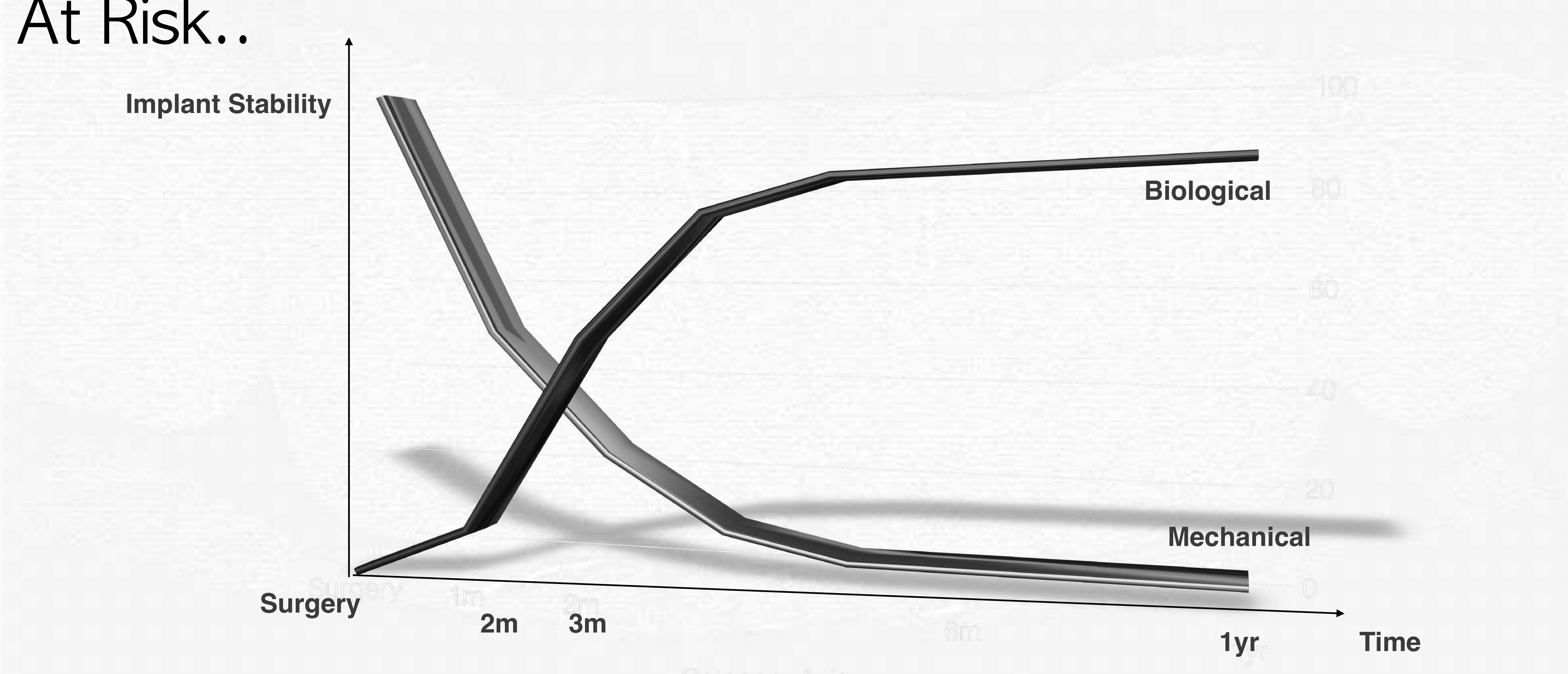
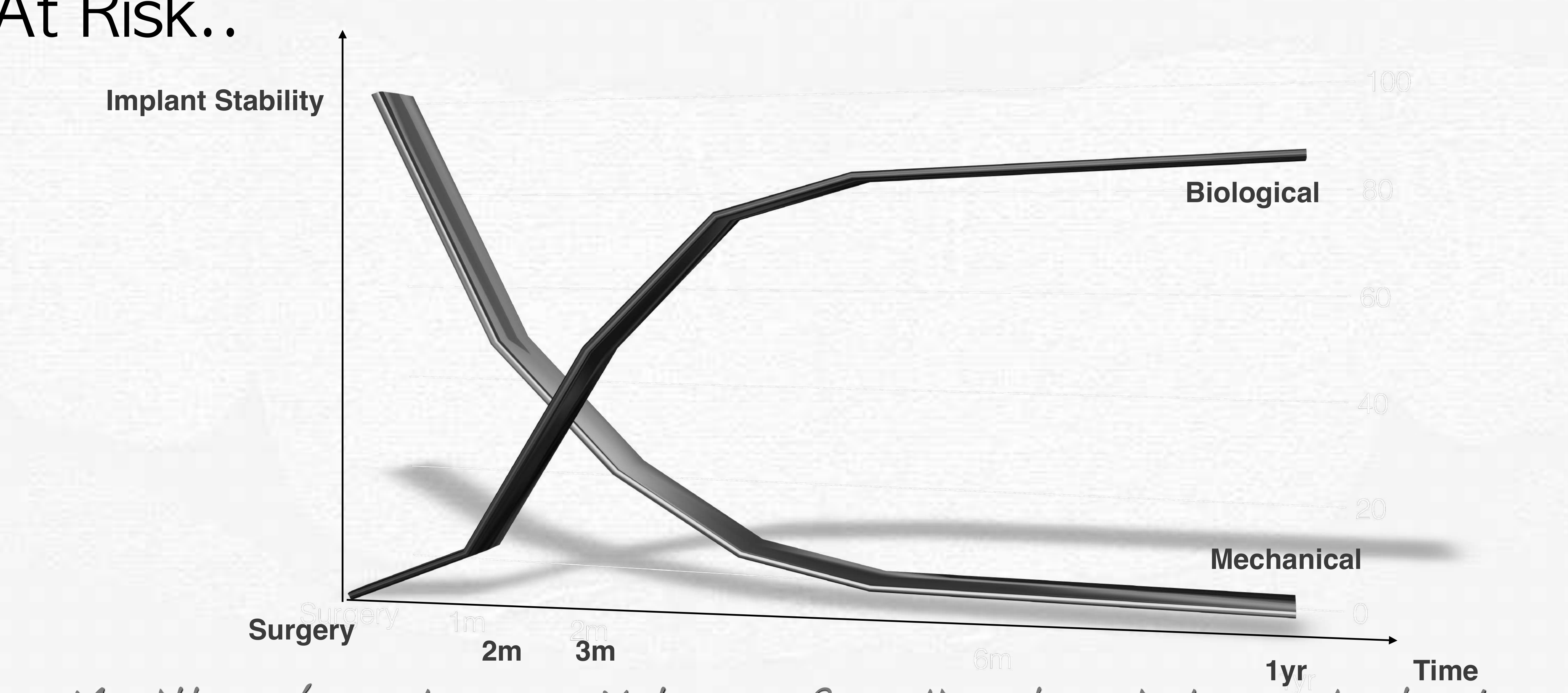
- Implant stability transitions from initial mechanical stability to biological stability through fibrin, collagen, osteoblast activity, and bone-to-implant contact.
- Biological stability takes approximately one year to reach full strength, although stability is already relatively good at approximately four or five months.
- Bone-to-implant contact is not 100%: the best cases may reach approximately 80%, while some functional implants remain at approximately 30%.
- Excessive loading during healing can interfere with osseous sufficiency.
Bruxing Patient With Immediate Loading
A heavily bruxing patient treated with four implants and loaded immediately may experience greater stress while biological stability is not yet complete. If plaque-related inflammation is added, the implant-supporting tissues may be further compromised.
Koka S, Zarb G. On osseointegration: The healing adaptation principle in the context of osseosufficiency, osseoseparation and dental implant failure. Int J Pros 2012;25:48-52.
Zarb G. Implantomania: Prosthodontics at a crossroads. Int J Pros 2012;25:180-185.
Albrektsson T, Buser D, Sennerby L. On crestal/marginal bone loss around dental implants. Int J Pros 2012;25:320-322.
Members — T Albrektsson, D Buser, S Chen, D Cochran, H De Bruyn, T Jemt, S Koka, M Nevins, L Sennerby, M Simion, T Taylor, A Wennerberg.
Patient Risk Factors and At-Risk Cases48
Patient Risk Factors49
| Primary Factors | Secondary Factors |
|---|---|
| Opposing natural dentition | Smoker |
| Poor bone density | Bone volume |
| Bruxer | |
| Male |
- The common factor among many of these risks, apart from smoking, is increased loading.
- Medical history, smoking, bruxism, bone quality, and the opposing dentition should be assessed before selecting an immediate-loading or graftless protocol. Patzelt, Bahat, Reynolds, Strub. The All-on-Four Treatment Concept: A Systematic Review. CIDRR 2014 16;6
Parel SM, Phillips WR. A risk assessment treatment planning protocol for the four implant immediately loaded maxilla: preliminary findings. J Prosthet Dent. 2011;106(6): 359-66.
74% of failures occurred within 12 months of placement
Patzelt, Bahat, Reynolds, Strub. The All-on-Four Treatment Concept: A Systematic Review. CIDRR 2014 16;6
Parel SM, Phillips WR. A risk assessment treatment planning protocol for the four implant immediately loaded maxilla: preliminary findings. J Prosthet Dent. 2011;106(6): 359-66.
Implant Stability in the Maxilla50
Implant Stability51
| Mechanical | Biological |
|---|---|
| Surgery | |
| 2m | |
| 3m | |
| 6m | |
| 1yr | 100 |
Mechanical stability of maxillary bone is 3–4× lower than functional resistance to load of mandibular bone. Implants are not as stable cf mandibular bone.
Lecturer — Maxillary Stability
Mechanical stability is present immediately after placement and is related to the torque required to place the implant, but it decreases as the initial forces relax.
- Biological stability develops as a fibrin network forms, collagen is produced, and osteoblasts and other cells create bone-to-implant contact.
- The transition between mechanical and biological stability is critical when implants are loaded immediately.
- The lower density of maxillary bone makes implant stability and healing more challenging than in the mandible.
Axis scale: 100, 80, 60, 40, 20, 0 — Surgery (Mechanical) vs Time (Biological).
Maxillary bone type 3–4× lower functional resistance to load — implants not as stable cf mandibular bone.
Patzelt, Bahat, Reynolds, Strub. The All-on-Four Treatment Concept: A Systematic Review. CIDRR 2014 16;6
Parel SM, Phillips WR. A risk assessment treatment planning protocol for the four implant immediately loaded maxilla: preliminary findings. J Prosthet Dent. 2011; 106(6): 359-66.
- Depending on the loading, four implants may be insufficient for complete stability, resulting in lower BIC and greater susceptibility to integration breakdown, opportunistic infection and bone loss.
- Exacerbated if other patient factors, e.g. smoking, diabetes, IL-1 phenotype, hx of perio, AI disease, etc.
- All-on-4 may not be appropriate for larger arch form and greater load.
Chung S, McCullagh A, Irinakis T. Immediate loading in the maxillary arch: evidence-based guidelines to improve success rates: A review. J Oral Implantol. 2011;37:610-21.
Management of the Atrophic Maxilla: Augmentation
Augmentation52
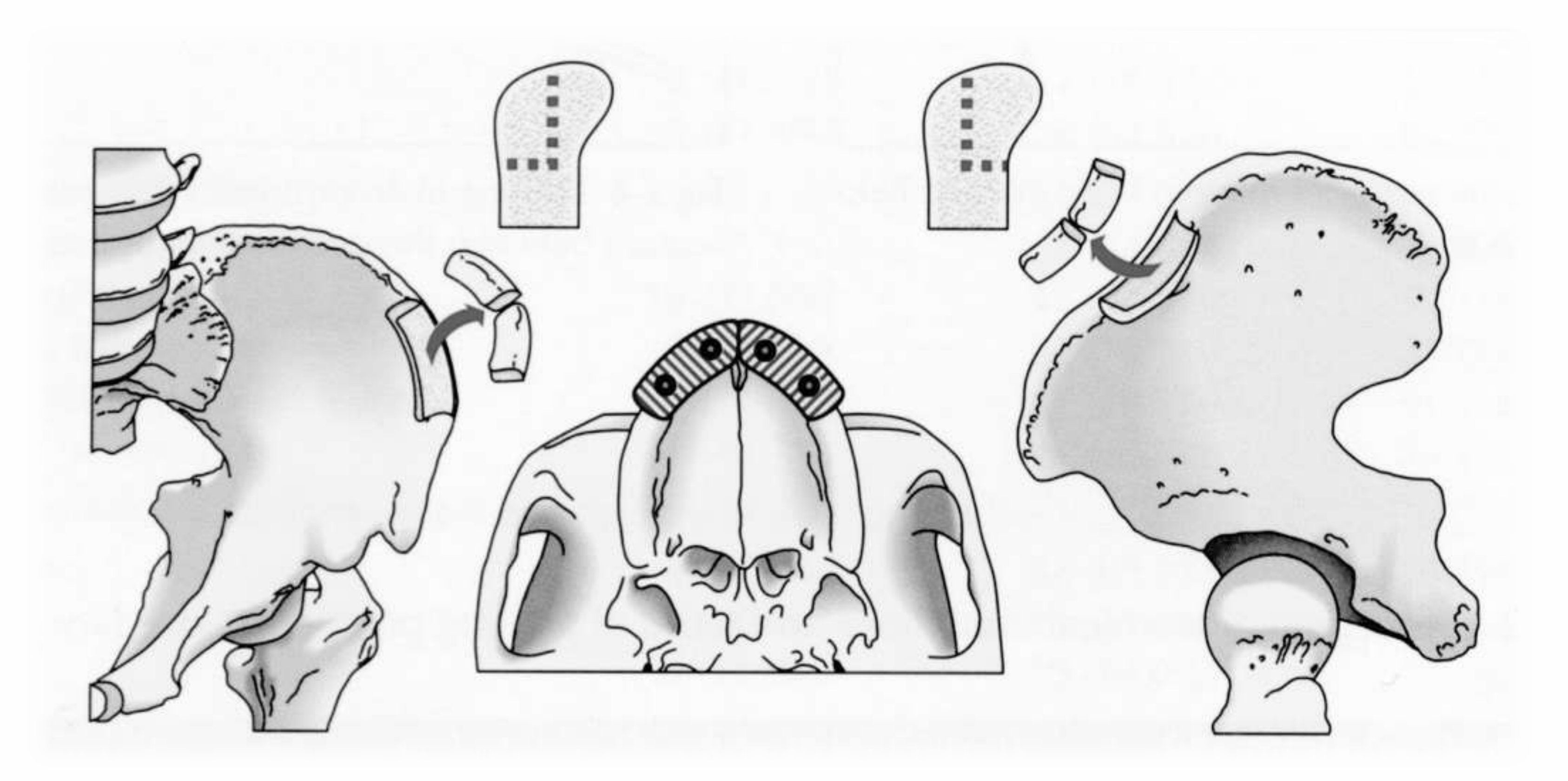
Iliac Crest Grafting Considerations53
Iliac Crest grafting
 | 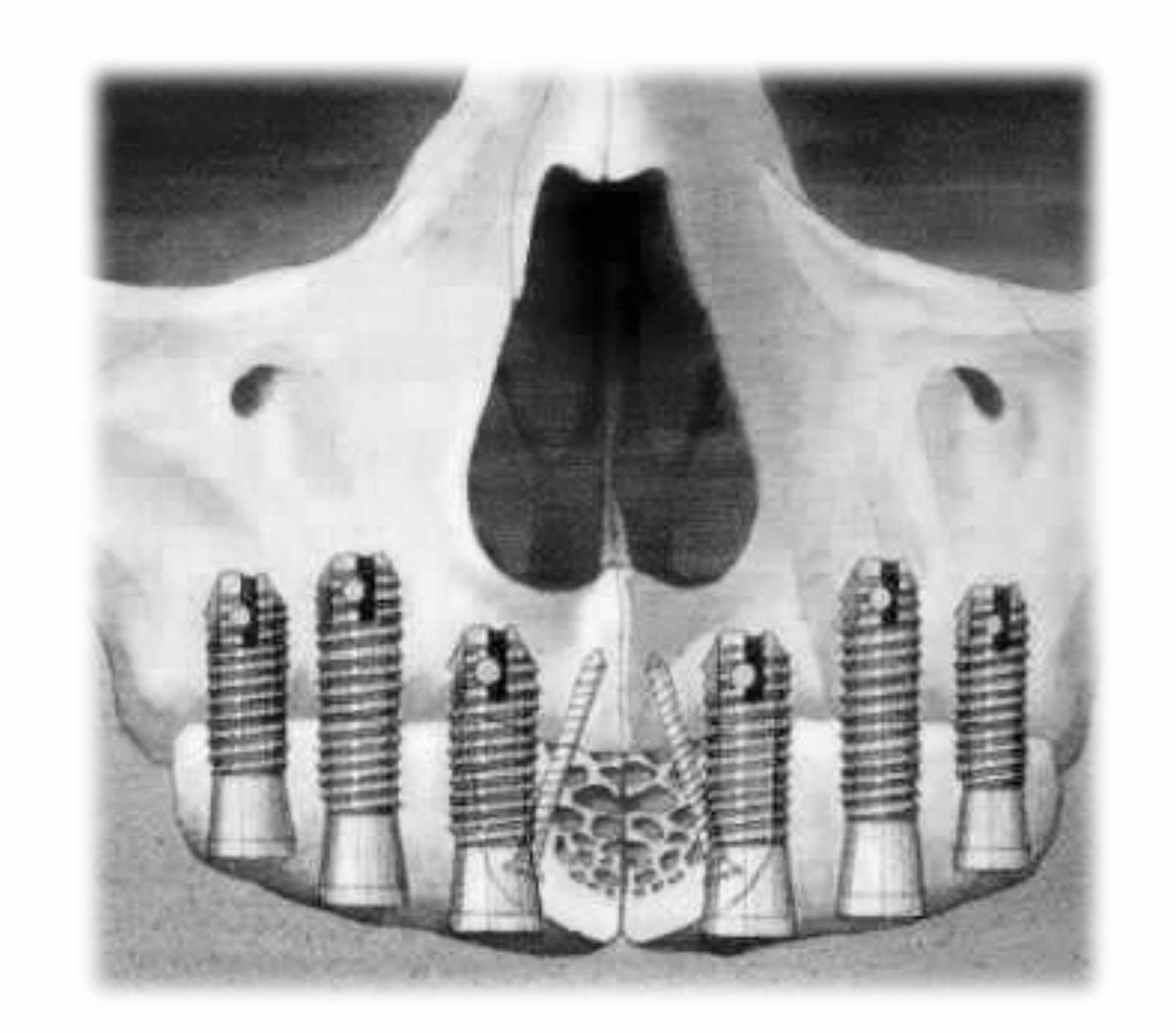 |
Maxillary Bone Graft Considerations54
-
Donor site
-
Recipient site preparation
-
Fixation
-
Implant installation delayed vs immediate
-
Post graft radiological requirements
-
Complications
-
Bone may be harvested from the lateral wall or lateral oblique ridge for anterior augmentation.
-
Grafted blocks may be fixed with screws and covered with membranes.
-
Grafting can combine autogenous bone, bovine bone, and other grafting materials.
MINIMUM 1 YEAR TREATMENT PLAN
Lecturer — Iliac Crest Grafting
Iliac crest grafting was described as a difficult and prolonged treatment for both patients and clinicians.
- Patients could be unable to wear their dentures for months.
- Some studies showed survival rates of approximately 75%, and the overall results discussed were approximately 80%, so this approach is no longer used in the same way.

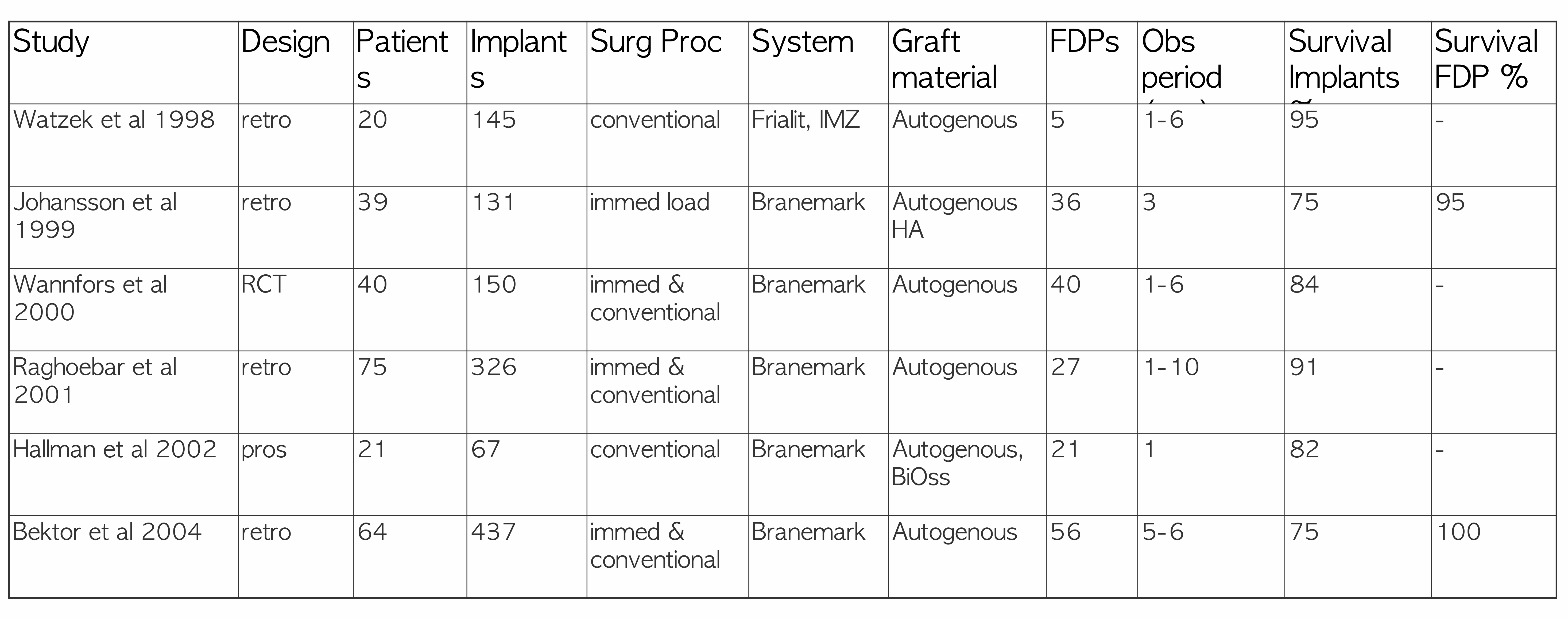
Sinus Lift Lateral Window Studies555657
Studies restoring the edentulous maxilla with fixed prostheses — Sinus Lift Lateral Window
| Study | Design | Patients | Implants | Surg Proc | System | Graft material | FDPs | Obs period | Survival Implants % | Survival FDP % |
|---|---|---|---|---|---|---|---|---|---|---|
| Watzek et al 1998 | retro | 20 | 145 | conventional | Frialit, IMZ | Autogenous | 5 | 1-6 | 95 | - |
| Johansson et al 1999 | retro | 39 | 131 | immed load | Branemark | Autogenous HA | 36 | 3 | 75 | 95 |
| Wannfors et al 2000 | RCT | 40 | 150 | immed & conventional | Branemark | Autogenous | 40 | 1-6 | 84 | - |
| Raghoebar et al 2001 | retro | 75 | 326 | immed & conventional | Branemark | Autogenous | 27 | 1-10 | 91 | - |
| Hallman et al 2002 | pros | 21 | 67 | conventional | Branemark | Autogenous, BioOss | 21 | 1 | 82 | - |
| Bektor et al 2004 | retro | 64 | 437 | immed & conventional | Branemark | Autogenous | 56 | 5-6 | 75 | 100 |
Lateral Window Treatment Course
In the example discussed, a lateral window was created and the Schneiderian membrane was carefully elevated to form the roof of the augmented space.
- The sinus was filled with a mixture of bovine and autogenous bone.
- Blocks were used to augment the anterior ridge, fixed with screws, and covered with membranes.
- Implants were placed after grafting and a bridge was subsequently fabricated.
- The complete process took approximately one and a half years, and the patient could not wear her denture for approximately four months.
Systematic reviews of studies restoring the edentulous maxilla with fixed prostheses — Sinus Lift Lateral Window
- Implant survival more variable in augmented sinuses (36–100%, avg ~90%)
- Amount of residual bone height is an important prognostic factor
- No evidence for the superiority of autogenous bone compared to substitutes
- Heterogeneous studies do not allow definitive statements
Graziani F, Donos N, Needleman I, Gabriele M, Tonetti M. Comparison of implant survival following sinus floor augmentation procedures with implants placed in pristine posterior maxillary bone: a systematic review. Clin. Oral Impl. Res. 15, 2004; 677–682.
Pjetursson BE, Tan WC, Zwahlen M, Lang NP. A systematic review of the success of sinus floor elevation and survival of implants inserted in combination with sinus floor elevation. J Clin Periodontol. 2008;35(8 Suppl): 216-40.
Nkenke E, Stelzle F. Clinical outcomes of sinus floor augmentation for implant placement using autogenous bone or bone substitutes: a systematic review. Clin Oral Implants Res. 2009;20 Suppl 4:124-33.
Rickert D, Slater JJ, Meijer HJ, Vissink A, Raghoebar GM. Maxillary sinus lift with solely autogenous bone compared to a combination of autogenous bone and growth factors or (solely) bone substitutes. A systematic review. Int J Oral Maxillofac Surg. 2012;41(2):160-7.
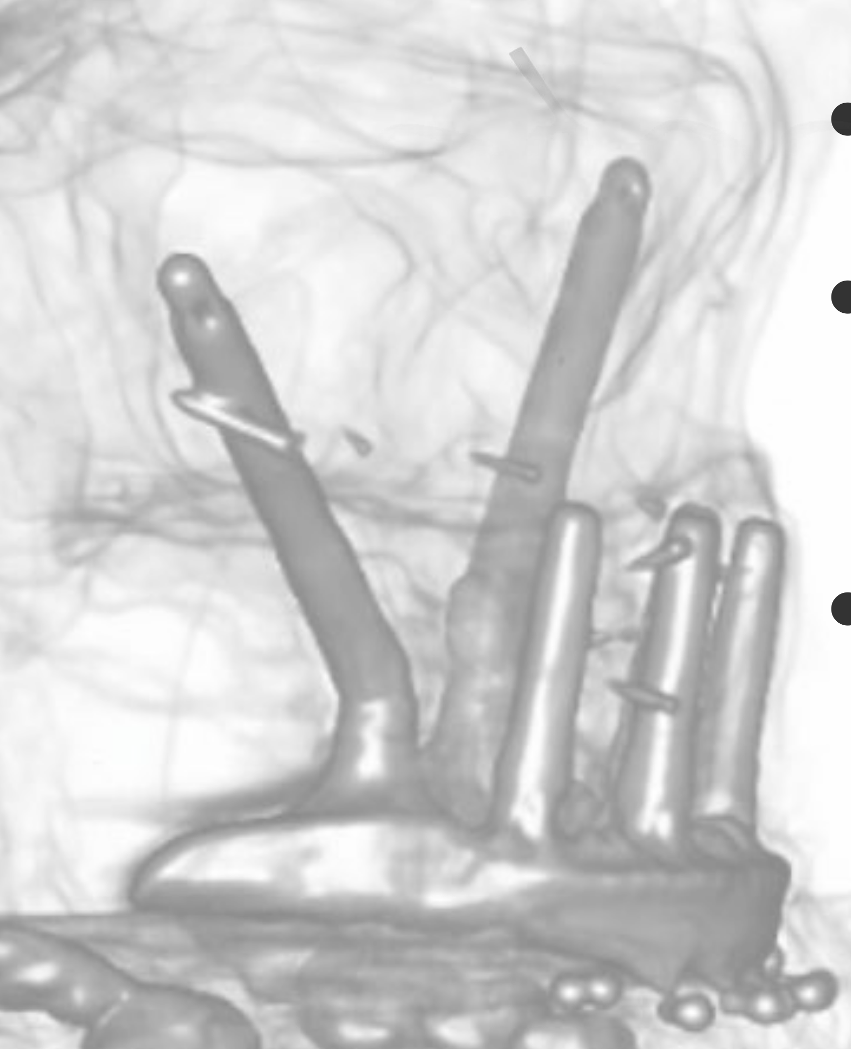
Zygoma Implants: Studies and Treatment Planning
Zygoma Implant Survival Studies58
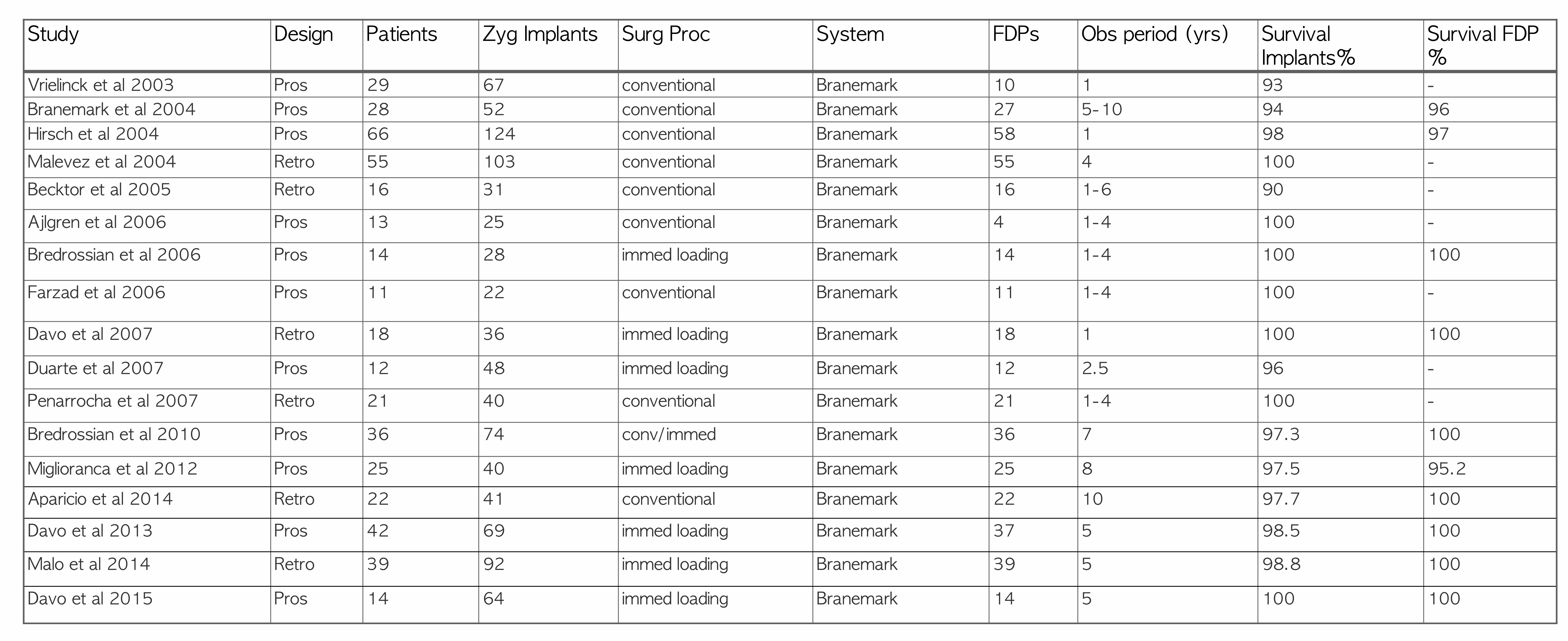
Studies Restoring the Edentulous Maxilla with Fixed Prostheses — Zygoma Implants59
| Study | Design | Patients | Zyg Implants | Surg Proc | System | FDPs | Obs period (yrs) | Survival Implants % | Survival FDP % |
|---|---|---|---|---|---|---|---|---|---|
| Vrielinck et al 2003 | Pros | 29 | 67 | conventional | Branemark | 10 | 1 | 93 | - |
| Branemark et al 2004 | Pros | 28 | 52 | conventional | Branemark | 27 | 5-10 | 94 | 96 |
| Hirsch et al 2004 | Pros | 66 | 124 | conventional | Branemark | 58 | 1 | 98 | 97 |
| Malevez et al 2004 | Retro | 55 | 103 | conventional | Branemark | 55 | 4 | 100 | - |
| Becktor et al 2005 | Retro | 16 | 31 | conventional | Branemark | 16 | 1-6 | 90 | - |
| Ajlgren et al 2006 | Pros | 13 | 25 | conventional | Branemark | 4 | 1-4 | 100 | - |
| Bredrossian et al 2006 | Pros | 14 | 28 | immed loading | Branemark | 14 | 1-4 | 100 | 100 |
| Farzad et al 2006 | Pros | 11 | 22 | conventional | Branemark | 11 | 1-4 | 100 | - |
| Davo et al 2007 | Retro | 18 | 36 | immed loading | Branemark | 18 | 1 | 100 | 100 |
| Duarte et al 2007 | Pros | 12 | 48 | immed loading | Branemark | 12 | 2.5 | 96 | - |
| Penarrocha et al 2007 | Retro | 21 | 40 | conventional | Branemark | 21 | 1-4 | 100 | - |
| Bredrossian et al 2010 | Pros | 36 | 74 | conv/immed | Branemark | 36 | 7 | 97.3 | 100 |
| Miglioranca et al 2012 | Pros | 25 | 40 | immed loading | Branemark | 25 | 8 | 97.5 | 95.2 |
| Aparicio et al 2014 | Retro | 22 | 41 | conventional | Branemark | 22 | 10 | 97.7 | 100 |
| Davo et al 2013 | Pros | 42 | 69 | immed loading | Branemark | 37 | 5 | 98.5 | 100 |
| Malo et al 2014 | Retro | 39 | 92 | immed loading | Branemark | 39 | 5 | 98.8 | 100 |
| Davo et al 2015 | Pros | 14 | 64 | immed loading | Branemark | 14 | 5 | 100 | 100 |
Lecturer — Zygoma Implant Context
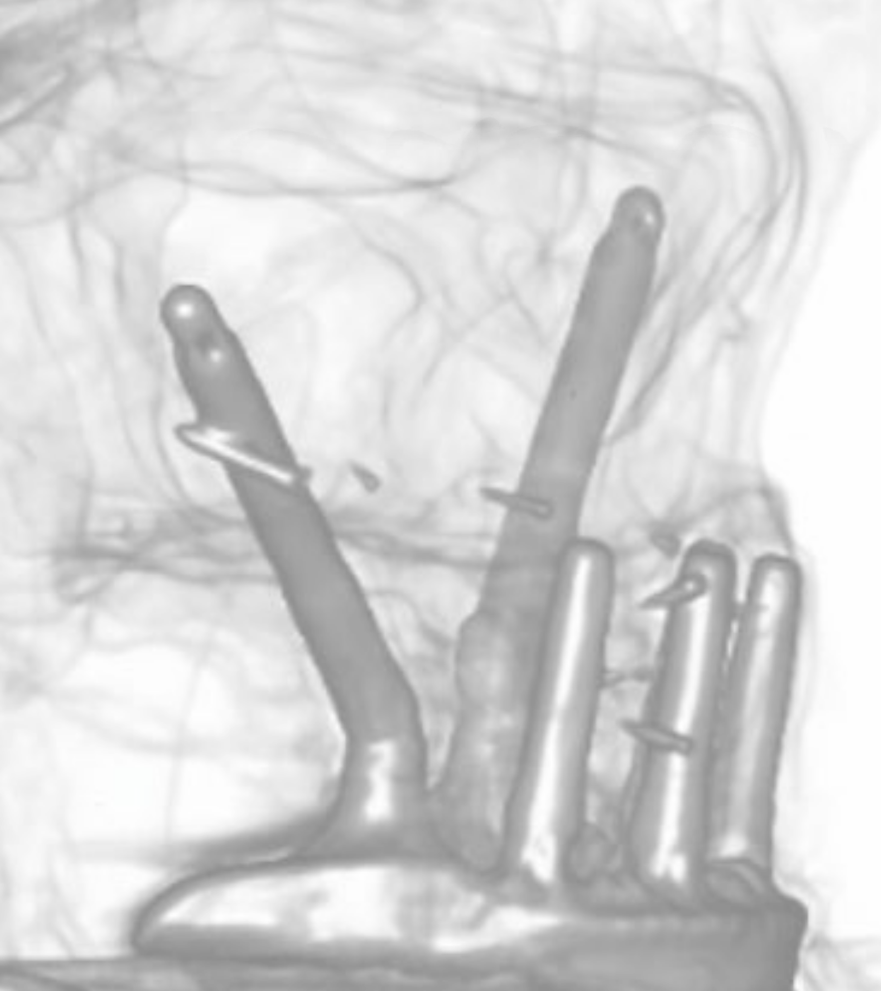
Zygoma implants engage the zygomatic arch and became an alternative to extensive grafting in severely atrophic maxillae.
- Early studies associated with Brånemark reported survival rates from approximately 95% to 100%.
- Systematic reviews also showed very high success rates.
Systematic Reviews60
- Overall survival rate 96.7% including cancer patients
- Intramaxillary immediate loaded (9 studies, 458 implants, 8 failures): ZI 98.3%
- 5 cases of sinusitis, all resolved
Chrcanovic BR, Abreu MH. Survival and complications of zygomatic implants: a systematic review. Oral Maxillofac Surg. 2013;17(2):81-93.
- 25 articles
- Overall survival rate 97.8% (1541 ZI, 33 failures)
- Failure generally in the first year due to infection and sinusitis
Goiato MC, Pellizzer EP, Moreno A, Gennari-Filho H, Dos Santos DM, Santiago JF Jr, Dos Santos EG. Implants in the zygomatic bone for maxillary prosthetic rehabilitation: a systematic review. Int J Oral Maxillofac Surg. 2014;43:748-757. Review.
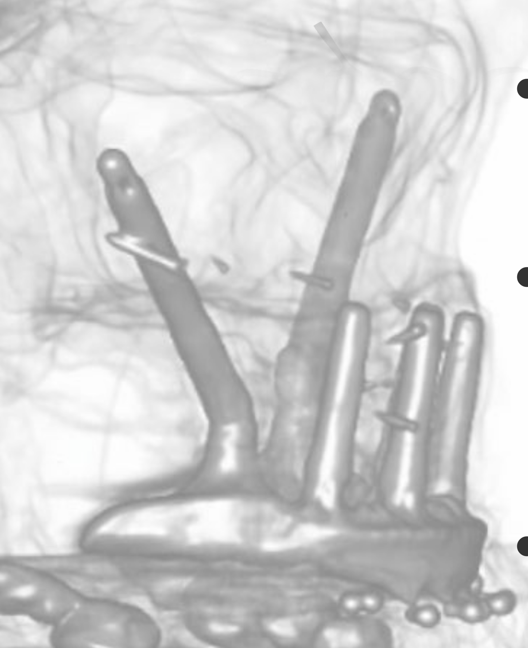
Patient Satisfaction With Zygoma Implants
Satisfaction Studies61
- 16 pts: all satisfied; 50% with some complaints (50% prosthesis, 38% implants)
- 22 pts: 84% satisfaction above 80% (32% had 100% satisfaction)
- Satisfaction (high) approaching the general population
Sartori et al. Evaluation of Patients Rehabilitated with Zygomatic Fixtures. J Oral Maxillofac Surg 2012;70:314-9.
Aparicio et al. The Long Term Use of Zygomatic Implants: A 10 Year Clinical and Radiographic Report. Clin Impl Dent Relat Res 2012.
Wang F, Monje A, Lin GH, Wu Y, Monje F, Wang HL, Davó R. Reliability of four zygomatic implant-supported prostheses for the rehabilitation of the atrophic maxilla: A systematic review. IJOMI 2015;30:293-298.
Lecturer — Patient Treatment Goals
Zygoma treatment can provide fixed teeth for patients who cannot tolerate or wear an upper denture.
- The approach addresses the need for a fixed prosthesis and improved facial support.
- It can be used in situations with little or no conventional implant bone.
- Prosthetic design and cleansability remain important even when zygoma implants provide the surgical foundation.
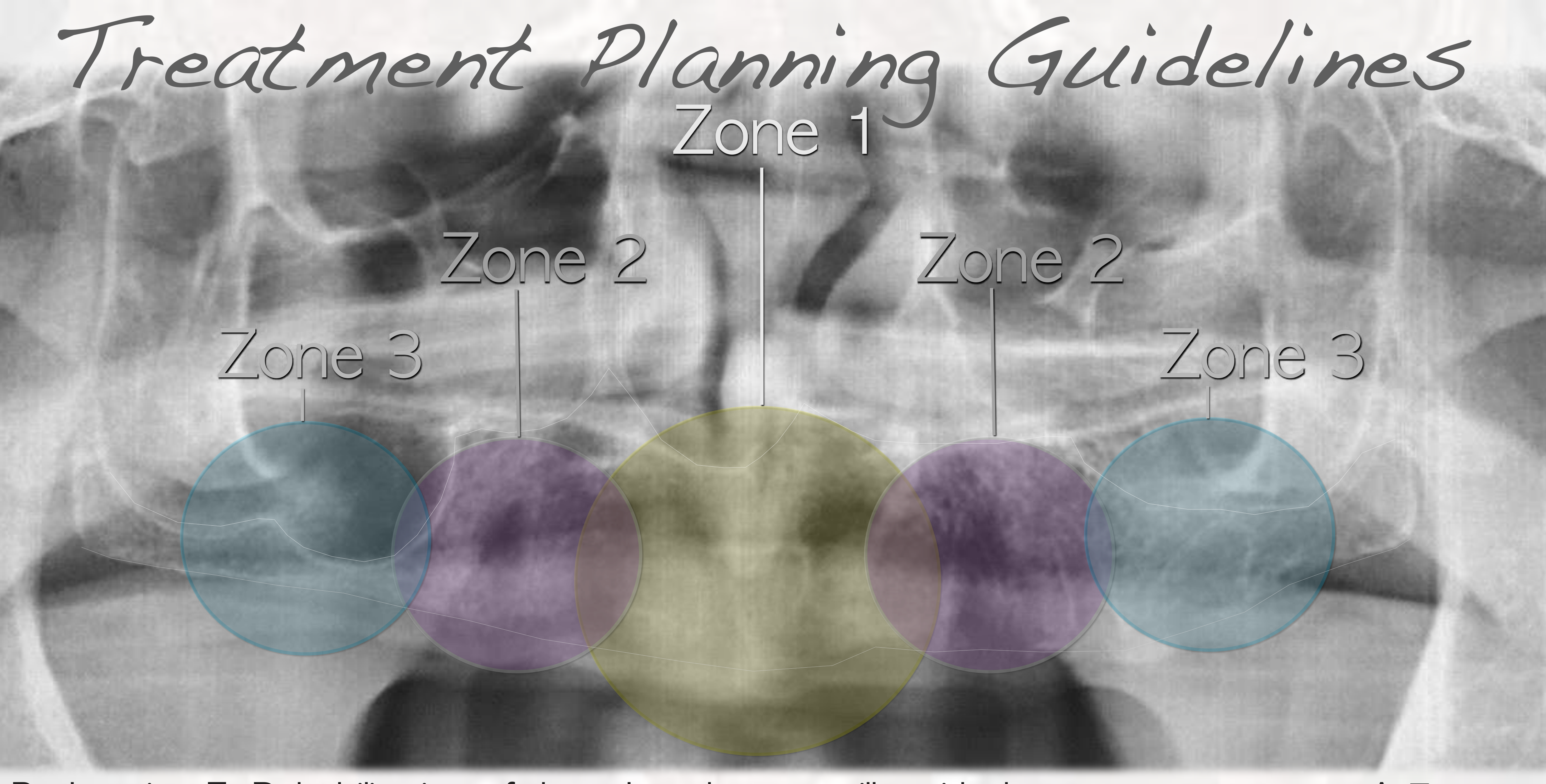
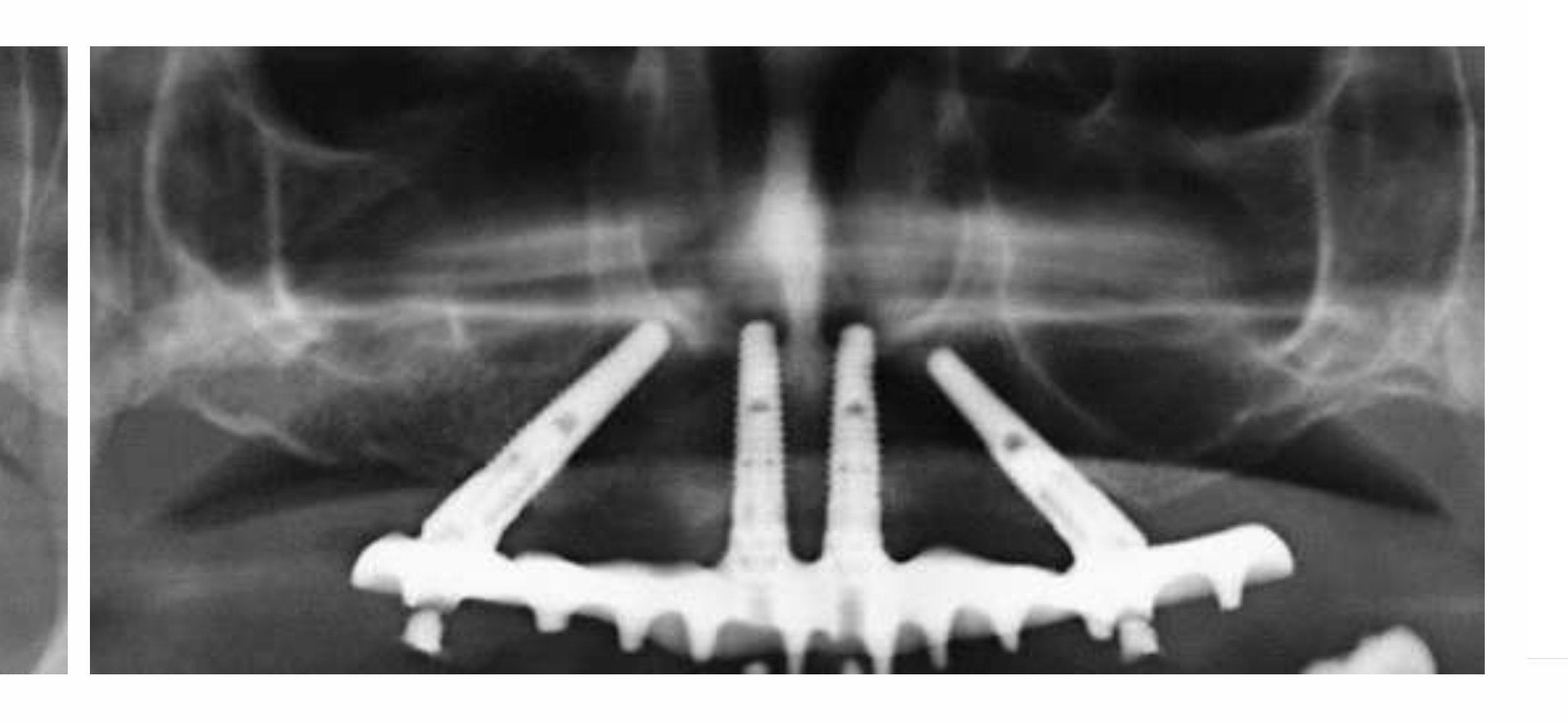
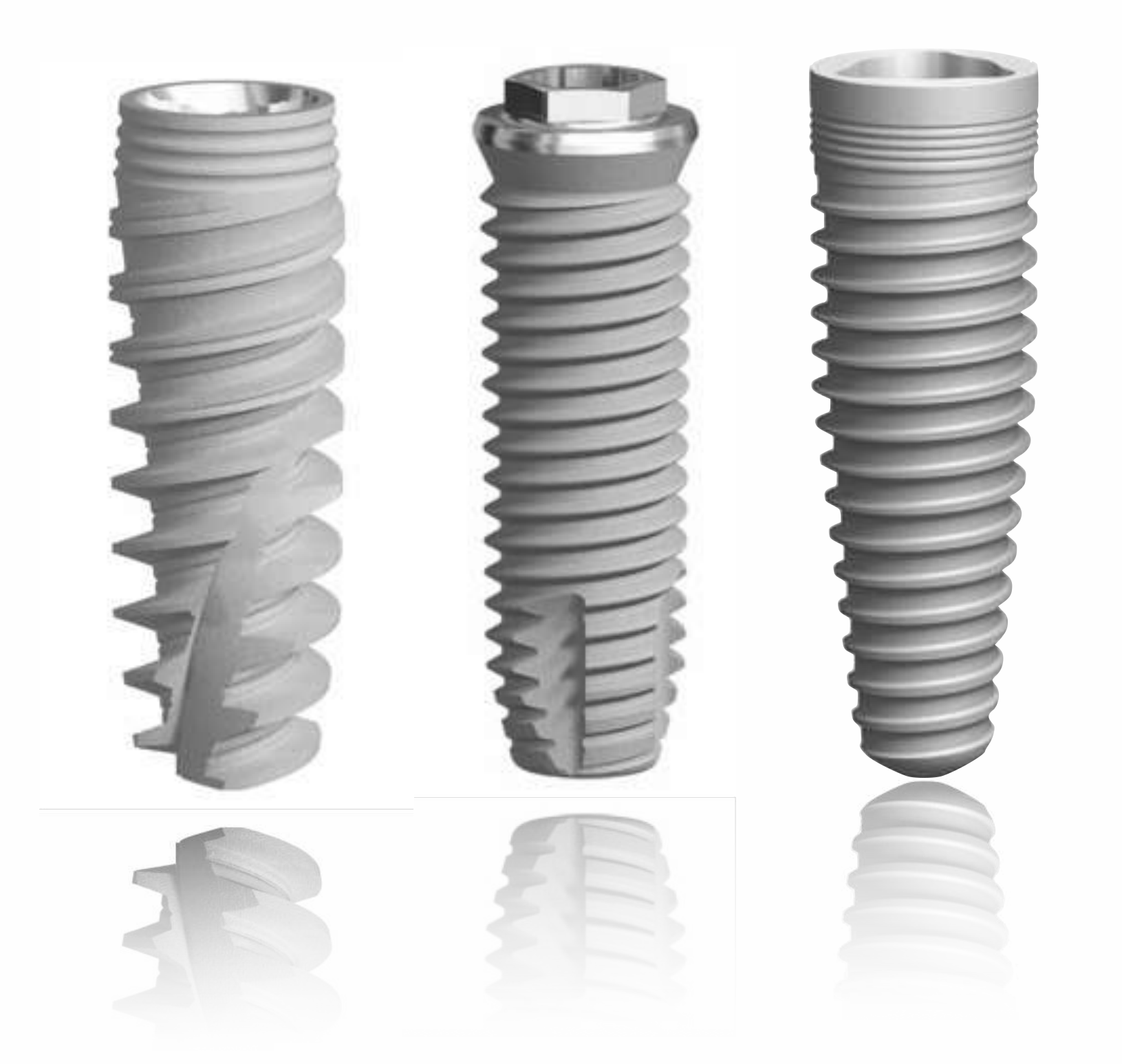
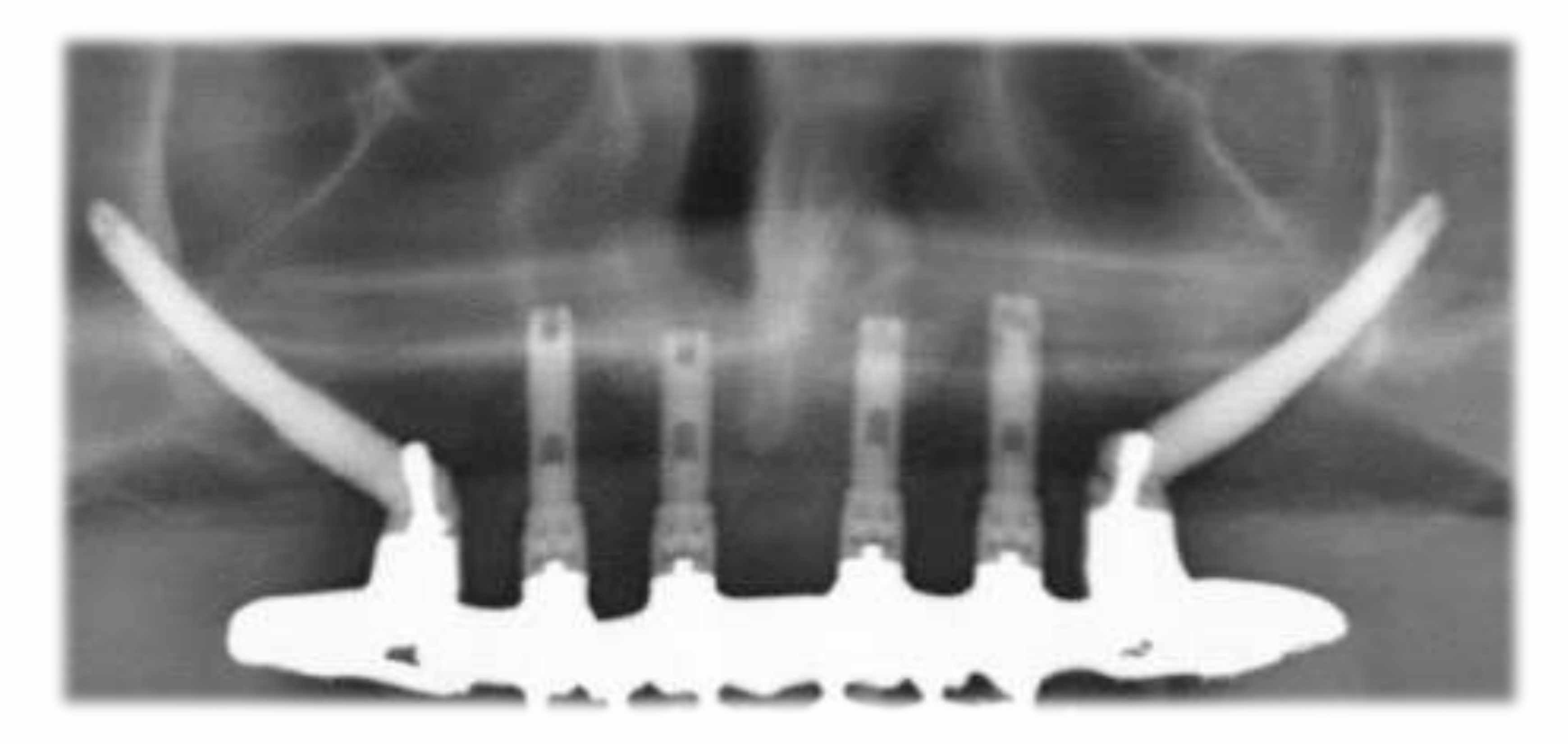
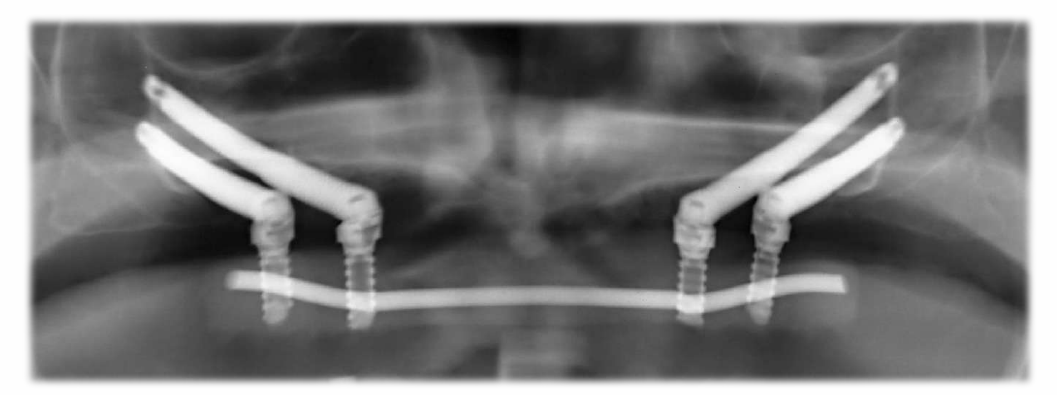
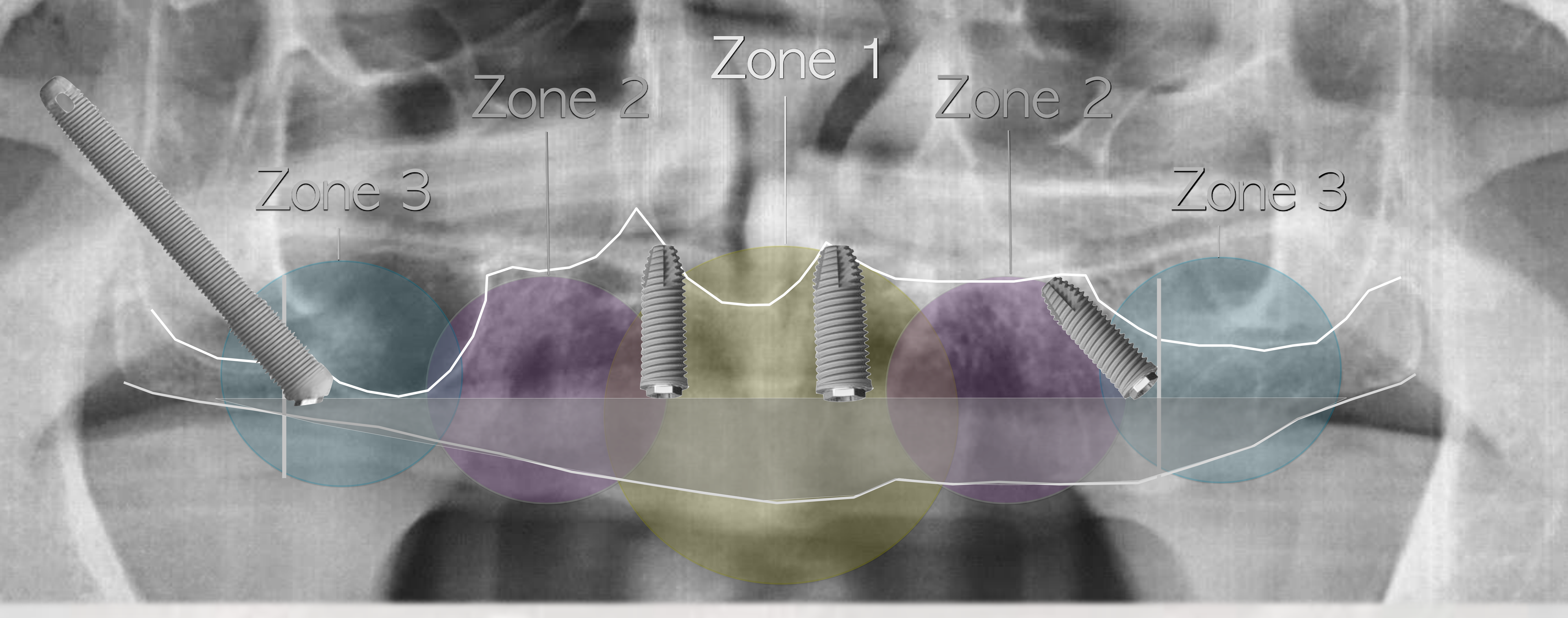
Bedrossian Treatment Planning Guidelines by Zone6263646566
Bedrossian E. Rehabilitation of the edentulous maxilla with the zygoma concept: A 7-year prospective study. IJOMI 2010;25:1213-1221.
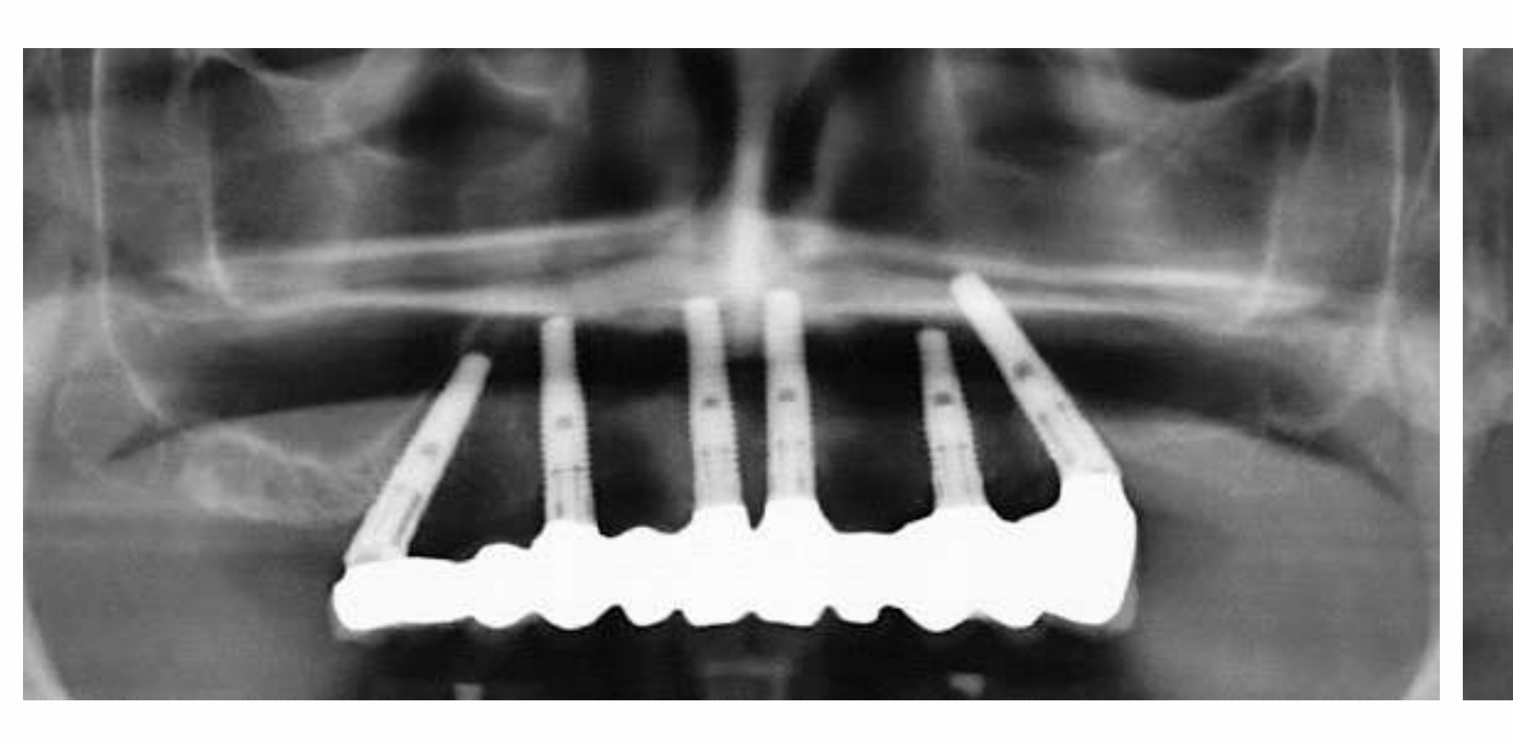
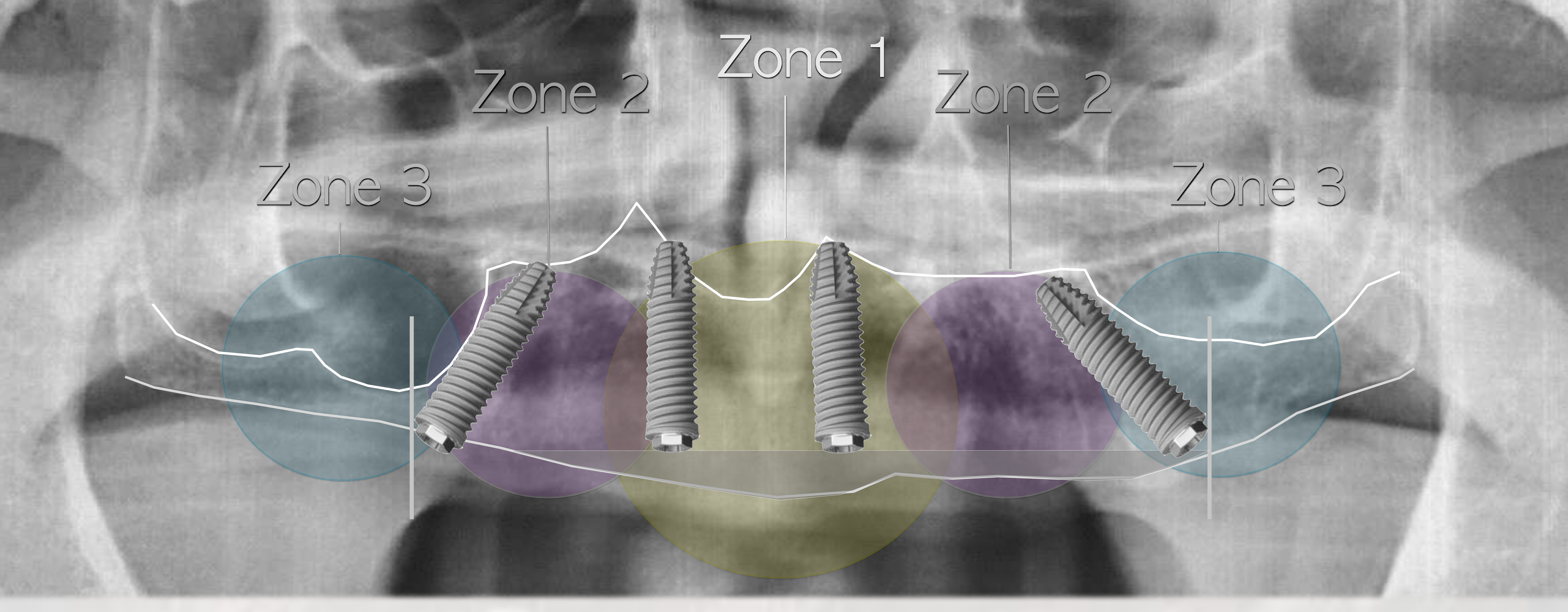
Treatment Planning Guidelines67
Zones 1, 2 & 3 — Traditional Axial implants
Bedrossian E. Rehabilitation of the edentulous maxilla with the zygoma concept: A 7-year prospective study. IJOMI 2010;25:1213-1221.
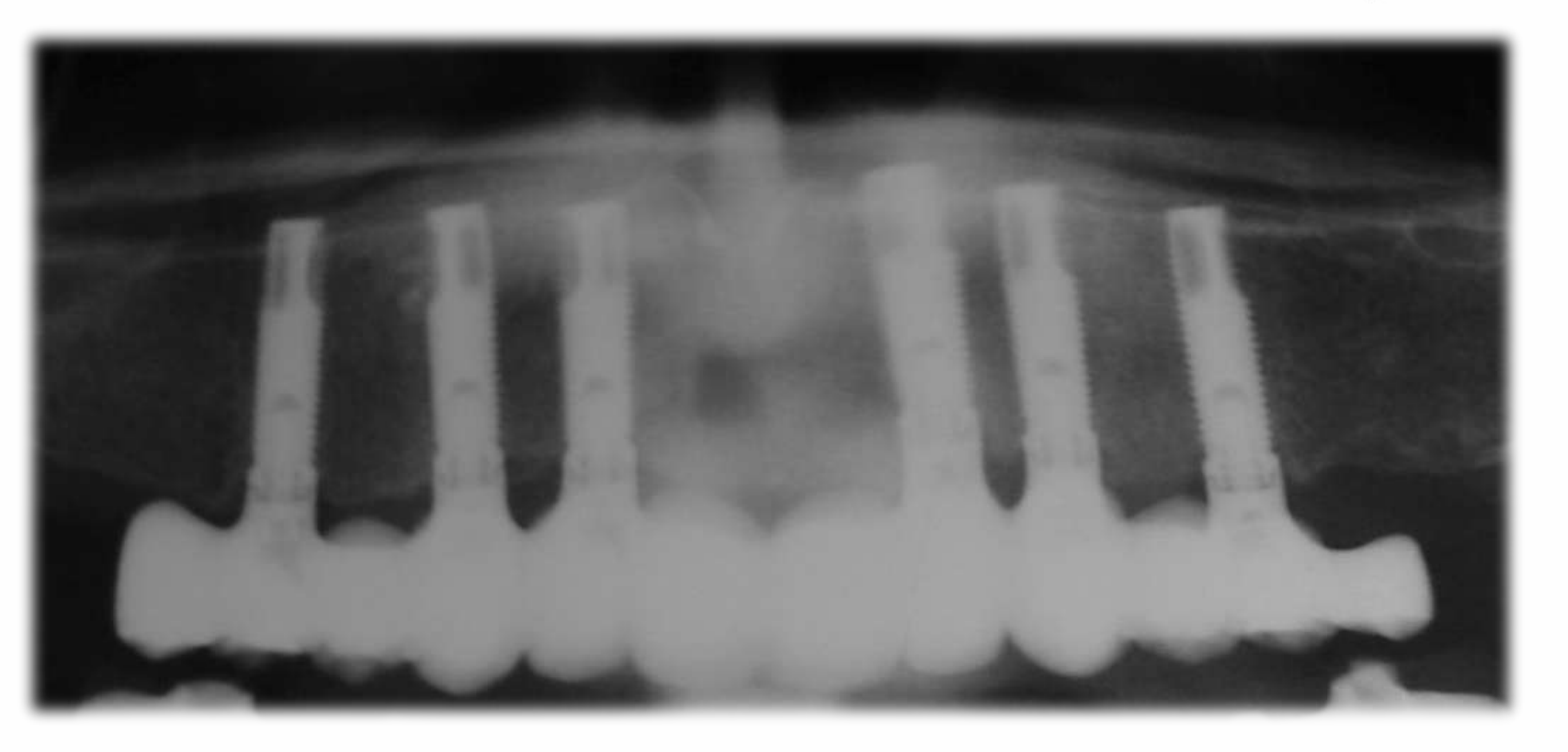
Zones 1, 2 — Traditional Axial and Tilted implants
Bedrossian E. Rehabilitation of the edentulous maxilla with the zygoma concept: A 7-year prospective study. IJOMI 2010;25:1213-1221.
Zone 1 only — Traditional Axial and Zygomatic implants
Bedrossian E. Rehabilitation of the edentulous maxilla with the zygoma concept: A 7-year prospective study. IJOMI 2010;25:1213-1221.
Insufficient bone in all zones — Quad Zygoma
Bedrossian E. Rehabilitation of the edentulous maxilla with the zygoma concept: A 7-year prospective study. IJOMI 2010;25:1213-1221.
Lecturer — Treatment Planning Zones
The maxilla was divided into three treatment-planning zones.
- The anterior zone generally has the best bone.
- The premolar or pre-canine zone and the molar zone complete the treatment-planning division.
- These zones help determine whether treatment should involve axial implants, tilted implants, zygoma implants, or combinations of these approaches.
Ridge reduction affects implant length and AP spread
Zones labeled on the diagram: Zone 3, Zone 2, Zone 1, Zone 2, Zone 3
Bedrossian E, Sullivan RM, Fortin Y, Malo P, Indresano T. Fixed-prosthetic implant restoration of the edentulous maxilla: a systematic pretreatment evaluation method. J Oral Maxillofac Surg. 2008;66(1):112-22.
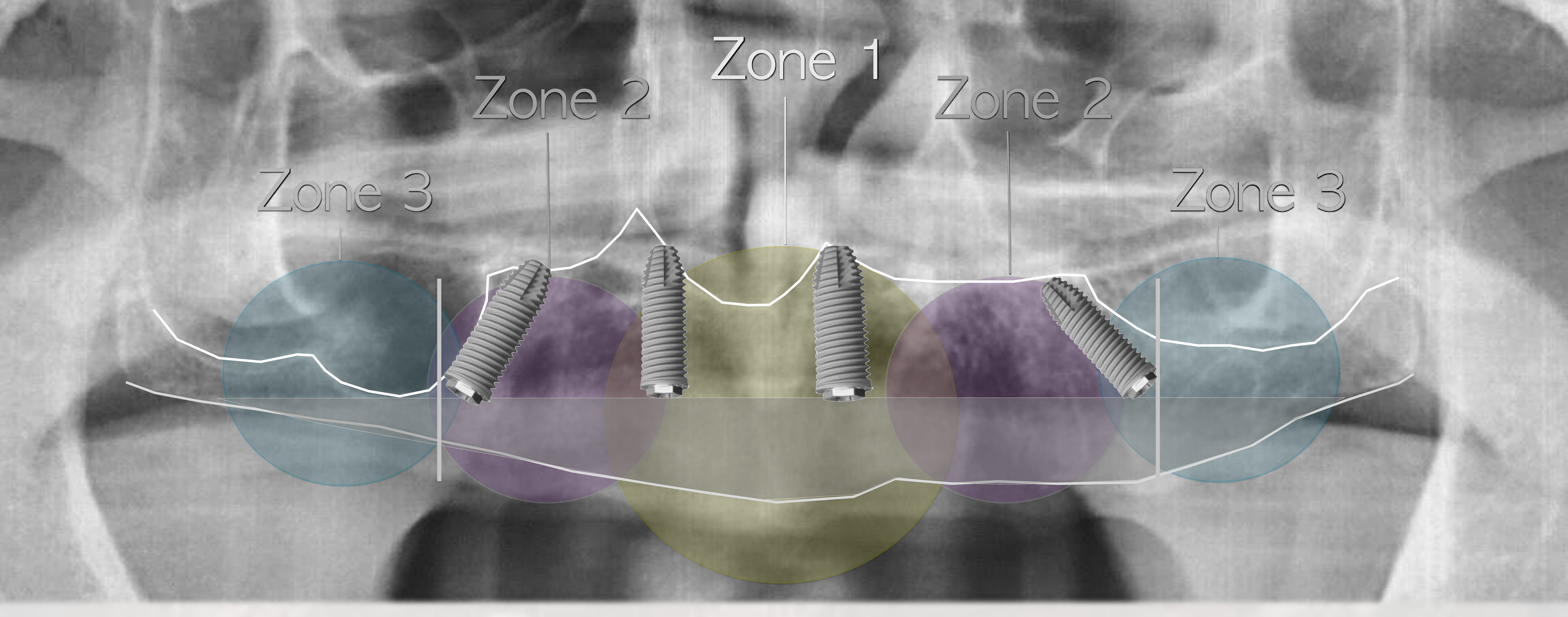
Ridge Reduction, Implant Length and AP Spread686970
Ridge reduction affects implant length and AP spread
Zones labeled on the diagram: Zone 3, Zone 2, Zone 1, Zone 2, Zone 3
Bedrossian E, Sullivan RM, Fortin Y, Malo P, Indresano T. Fixed-prosthetic implant restoration of the edentulous maxilla: a systematic pretreatment evaluation method. J Oral Maxillofac Surg. 2008;66(1):112-22.
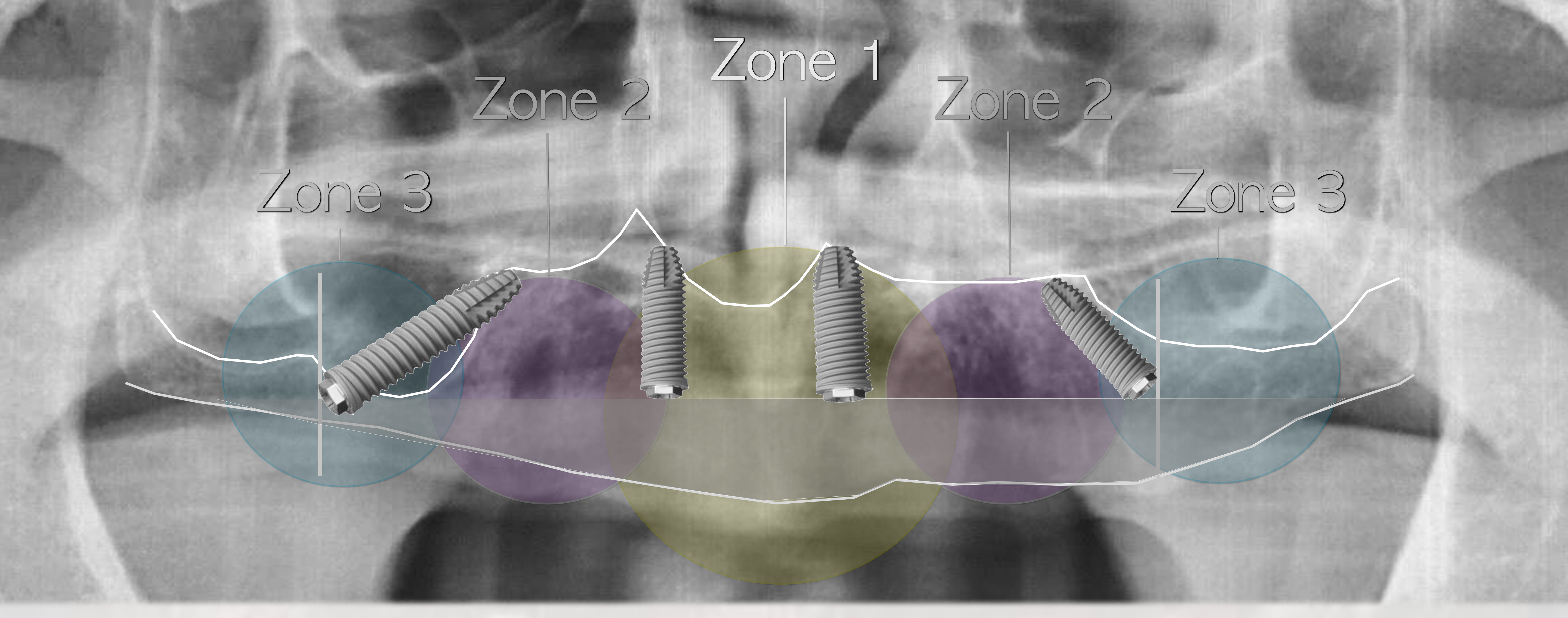
Ridge reduction affects implant length and AP spread
Zones labeled on the diagram: Zone 3, Zone 2, Zone 1, Zone 2, Zone 3
Maló P, Nobre Md, Lopes A. Immediate loading of “All-on-4” maxillary prostheses using trans-sinus tilted implants without sinus bone grafting: a retrospective study reporting the 3-year outcome. Eur J Oral Implantol. 2013;6(3):273-83.
Ridge reduction affects implant length and AP spread
Zones labeled on the diagram: Zone 3, Zone 2, Zone 1, Zone 2, Zone 3
Goiato MC, Pellizzer EP, Moreno A, Gennari-Filho H, dos Santos DM, Santiago JF Jr, dos Santos EG. Implants in the zygomatic bone for maxillary prosthetic rehabilitation: a systematic review. Int J Oral Maxillofac Surg. 2014;43(6):748-57.
Lecturer — Ridge Reduction Effects
Ridge reduction affects both restorative space and implant placement.
- If only a small amount of ridge reduction is needed, treatment may remain straightforward.
- With greater reduction, smaller implants may be required, implants may need to be positioned trans-sinus, and implants may need to extend into the zygoma.
- Implant length and position must be evaluated after determining the amount of ridge reduction needed for the prosthesis.
- The final decision depends on the residual ridge, required facial support, planned tooth position, available implant bone, and the need for an acceptable anteroposterior spread.
Home Care and Prosthetic Design
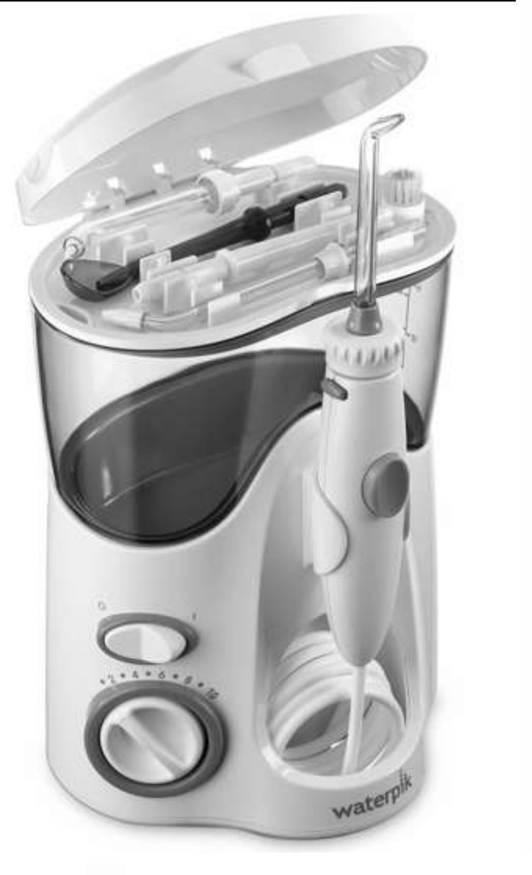
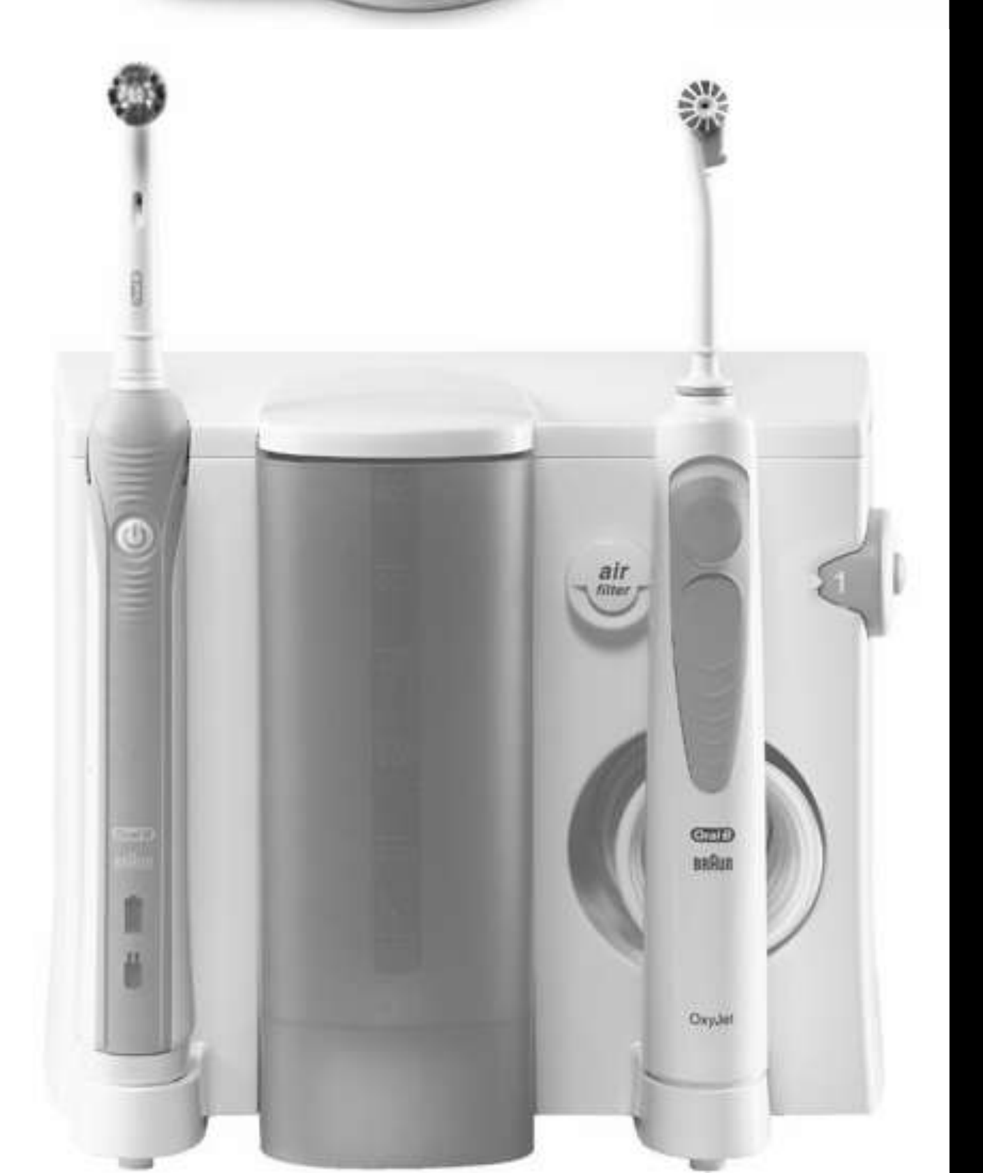
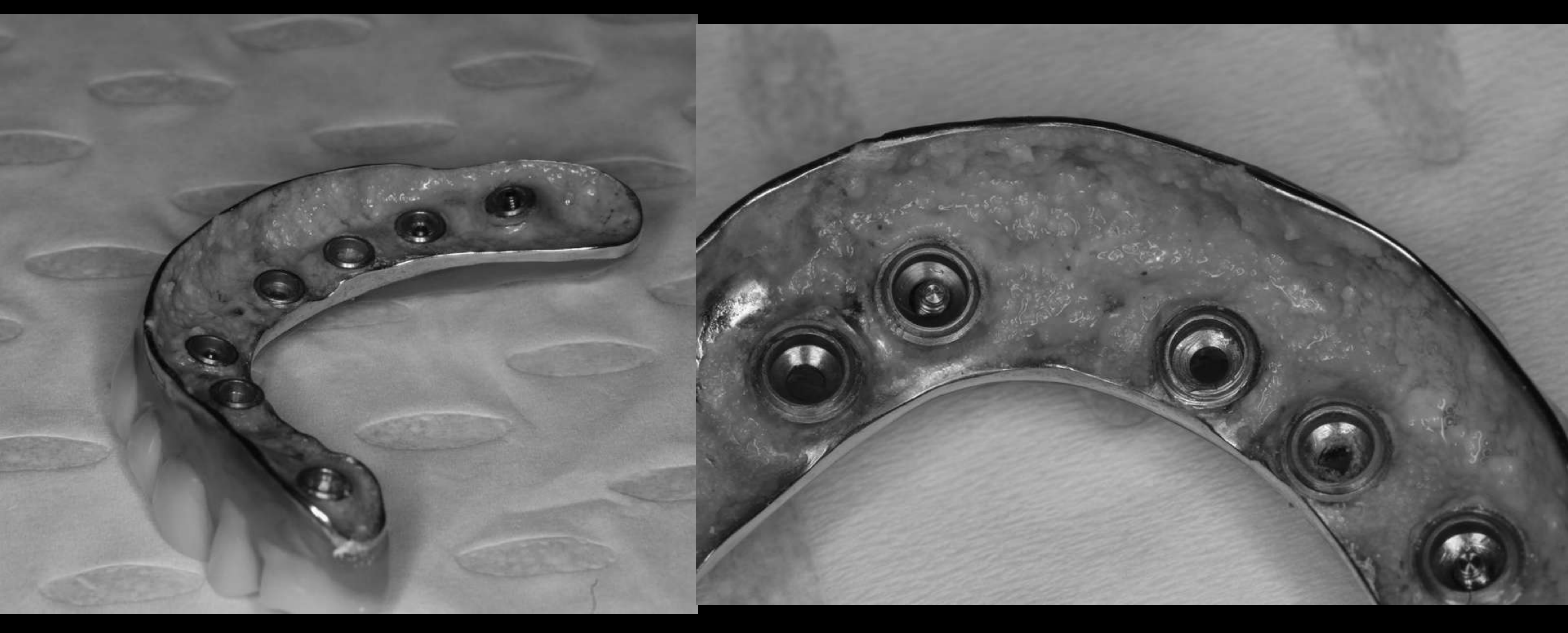
Home Care Patient Aids and Cleansibility717273
Home Care Patient Aids
Cleansibility
Lecturer — Home Care Access
Cleansability is particularly important in the maxilla, and the prosthesis should balance facial support with access for home care.
- Useful aids include water jets, interdental brushes or “pixters,” and floss where access permits.
- Concave or bulky surfaces can trap plaque, spirochetes, and other deposits, contributing to peri-implant inflammation.
- If implants are not placed deeply enough or the prosthesis is poorly designed, excessive tissue contours may require surgical reduction to restore cleansability.
-
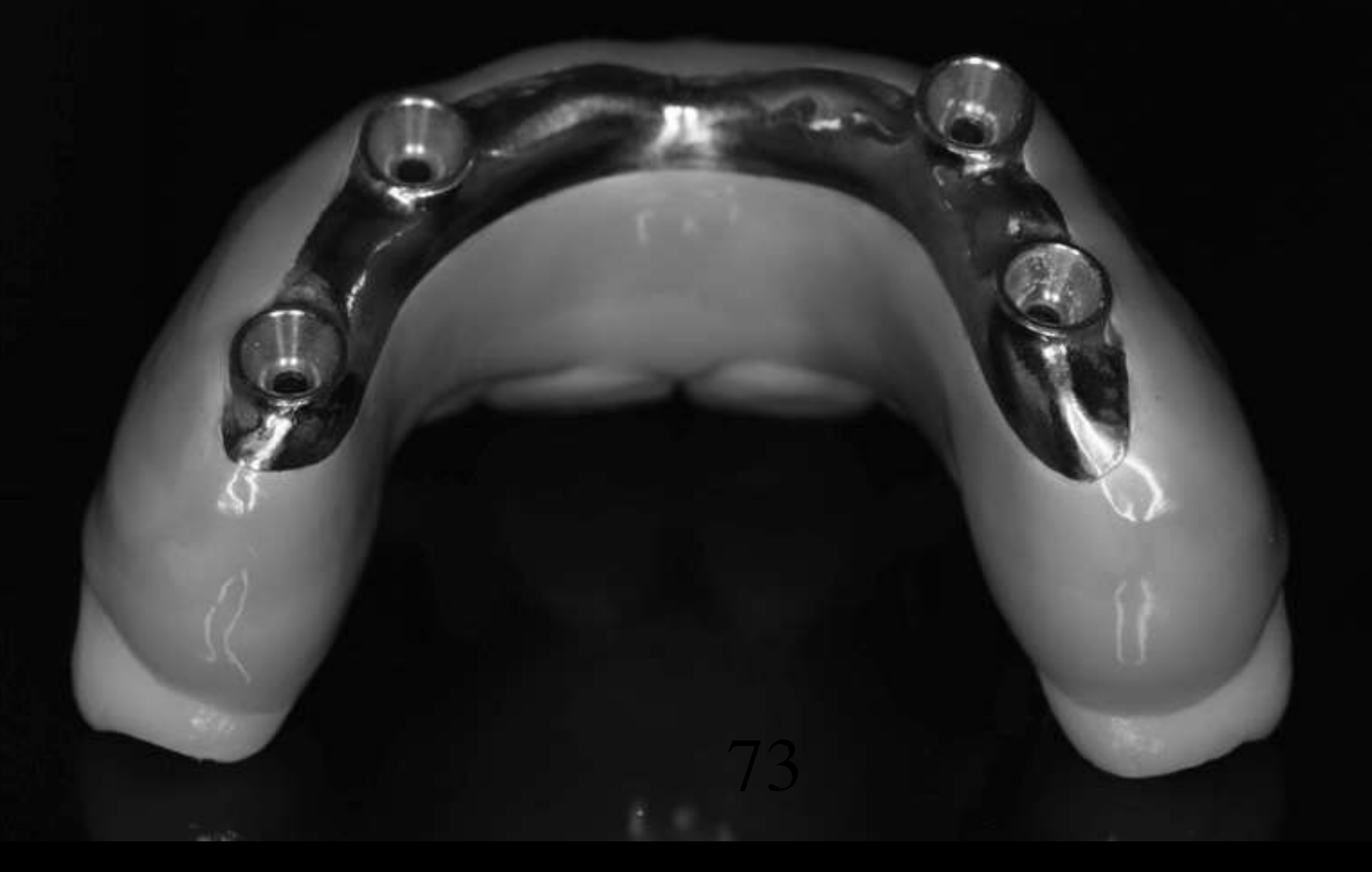
Convex tissue fitting surface
-
A convex surface allows a water jet to pass along and clean the entire tissue-fitting area
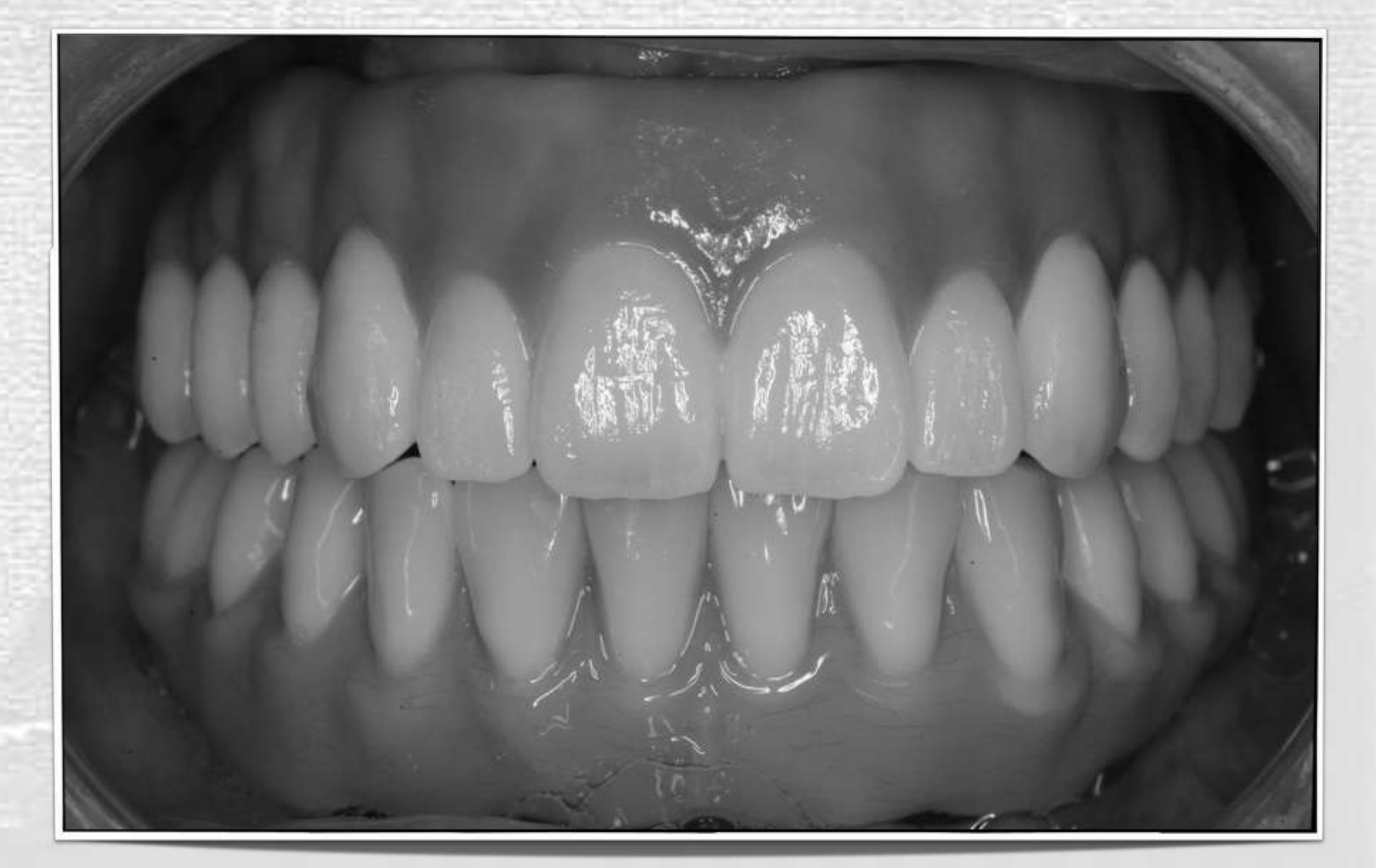
Definitive Bridge Options for Both Jaws74
Definitive Bridge Options — Both Jaws
- Mandible — acrylic resin
- Maxilla — more wear resistant
- Ceramic if possible
Lecturer — Opposing Materials
Using acrylic on the lower jaw and ceramic on the upper jaw allows one material to wear rather than having both arches wear against each other in the same way.
- Material selection should account for the opposing dentition, bruxism, wear, fracture risk, patient comfort, and cleansability.
Maxillary Bridge Options: Framework Materials75
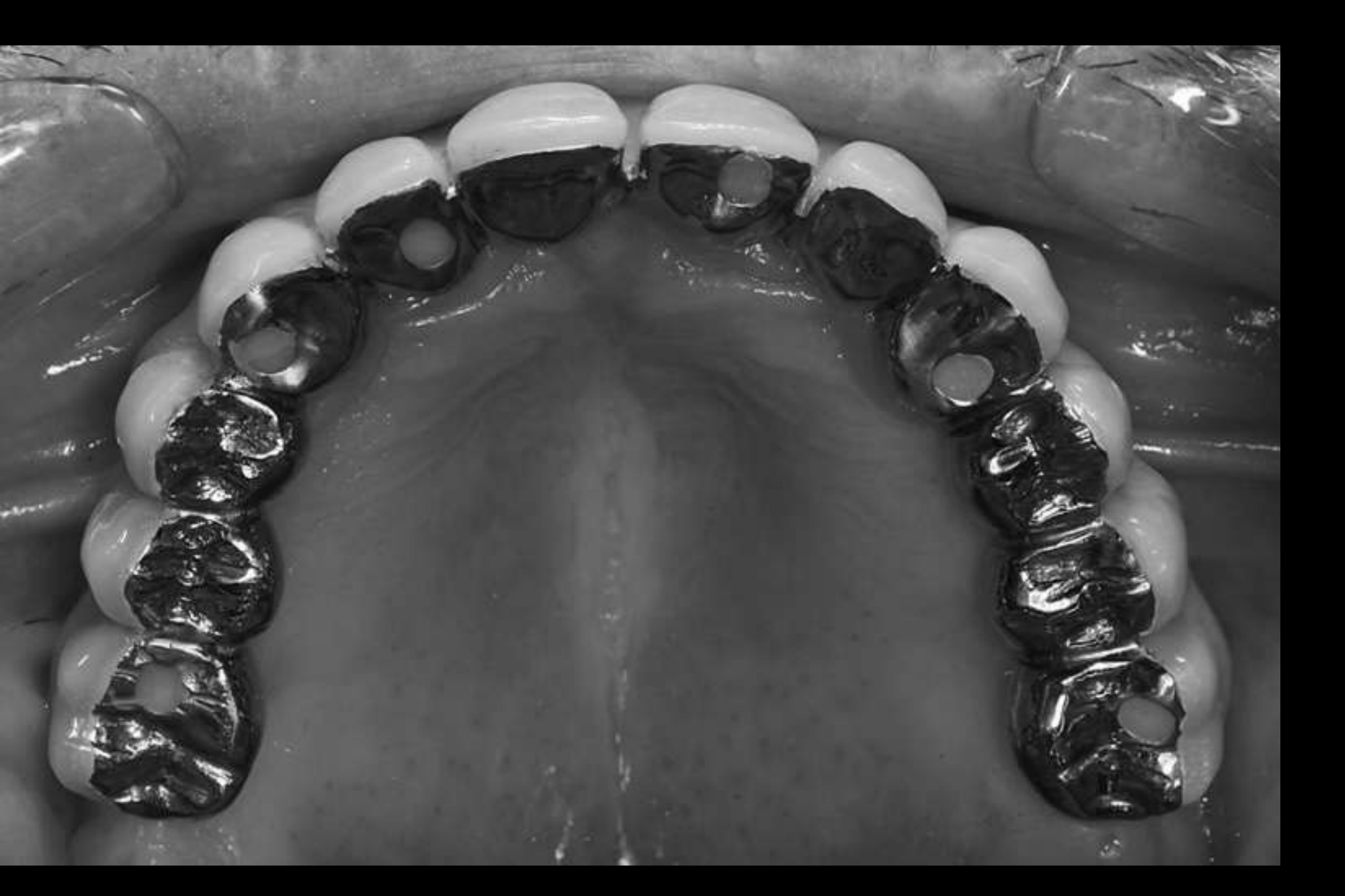
Procera Titanium Hybrid with Acrylic Teeth76
Need occlusal splint.
- Maintenance medium but relatively easy to fix
- Retread 5–7 years for wear
- Staining around margins
- Acrylic resin fractures
- Tooth fractures
Torsello F, di Torresanto VM, Ercoli C, Cordaro L. Evaluation of the marginal precision of one-piece complete arch titanium frameworks fabricated using five different methods for implant-supported restorations. Clin Oral Implants Res. 2008;19(8):772-9.
Hjalmarsson L, Örtorp A, Smedberg JI, Jemt T. Precision of fit to implants: a comparison of Cresco™ and Procera® implant bridge frameworks. Clin Implant Dent Relat Res. 2010;12(4): 271-80.
Lecturer — Acrylic Maintenance
Titanium-reinforced acrylic bridges may be used for full-arch treatment, but acrylic teeth and prosthetic materials commonly require replacement or refurbishment approximately every five to seven years.
- Typical maintenance includes staining, fracture, replacement of denture teeth, and retreatment or refurbishment of the prosthesis.
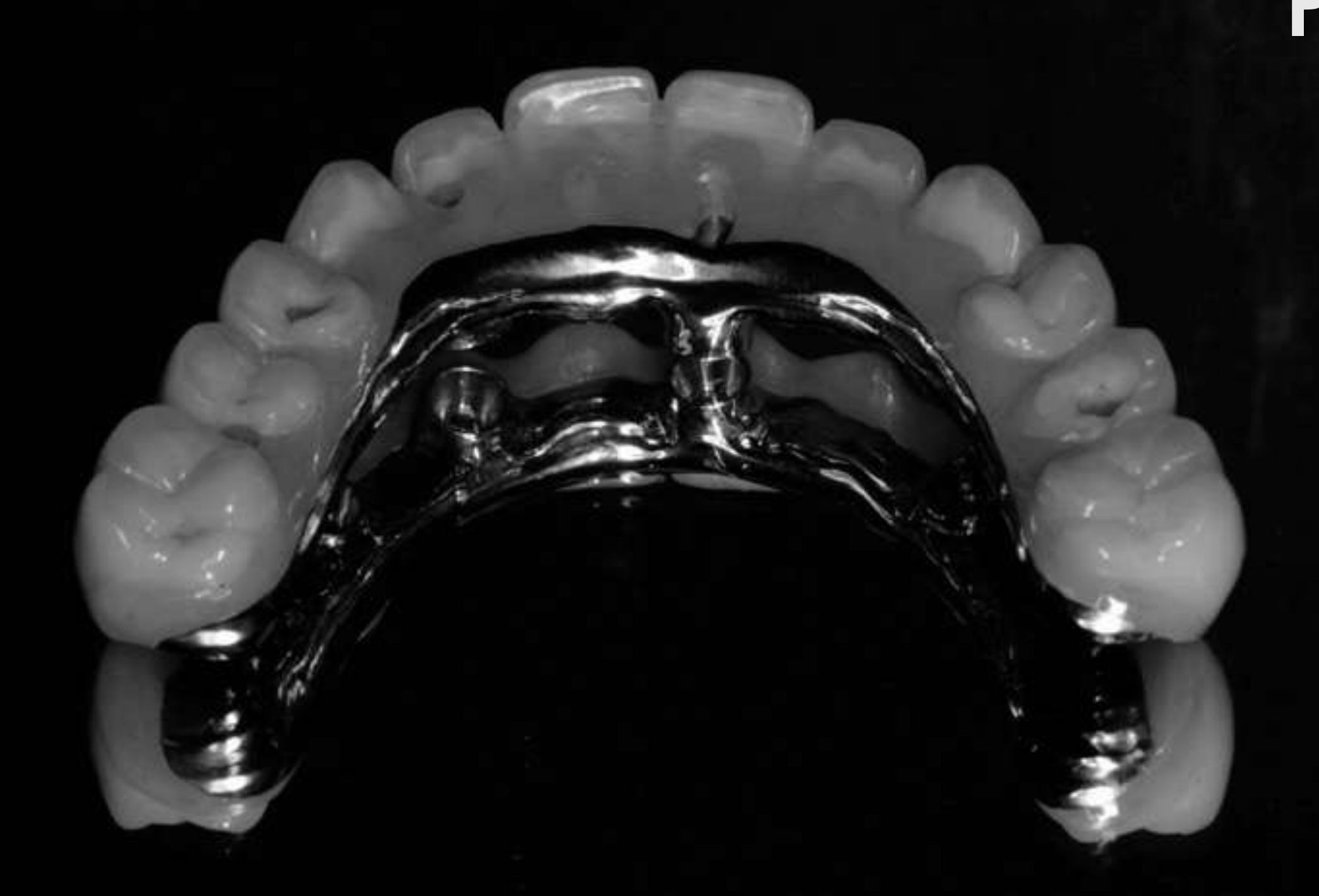
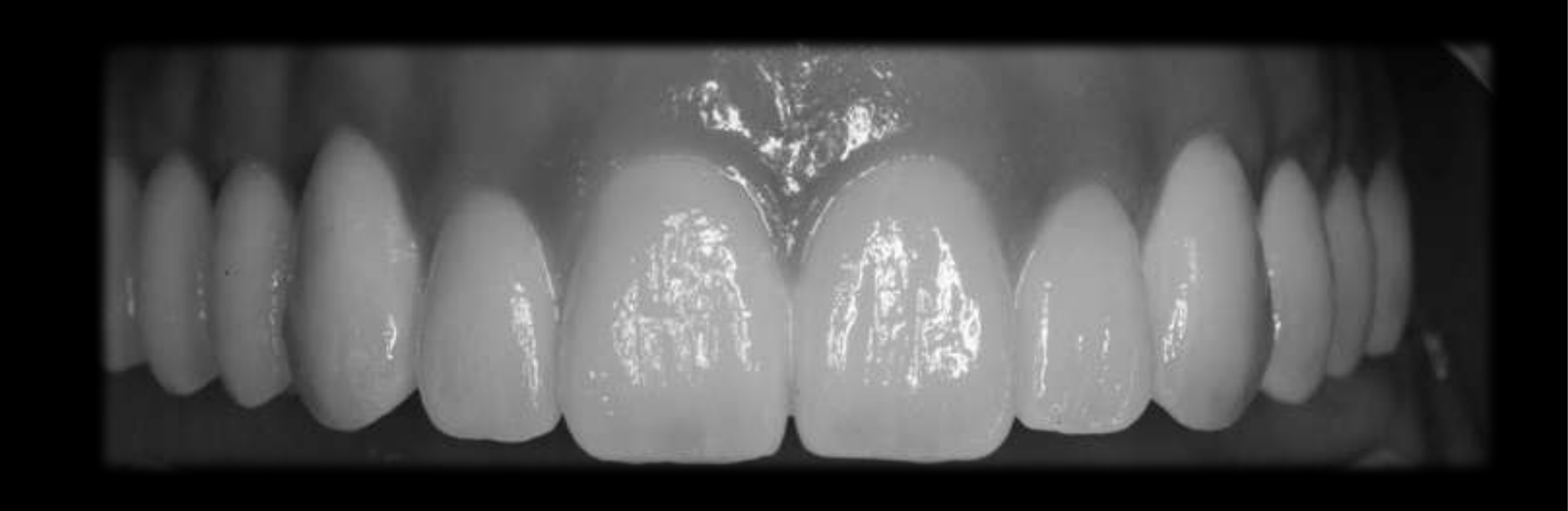
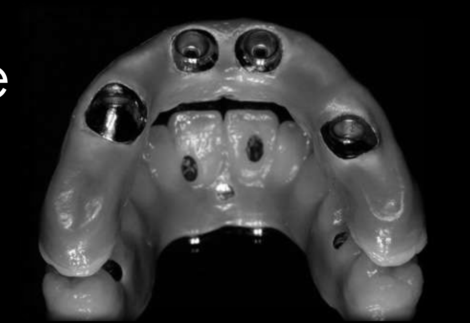
PFM — Gold Cemented Framework77
Gold prices are getting crazy — US$11K for this one!
Lecturer — Gold Frameworks
Gold porcelain-fused-to-metal frameworks were used for severe bruxers, but gold was described as heavy and increasingly unaffordable.
- The lecturer noted that the gold cost had been approximately 11,000 US dollars even about 10 years earlier, and that gold frameworks are no longer commonly used in the described treatment approach.
Maintenance low but expensive and difficult to fix.
 |  |
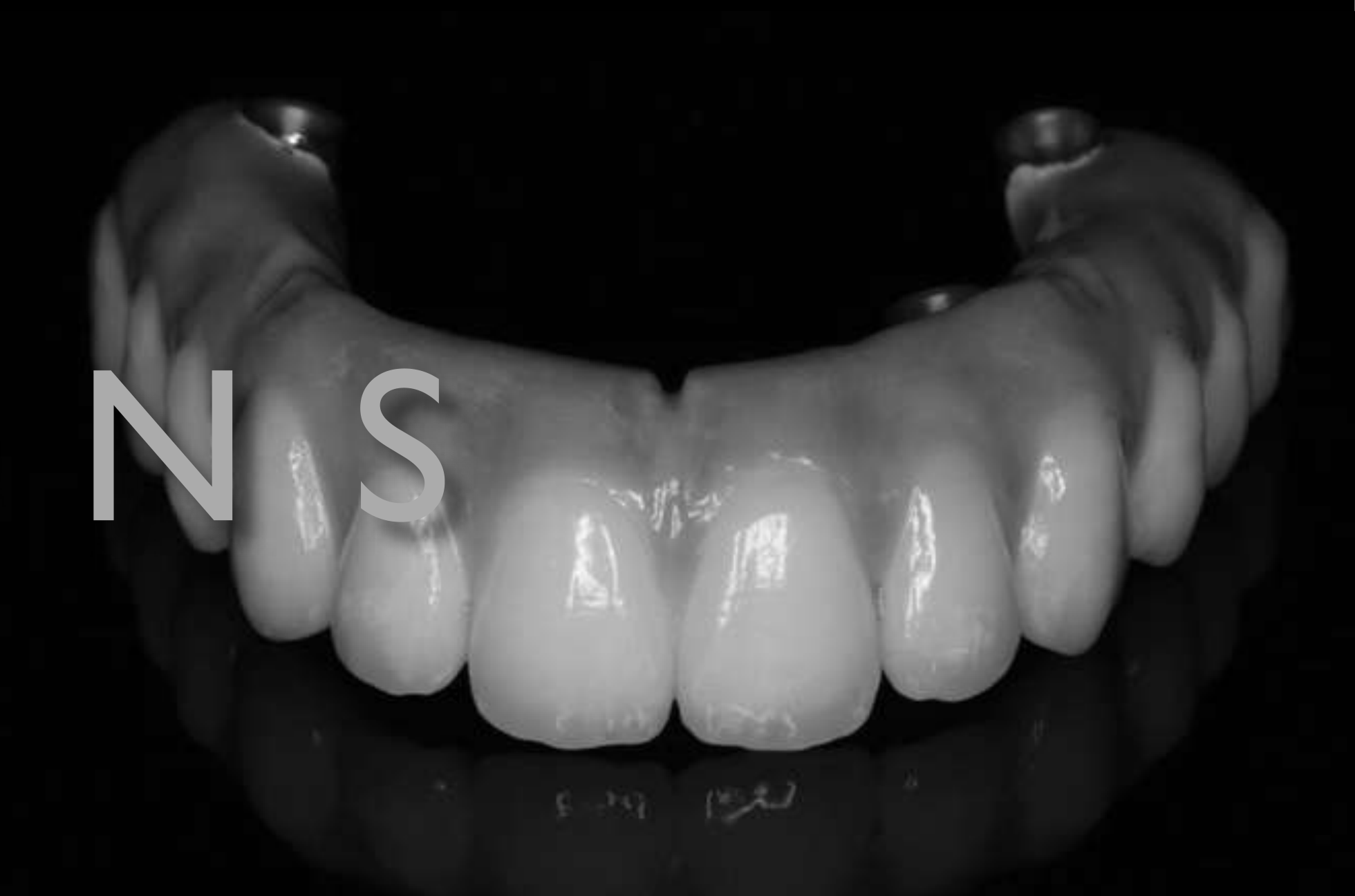
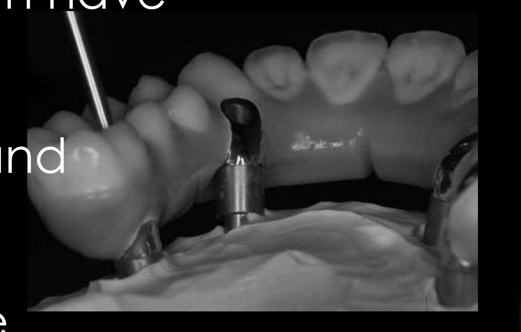
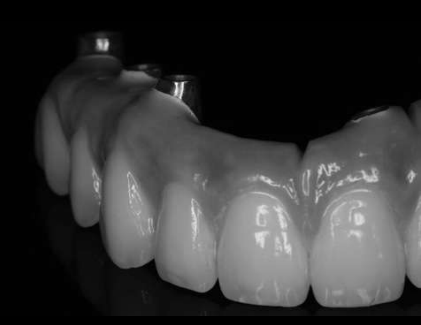
PFM — CrCo Framework78
- CadCam framework — low cost, can have reasonable fit
- Need occlusal splint
- Maintenance low but expensive and difficult to fix
- Gingival replacement — composite (marginal staining), porcelain (aesthetics getting better)
 |  |
 |
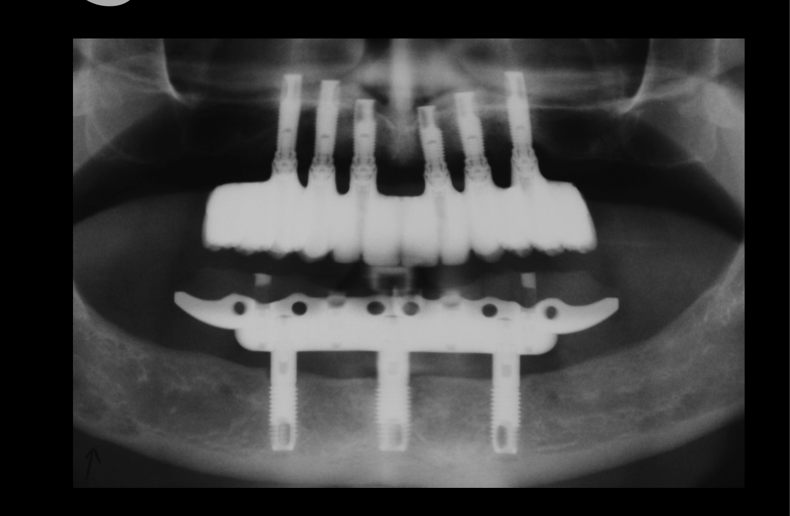
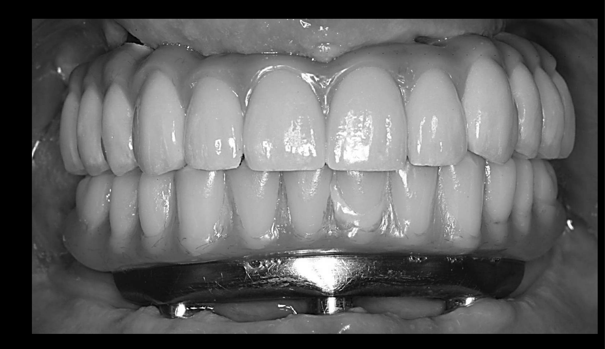
PFM — Porcelain Fused to Titanium79
- CadCam Procera framework — low cost, excellent fit
- Maintenance low but expensive and difficult to fix
- Aesthetics of Ti porcelain ordinary
 |  |
Flexural Strength of Dental Materials80818283
Flexural Strength — Current Dental Materials
| Material | Examples | Flexural Strength |
|---|---|---|
| Feldspathic | veneering porcelain, veneers | ~90 MPa |
| Pressable ceramics | Empress, Cergo, Optimal | ~150 MPa |
| Indirect composites | Belleglass, Critobal | ~200 MPa |
| Pressable lithium disilicate | Empr.2, e-Max | ~300 MPa |
| Glass infiltrated ceramics | In-Ceram Alum. | ~400 MPa |
| Alumina | Procera | ~650 MPa |
| (Y-TPZ) Zirconia | Procera, Lava, Allzirron | ~1200 MPa |
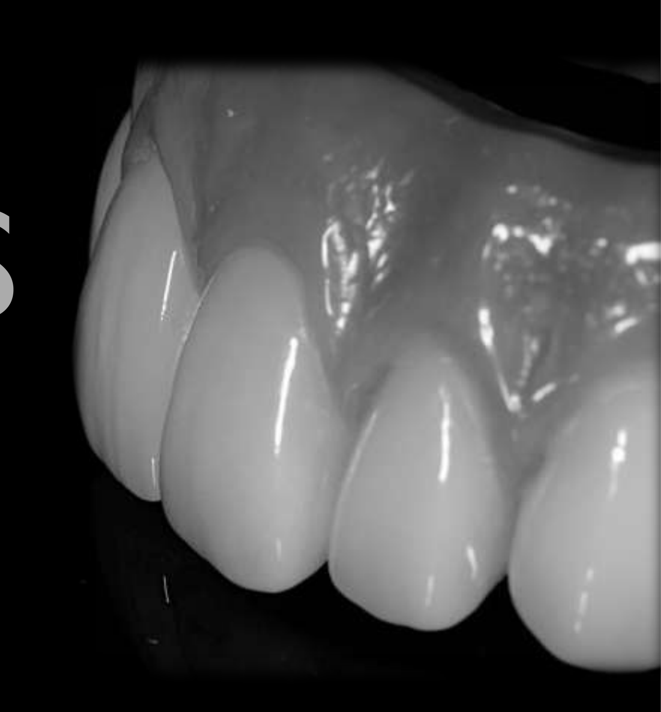
eMax Ceramic Crowns84
- Layered anteriorly, monolithic posteriorly (300 MPa vs 90 MPa veneering porcelain)
- No gold component in Procera framework and no difference in cost for size of framework
- Maintenance low and generally just replacement of one crown
- Gingival replacement — composite (marginal staining) — can be retreaded relatively easily
Lecturer — Zirconia Strength
Some more translucent zirconias may have strength values around the level of E.max, approximately 350 MPa, rather than the approximately 1,200 MPa associated with stronger 3Y zirconia.
- The material selected must therefore be appropriate for the functional demands of the restoration.
Flexural Strength — Current Dental Materials
Lecturer — Material Selection
Material selection should account for the patient and the clinical situation rather than relying only on appearance.
- The lecturer contrasted aesthetic, more translucent materials with stronger, less translucent materials.
| Material | Flexural Strength |
|---|---|
| Feldspathic — eg veneering porcelain, veneers | ~90 MPa |
| Pressable ceramics — eg: Empress, Cergo, Optimal | ~150 MPa |
| Indirect composites — eg: Belleglass, Critobal | ~200 MPa |
| Pressable lithium disilicate — eg: Empr.2, e-Max | ~300 MPa |
| Glass infiltrated ceramics — eg: In-Ceram Alum. | ~400 MPa |
| Alumina — Procera | ~650 MPa |
| (Y-TPZ) Zirconia — eg: Procera, Lava, Allzirkon | ~1200 MPa |
 | 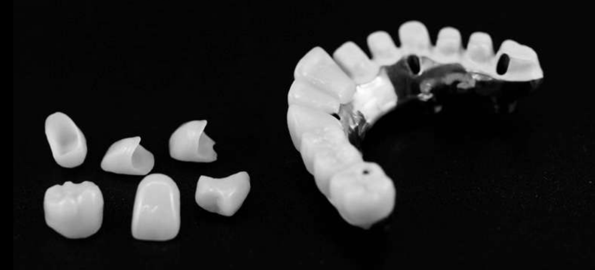 |
Zirconia and Hybrid Bridge Options8586
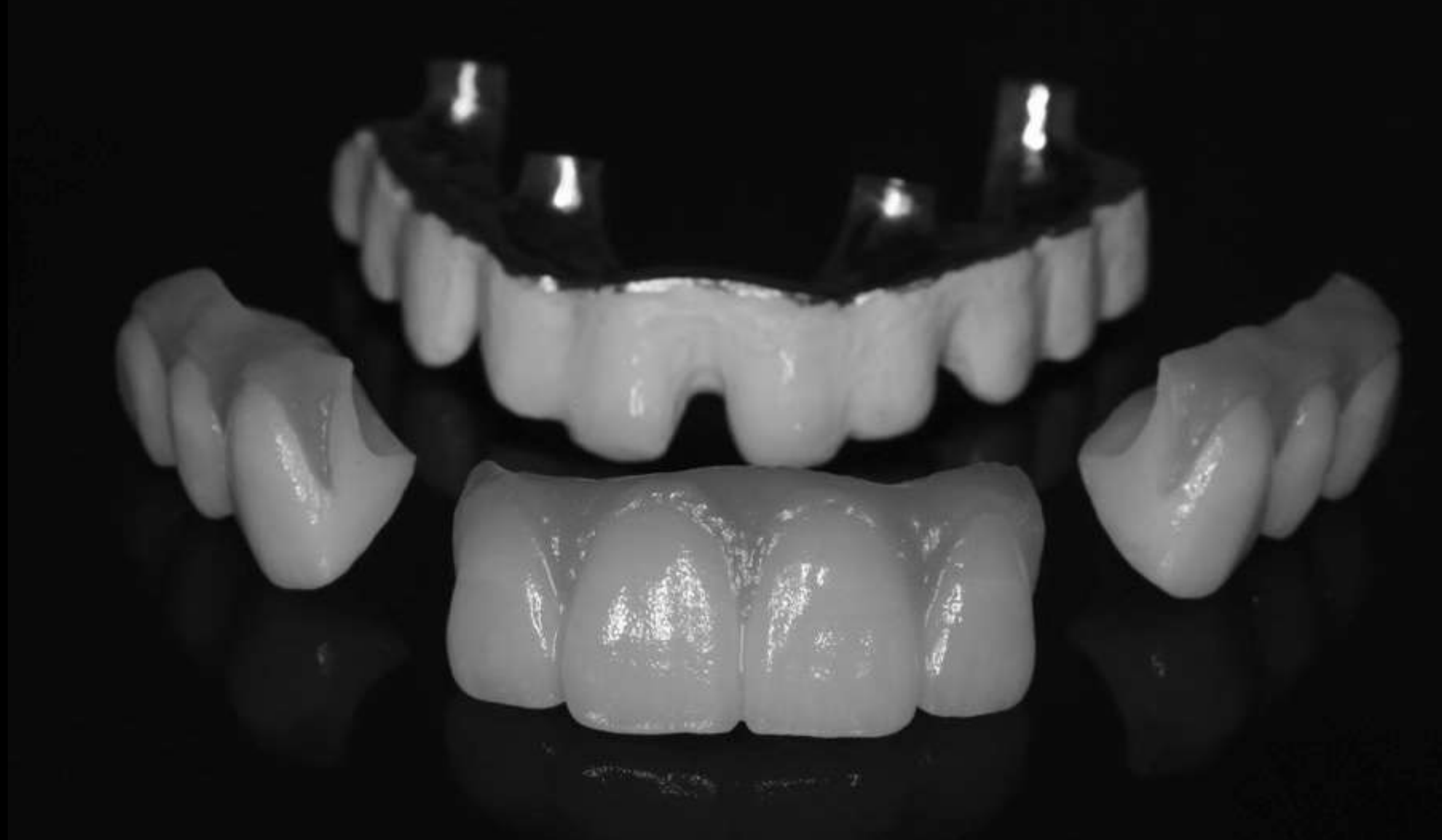
Full Zirconia87
- Massive downside if framework fracture
- Industrial fabrication essential
- Need highly experienced technician → need occlusal splint
- Attention to detail
- More long term clinical research required
Ti CAD CAM framework / stained monolithic zirconia segments
Lecturer — Zirconia Selection
Full zirconia has improved aesthetically, but zirconia-to-zirconia contact provides little cushioning and can create a clacking sound that resonates through the patient’s head and may disturb family members.
- 3Y zirconia is stronger and less translucent, whereas higher-yttria and cubic zirconias are more translucent but weaker.
- The clinician should know the yttria content, zirconia phase, strength, translucency, manufacturer, milling process, and staining and glazing process.
- Multi-layered pucks may contain dentine-like gingival, graduated middle, and enamel-like incisal regions; strength depends partly on where the restoration is milled within the puck.
- Newer multi-layered formulations lack fully established five-, ten-, and twenty-year outcomes, and interfaces between layers may contain weaknesses through which cracks can propagate.
- Patients with little wear and minimal bruxism may tolerate a wider range of materials, whereas heavy bruxers may require monolithic 3Y zirconia with strength prioritized.
- Strength can be affected by staining, glazing, clinical adjustment, milling, water cooling, zirconia composition, and milling-unit quality. Glaze may be lost through function and may be rough against the opposing arch.
- The laboratory’s material, milling equipment, and manufacturing controls are important; zirconia printing was described as a developing technology that had not yet fully matured.
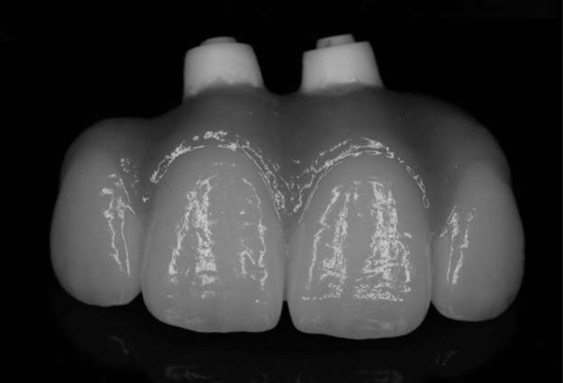
Maintenance Protocol and Clinical Preferences
Maintenance Protocol88
- Follow-up weekly for first 4 weeks, then 6-weekly until 6 months
- Then 3–6 monthly according to need
- OPG radiographs at bridge placement, 6 months, then yearly for 5 years
- Then every 2–3 years depending on history
Lecturer — Maintenance Requirements
Maintenance also depends on how the prosthesis is designed and fabricated.
- Regular attention is needed for prosthetic cleansability, plaque control, home-care technique, prosthetic wear, fracture, staining, and replacement of acrylic teeth when required.
- Patients should be instructed in the use of water jets and interdental cleaning aids, and the tissue-fitting surfaces should remain accessible.
- Requirements are affected by the material used, the opposing dentition, bruxism, prosthesis design, implant position, and laboratory fabrication quality.
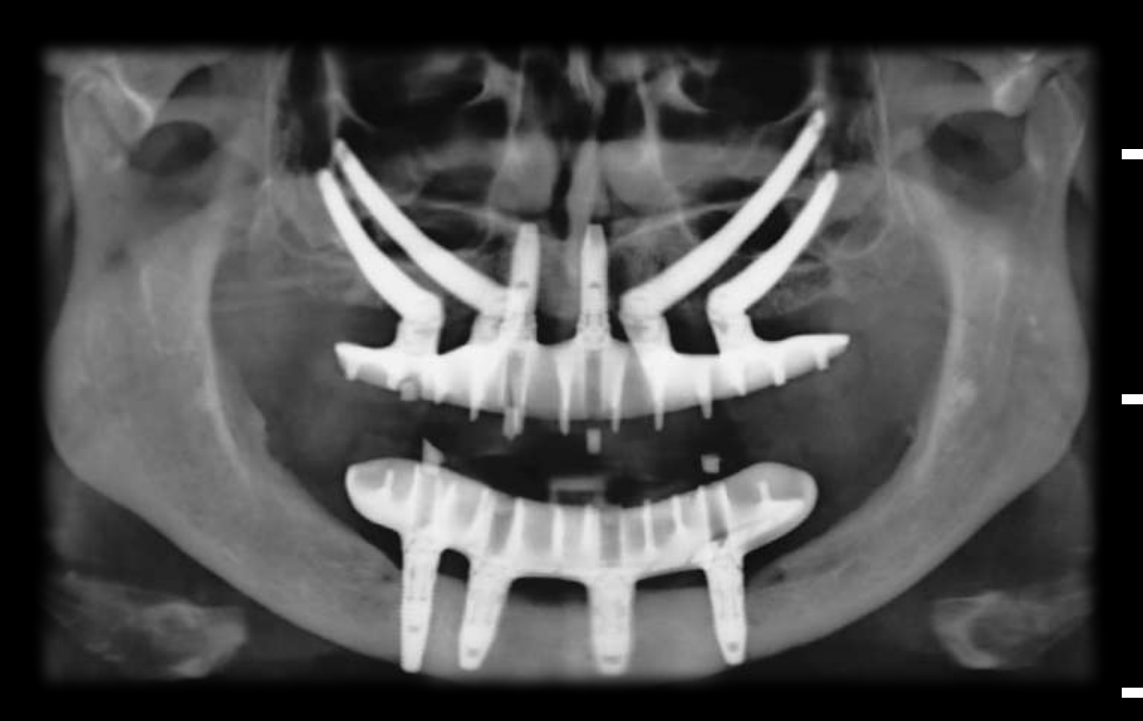
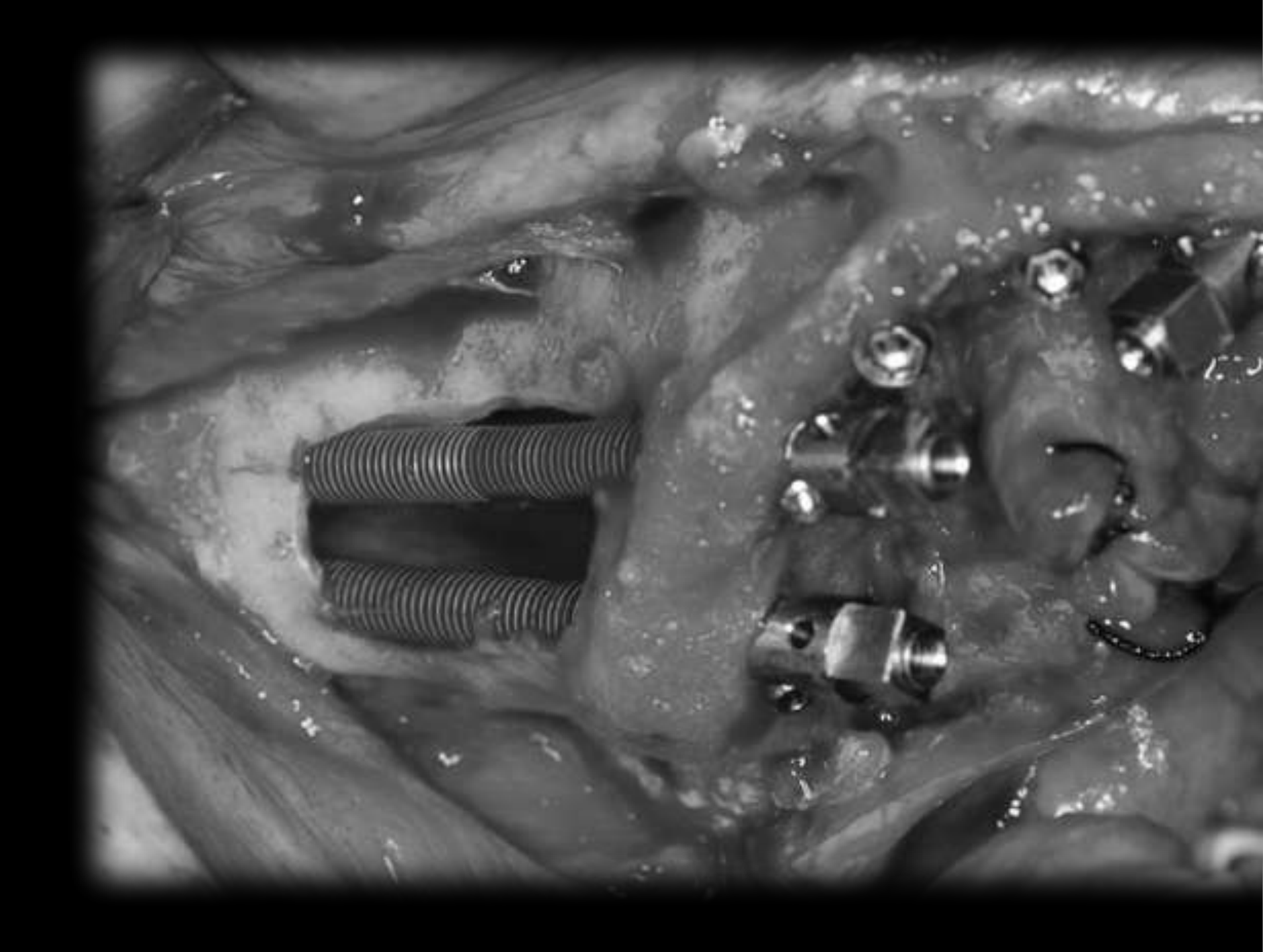
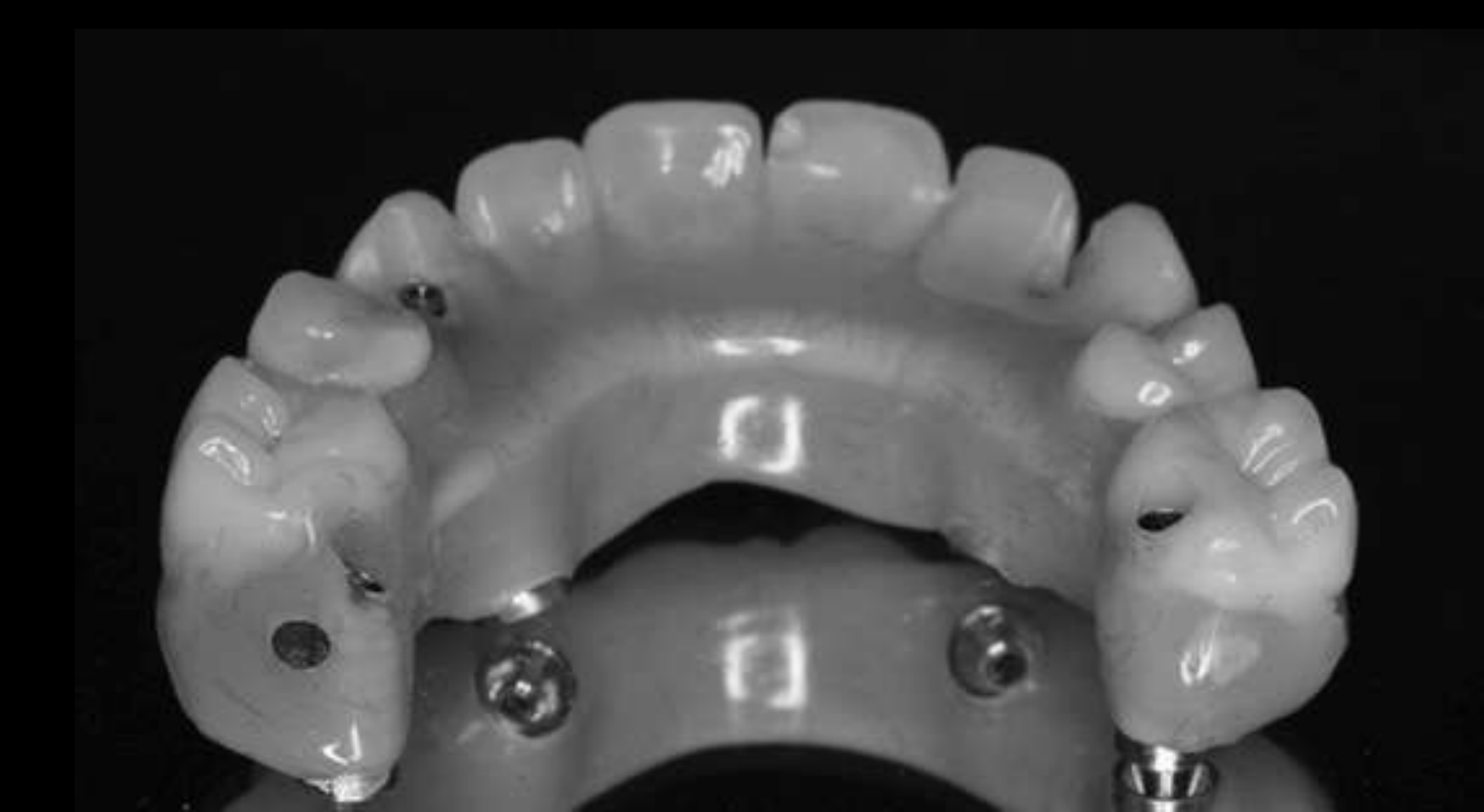
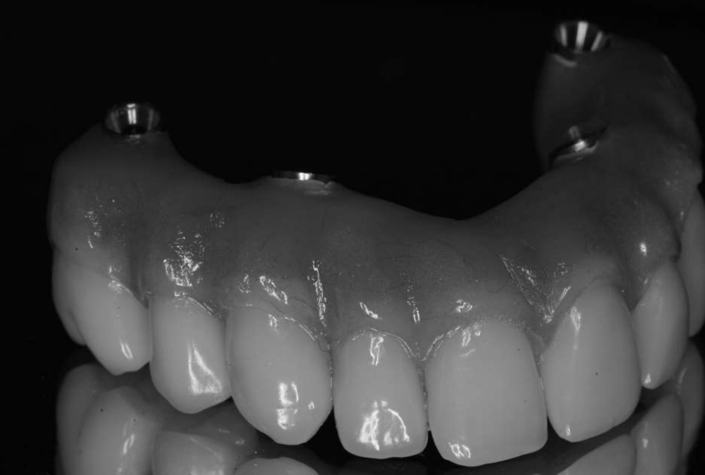
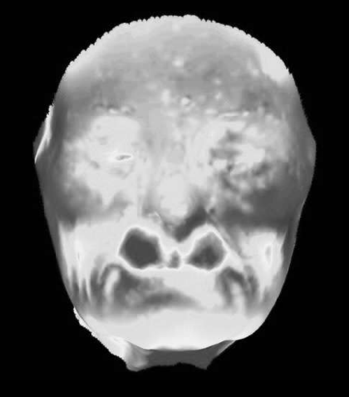
Our Preferences in Treatment899091
No Pre-implant Grafting
- Utilize available native bone
Cross arch splinting
Rigid prosthesis shows cross arch splinting
Prosthetic replacement of soft tissue and facial support
Lecturer — Treatment Preferences
The lecturer favored fixed treatment in the maxilla when patients do not want a removable prosthesis and require facial support. The preferred pathway depends on the available bone and the patient’s functional and restorative requirements.
- Use axial implants when sufficient bone exists.
- Use tilted implants to avoid the sinus or engage denser bone.
- Consider grafting when indicated and zygoma implants in severely atrophic cases.
- Prostheses should be prosthetically directed, anatomical in contour, cleansable, and designed around the patient’s facial support requirements.
- For high functional loads, stronger and less layered materials may be preferred; material selection should reflect bruxism, wear, aesthetics, and function rather than simply choosing the most translucent option.
Patient and Clinician Benefits9293
Optimal Comfort
- Less invasive surgery
- Less pain and swelling
- Reduced chairtime
Cost Saving
- Much earlier return to work and social life
Fast Treatment
-
Immediate function
-
Increased Predictability & Safety
-
Reduction in Interim Management
-
Predictable Prosthetic Outcome
Lecturer — Fixed Treatment Benefits
Fixed treatment can restore teeth without covering the palate, avoid the inconvenience of removability, improve speech and taste compared with a palatal denture, and restore facial support. When immediate loading is appropriate, it can also provide teeth more quickly.
- Graftless or zygomatic pathways may reduce treatment time, the period without a wearable denture, the need for extensive grafting, and the complexity associated with hip-harvested bone.
- These benefits must be balanced against implant stability, loading risk, bone quality, cleansability, long-term prosthetic maintenance, and the limitations of newer restorative materials.
Closing and Contact Information94
Eradication of Pathology
Thank you!
BRÅNEMARK CENTRE, PERTH, AUSTRALIA
Clinical Cases
Case: Female patient with FP2 prosthesis and facial aesthetics evaluation
Question
Scenario: A female patient is presented to evaluate her facial aesthetics, lip support, and the transition line of her prosthesis.
What’s shown: Before and after facial images showing creases and vermilion support. An intraoral view shows pink reproduction (FP2) and the patient’s smile line.
Consider: How to manage the transition line between the prosthesis and natural gingiva in relation to the smile line, and what type of prosthesis is appropriate.
Answer
Observations:
- The patient has an FP2 prosthesis with some pink reproduction.
- The color match between the natural gum and the prosthesis is not perfect.
- The transition line is positioned above the patient’s smile line.
Reasoning: If the smile line is above the transition line, the mismatch between the translucent natural gingiva and the porcelain or acrylic prosthesis will look terrible. By keeping the transition line above the smile line, the aesthetic discrepancy is hidden when the patient smiles.
Takeaway: The transition line between a prosthesis and natural gingiva must be kept above the smile line to avoid visible aesthetic mismatches.
Case: Male patient with a 15-year follow-up of an implant-supported bridge
Question
Scenario: A male patient who had teeth extracted and implants placed is presented to show long-term outcomes.
What’s shown: The patient’s provisional bridge and a follow-up image 15 years later.
Consider: What the long-term outcome and success rate of this type of implant-supported fixed bridge treatment is.
Answer
Observations:
- The patient received a provisional bridge after implant placement.
- The 15-year follow-up shows the final result and long-term stability.
Reasoning: The lecturer uses this case to demonstrate that immediate loading and provisionalization in the maxilla can yield highly successful long-term results, as the patient has maintained the restoration for 15 years without issue.
Takeaway: Immediate loading and provisional bridges in the maxilla can provide excellent, long-lasting outcomes.
Case: Female patient with an atrophic maxilla treated with a lateral window sinus lift
Question
Scenario: A female patient with an atrophic maxilla requires full arch reconstruction but has minimal bone.
What’s shown: Intraoperative images of a lateral window sinus lift, elevation of the Schneiderian membrane, placement of bovine bone and autogenous bone blocks fixated with screws, and covered with membranes. Post-operative images show the final bridge.
Consider: What surgical techniques are used to augment the atrophic maxilla, and what are the clinical implications and patient management challenges of this approach.
Answer
Observations:
- A lateral window sinus lift was performed to elevate the Schneiderian membrane.
- The sinus was filled with a mixture of bovine bone and autogenous bone blocks fixated with screws and covered with membranes.
- Implants were placed, and a final bridge was fabricated.
- The treatment took a year and a half, and the patient could not wear her denture for about four months.
Reasoning: While this traditional sinus lift and block grafting approach can successfully restore an atrophic maxilla, it requires a long treatment time and a prolonged period without a functional denture. This creates significant patient management issues and has a lower success rate compared to newer techniques like zygomatic implants.
Takeaway: Traditional lateral window sinus lifts with block grafting for atrophic maxillae are highly demanding, require long treatment times, and cause significant patient discomfort due to prolonged periods without a denture.
Case: Female patient with severe maxillary atrophy treated with semi-guided zygomatic implants
Question
Scenario: A female patient who has not been able to wear an upper denture for 10 years and has failing anterior mandibular teeth presents with severe maxillary atrophy.
What’s shown: Radiographic and clinical images showing a very thin, shallow ridge. Intraoperative images show a semi-guided zygomatic implant placement via a lateral wall approach, with 50mm long implants engaging the zygoma. Follow-up images at 6 years and 18 years show bone growth along the implant and the final restoration.
Consider: How to manage a patient with an extremely atrophic maxilla who cannot tolerate a denture, and what the long-term outcomes of this specific surgical approach are.
Answer
Observations:
- The patient had a very thin, shallow maxillary ridge and had not worn an upper denture for 10 years.
- Semi-guided zygomatic implants (approximately 50mm long) were placed, engaging the zygomatic arch, along with a lateral wall sinus lift.
- Follow-up at 6 and 18 years shows successful bone growth along the length of the implants into the sinus and a stable final restoration.
Reasoning: In cases of extreme maxillary atrophy where traditional grafting is insufficient or too burdensome, zygomatic implants bypass the need for extensive grafting by anchoring in the zygomatic bone. The semi-guided approach and lateral wall sinus lift allow for precise placement. The long-term follow-up demonstrates the high success and stability of this technique.
Takeaway: Zygomatic implants are a highly successful, long-term solution for patients with severe maxillary atrophy who cannot tolerate conventional dentures, avoiding the need for extensive bone grafting.
Footnotes
-
Original PDF page 1: L7 Management of edentulism and the terminal dentitionl Maxilla fixed notes compressed (1), p.1 ↩
-
Original PDF page 2: L7 Management of edentulism and the terminal dentitionl Maxilla fixed notes compressed (1), p.2 ↩
-
Original PDF page 3: L7 Management of edentulism and the terminal dentitionl Maxilla fixed notes compressed (1), p.3 ↩
-
Original PDF page 4: L7 Management of edentulism and the terminal dentitionl Maxilla fixed notes compressed (1), p.4 ↩
-
Original PDF page 5: L7 Management of edentulism and the terminal dentitionl Maxilla fixed notes compressed (1), p.5 ↩
-
Original PDF page 6: L7 Management of edentulism and the terminal dentitionl Maxilla fixed notes compressed (1), p.6 ↩
-
Original PDF page 7: L7 Management of edentulism and the terminal dentitionl Maxilla fixed notes compressed (1), p.7 ↩
-
Original PDF page 8: L7 Management of edentulism and the terminal dentitionl Maxilla fixed notes compressed (1), p.8 ↩
-
Original PDF page 9: L7 Management of edentulism and the terminal dentitionl Maxilla fixed notes compressed (1), p.9 ↩
-
Original PDF page 10: L7 Management of edentulism and the terminal dentitionl Maxilla fixed notes compressed (1), p.10 ↩
-
Original PDF page 11: L7 Management of edentulism and the terminal dentitionl Maxilla fixed notes compressed (1), p.11 ↩
-
Original PDF page 14: L7 Management of edentulism and the terminal dentitionl Maxilla fixed notes compressed (1), p.14 ↩
-
Original PDF page 12: L7 Management of edentulism and the terminal dentitionl Maxilla fixed notes compressed (1), p.12 ↩
-
Original PDF page 13: L7 Management of edentulism and the terminal dentitionl Maxilla fixed notes compressed (1), p.13 ↩
-
Original PDF page 15: L7 Management of edentulism and the terminal dentitionl Maxilla fixed notes compressed (1), p.15 ↩
-
Original PDF page 16: L7 Management of edentulism and the terminal dentitionl Maxilla fixed notes compressed (1), p.16 ↩
-
Original PDF page 17: L7 Management of edentulism and the terminal dentitionl Maxilla fixed notes compressed (1), p.17 ↩
-
Original PDF page 18: L7 Management of edentulism and the terminal dentitionl Maxilla fixed notes compressed (1), p.18 ↩
-
Original PDF page 19: L7 Management of edentulism and the terminal dentitionl Maxilla fixed notes compressed (1), p.19 ↩
-
Original PDF page 20: L7 Management of edentulism and the terminal dentitionl Maxilla fixed notes compressed (1), p.20 ↩
-
Original PDF page 21: L7 Management of edentulism and the terminal dentitionl Maxilla fixed notes compressed (1), p.21 ↩
-
Original PDF page 22: L7 Management of edentulism and the terminal dentitionl Maxilla fixed notes compressed (1), p.22 ↩
-
Original PDF page 23: L7 Management of edentulism and the terminal dentitionl Maxilla fixed notes compressed (1), p.23 ↩
-
Original PDF page 24: L7 Management of edentulism and the terminal dentitionl Maxilla fixed notes compressed (1), p.24 ↩
-
Original PDF page 25: L7 Management of edentulism and the terminal dentitionl Maxilla fixed notes compressed (1), p.25 ↩
-
Original PDF page 26: L7 Management of edentulism and the terminal dentitionl Maxilla fixed notes compressed (1), p.26 ↩
-
Original PDF page 27: L7 Management of edentulism and the terminal dentitionl Maxilla fixed notes compressed (1), p.27 ↩
-
Original PDF page 28: L7 Management of edentulism and the terminal dentitionl Maxilla fixed notes compressed (1), p.28 ↩
-
Original PDF page 29: L7 Management of edentulism and the terminal dentitionl Maxilla fixed notes compressed (1), p.29 ↩
-
Original PDF page 30: L7 Management of edentulism and the terminal dentitionl Maxilla fixed notes compressed (1), p.30 ↩
-
Original PDF page 31: L7 Management of edentulism and the terminal dentitionl Maxilla fixed notes compressed (1), p.31 ↩
-
Original PDF page 32: L7 Management of edentulism and the terminal dentitionl Maxilla fixed notes compressed (1), p.32 ↩
-
Original PDF page 34: L7 Management of edentulism and the terminal dentitionl Maxilla fixed notes compressed (1), p.34 ↩
-
Original PDF page 35: L7 Management of edentulism and the terminal dentitionl Maxilla fixed notes compressed (1), p.35 ↩
-
Original PDF page 36: L7 Management of edentulism and the terminal dentitionl Maxilla fixed notes compressed (1), p.36 ↩
-
Original PDF page 37: L7 Management of edentulism and the terminal dentitionl Maxilla fixed notes compressed (1), p.37 ↩
-
Original PDF page 38: L7 Management of edentulism and the terminal dentitionl Maxilla fixed notes compressed (1), p.38 ↩
-
Original PDF page 39: L7 Management of edentulism and the terminal dentitionl Maxilla fixed notes compressed (1), p.39 ↩
-
Original PDF page 40: L7 Management of edentulism and the terminal dentitionl Maxilla fixed notes compressed (1), p.40 ↩
-
Original PDF page 41: L7 Management of edentulism and the terminal dentitionl Maxilla fixed notes compressed (1), p.41 ↩
-
Original PDF page 42: L7 Management of edentulism and the terminal dentitionl Maxilla fixed notes compressed (1), p.42 ↩
-
Original PDF page 43: L7 Management of edentulism and the terminal dentitionl Maxilla fixed notes compressed (1), p.43 ↩
-
Original PDF page 44: L7 Management of edentulism and the terminal dentitionl Maxilla fixed notes compressed (1), p.44 ↩
-
Original PDF page 45: L7 Management of edentulism and the terminal dentitionl Maxilla fixed notes compressed (1), p.45 ↩
-
Original PDF page 46: L7 Management of edentulism and the terminal dentitionl Maxilla fixed notes compressed (1), p.46 ↩
-
Original PDF page 47: L7 Management of edentulism and the terminal dentitionl Maxilla fixed notes compressed (1), p.47 ↩
-
Original PDF page 48: L7 Management of edentulism and the terminal dentitionl Maxilla fixed notes compressed (1), p.48 ↩
-
Original PDF page 50: L7 Management of edentulism and the terminal dentitionl Maxilla fixed notes compressed (1), p.50 ↩
-
Original PDF page 49: L7 Management of edentulism and the terminal dentitionl Maxilla fixed notes compressed (1), p.49 ↩
-
Original PDF page 52: L7 Management of edentulism and the terminal dentitionl Maxilla fixed notes compressed (1), p.52 ↩
-
Original PDF page 51: L7 Management of edentulism and the terminal dentitionl Maxilla fixed notes compressed (1), p.51 ↩
-
Original PDF page 53: L7 Management of edentulism and the terminal dentitionl Maxilla fixed notes compressed (1), p.53 ↩
-
Original PDF page 54: L7 Management of edentulism and the terminal dentitionl Maxilla fixed notes compressed (1), p.54 ↩
-
Original PDF page 55: L7 Management of edentulism and the terminal dentitionl Maxilla fixed notes compressed (1), p.55 ↩
-
Original PDF page 56: L7 Management of edentulism and the terminal dentitionl Maxilla fixed notes compressed (1), p.56 ↩
-
Original PDF page 57: L7 Management of edentulism and the terminal dentitionl Maxilla fixed notes compressed (1), p.57 ↩
-
Original PDF page 58: L7 Management of edentulism and the terminal dentitionl Maxilla fixed notes compressed (1), p.58 ↩
-
Original PDF page 61: L7 Management of edentulism and the terminal dentitionl Maxilla fixed notes compressed (1), p.61 ↩
-
Original PDF page 59: L7 Management of edentulism and the terminal dentitionl Maxilla fixed notes compressed (1), p.59 ↩
-
Original PDF page 60: L7 Management of edentulism and the terminal dentitionl Maxilla fixed notes compressed (1), p.60 ↩
-
Original PDF page 62: L7 Management of edentulism and the terminal dentitionl Maxilla fixed notes compressed (1), p.62 ↩
-
Original PDF page 63: L7 Management of edentulism and the terminal dentitionl Maxilla fixed notes compressed (1), p.63 ↩
-
Original PDF page 65: L7 Management of edentulism and the terminal dentitionl Maxilla fixed notes compressed (1), p.65 ↩
-
Original PDF page 66: L7 Management of edentulism and the terminal dentitionl Maxilla fixed notes compressed (1), p.66 ↩
-
Original PDF page 67: L7 Management of edentulism and the terminal dentitionl Maxilla fixed notes compressed (1), p.67 ↩
-
Original PDF page 68: L7 Management of edentulism and the terminal dentitionl Maxilla fixed notes compressed (1), p.68 ↩
-
Original PDF page 64: L7 Management of edentulism and the terminal dentitionl Maxilla fixed notes compressed (1), p.64 ↩
-
Original PDF page 69: L7 Management of edentulism and the terminal dentitionl Maxilla fixed notes compressed (1), p.69 ↩
-
Original PDF page 70: L7 Management of edentulism and the terminal dentitionl Maxilla fixed notes compressed (1), p.70 ↩
-
Original PDF page 71: L7 Management of edentulism and the terminal dentitionl Maxilla fixed notes compressed (1), p.71 ↩
-
Original PDF page 72: L7 Management of edentulism and the terminal dentitionl Maxilla fixed notes compressed (1), p.72 ↩
-
Original PDF page 73: L7 Management of edentulism and the terminal dentitionl Maxilla fixed notes compressed (1), p.73 ↩
-
Original PDF page 74: L7 Management of edentulism and the terminal dentitionl Maxilla fixed notes compressed (1), p.74 ↩
-
Original PDF page 75: L7 Management of edentulism and the terminal dentitionl Maxilla fixed notes compressed (1), p.75 ↩
-
Original PDF page 77: L7 Management of edentulism and the terminal dentitionl Maxilla fixed notes compressed (1), p.77 ↩
-
Original PDF page 76: L7 Management of edentulism and the terminal dentitionl Maxilla fixed notes compressed (1), p.76 ↩
-
Original PDF page 78: L7 Management of edentulism and the terminal dentitionl Maxilla fixed notes compressed (1), p.78 ↩
-
Original PDF page 79: L7 Management of edentulism and the terminal dentitionl Maxilla fixed notes compressed (1), p.79 ↩
-
Original PDF page 80: L7 Management of edentulism and the terminal dentitionl Maxilla fixed notes compressed (1), p.80 ↩
-
Original PDF page 81: L7 Management of edentulism and the terminal dentitionl Maxilla fixed notes compressed (1), p.81 ↩
-
Original PDF page 82: L7 Management of edentulism and the terminal dentitionl Maxilla fixed notes compressed (1), p.82 ↩
-
Original PDF page 84: L7 Management of edentulism and the terminal dentitionl Maxilla fixed notes compressed (1), p.84 ↩
-
Original PDF page 85: L7 Management of edentulism and the terminal dentitionl Maxilla fixed notes compressed (1), p.85 ↩
-
Original PDF page 83: L7 Management of edentulism and the terminal dentitionl Maxilla fixed notes compressed (1), p.83 ↩
-
Original PDF page 87: L7 Management of edentulism and the terminal dentitionl Maxilla fixed notes compressed (1), p.87 ↩
-
Original PDF page 88: L7 Management of edentulism and the terminal dentitionl Maxilla fixed notes compressed (1), p.88 ↩
-
Original PDF page 86: L7 Management of edentulism and the terminal dentitionl Maxilla fixed notes compressed (1), p.86 ↩
-
Original PDF page 89: L7 Management of edentulism and the terminal dentitionl Maxilla fixed notes compressed (1), p.89 ↩
-
Original PDF page 90: L7 Management of edentulism and the terminal dentitionl Maxilla fixed notes compressed (1), p.90 ↩
-
Original PDF page 91: L7 Management of edentulism and the terminal dentitionl Maxilla fixed notes compressed (1), p.91 ↩
-
Original PDF page 92: L7 Management of edentulism and the terminal dentitionl Maxilla fixed notes compressed (1), p.92 ↩
-
Original PDF page 93: L7 Management of edentulism and the terminal dentitionl Maxilla fixed notes compressed (1), p.93 ↩
-
Original PDF page 94: L7 Management of edentulism and the terminal dentitionl Maxilla fixed notes compressed (1), p.94 ↩
-
Original PDF page 95: L7 Management of edentulism and the terminal dentitionl Maxilla fixed notes compressed (1), p.95 ↩