Maxillofacial Trauma1
The University of Western Australia — Oral Health Centre of Western Australia
MAXILLOFACIAL TRAUMA
Magdalen Foo Discipline Lead, Oral Maxillofacial Surgery
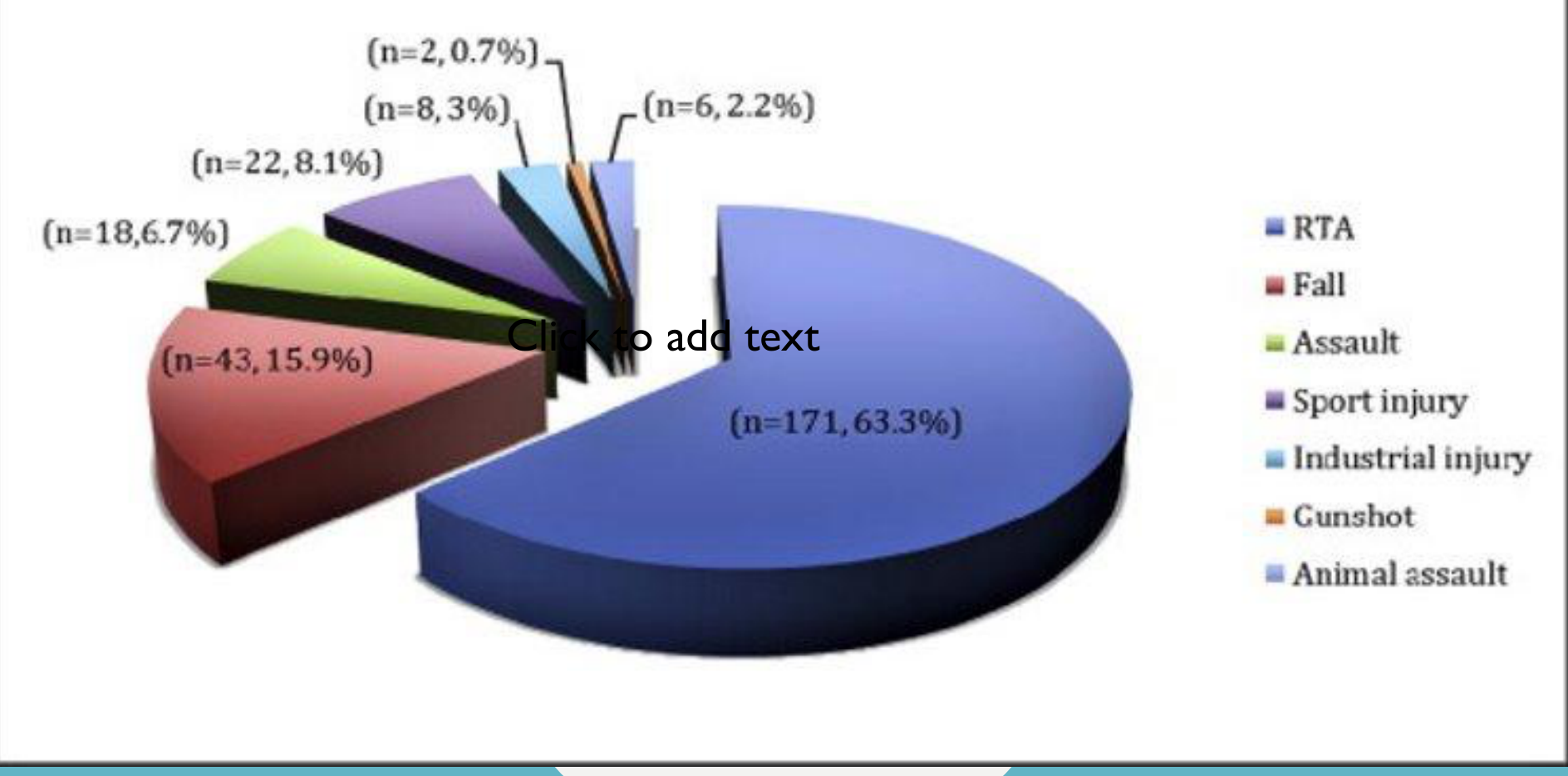
Epidemiology234
-
RTA (n=171, 63.3%)
-
Fall (n=43, 15.9%)
-
Assault (n=22, 8.1%)
-
Sport injury (n=18, 6.7%)
-
Industrial injury (n=8, 3%)
-
Gunshot (n=6, 2.2%)
-
Animal assault (n=2, 0.7%)
-
Nasal bones is the most common isolated facial bone fracture (largely due to the prominence and central location of the nose on the face and weakness of the nasal cartilage), followed by mandibular fracture which is the second most common
-
Most common causes of facial fractures are interpersonal violence and motor vehicle accidents
-
Other causes are falls, sporting injuries, and industrial injuries
-
Additional causes include dog bites, horse kicks, stab wounds, and firearm injuries; assaults may involve punches or group assaults.
-
Alcohol is an aggravating factor
-
80% are males, majority aged 20–35
Lecturer — Alcohol and Trauma
Alcohol is particularly associated with injuries involving young males.
 |  |
Biomechanics of Trauma
Principles of Biomechanics56
| Injury identification | Injury classification | Injury prevention |
|---|---|---|
| Nature of injury – degree of force, resistance to force | Severity of injury – direction of force, point of application of force | Pattern of injury – |
Trauma can be defined as the application of excessive energy to the human body above its tolerance, resulting in damage.
KE = ½ m v²
- Kinetic Energy is directly related to injury severity, and is predicted by ½ mass and (velocity)²
Lecturer — Injury History
Trauma is the transfer of energy, and injury severity depends on how much energy is transferred, how quickly it is transferred, how it is absorbed, and the direction and pattern of injury.
- The history helps estimate the likely severity and pattern of injury.
- Important details include the mechanism, fall height, vehicle position and speed, vehicle damage, seat-belt use, whether the patient was a driver, passenger, or pedestrian, and associated injuries.
Types of Injuries7
Blunt
- Motor vehicle accidents
- Falls
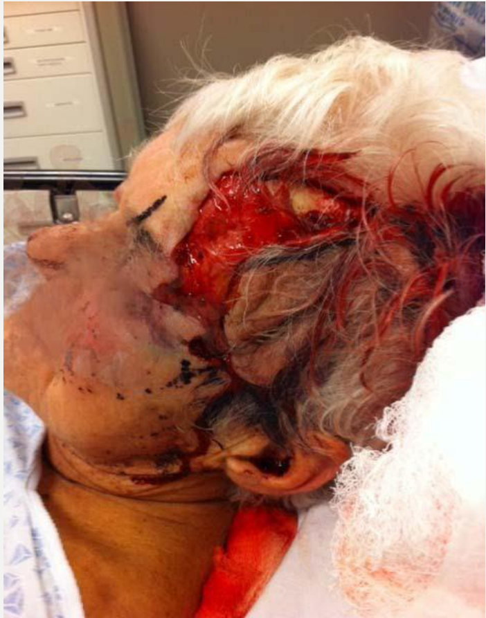
Lecturer — Falls in Elderly Patients
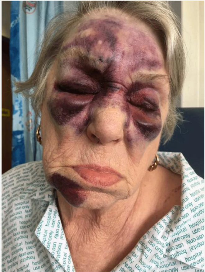
Elderly patients may fall, forget the incident, and later present with altered occlusion, denture problems, or other delayed signs.
- A fall in an elderly patient taking aspirin or warfarin may be associated with a slowly developing subdural hematoma.
Penetrating
- Stab wounds
- Firearm injuries
Lecturer — Fracture Patterns
Facial fractures may be clean breaks or comminuted fractures, in which the bone is broken into multiple pieces and exposed to the air. Greenstick fractures can occur in children because their bones are more malleable.
- An undisplaced fracture has bone ends that have not moved; a displaced fracture may involve movement, overlap, or shortening of the involved bone segment.
Causes of Injury
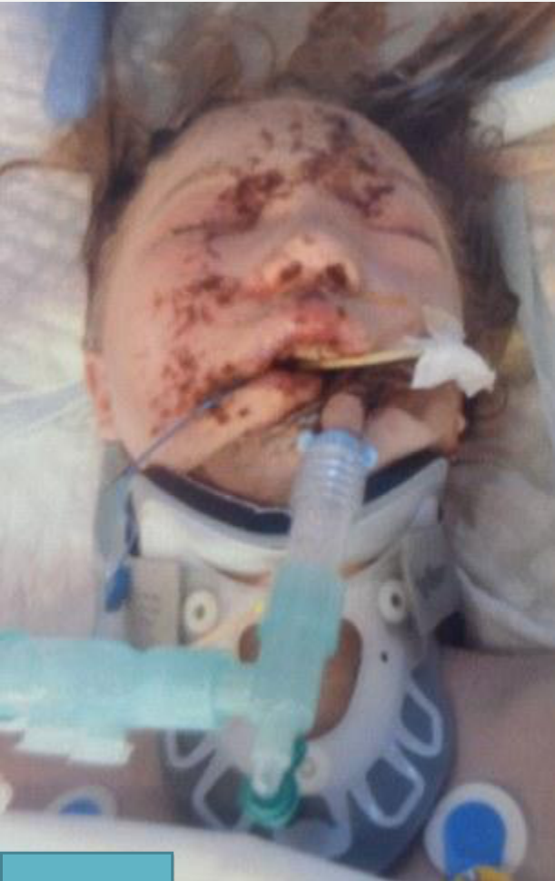
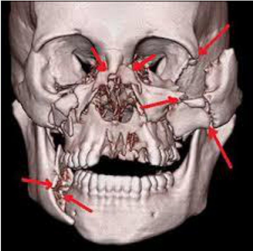
Motor Vehicle Accidents8
Lecturer — Motor Vehicle Trauma
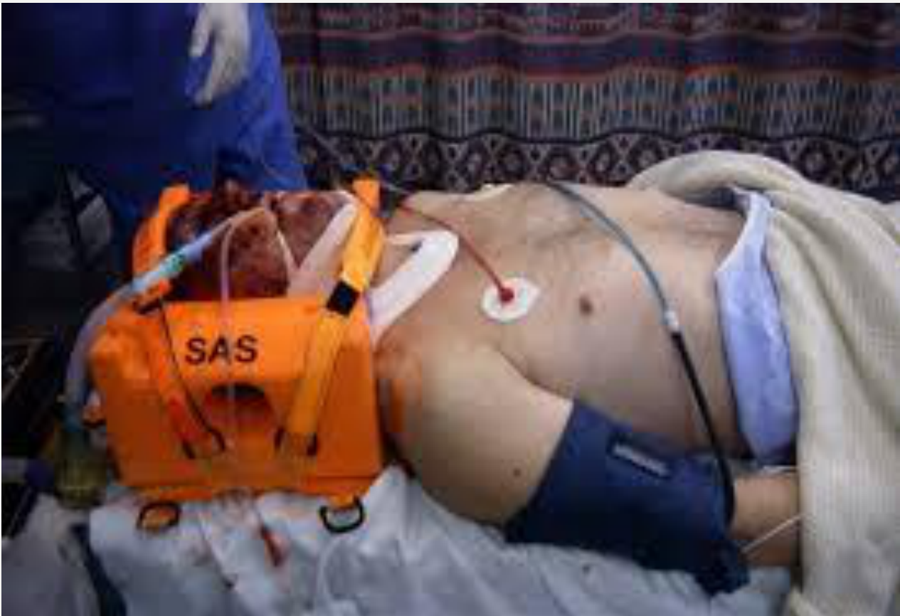
Severe facial injury should raise concern for an associated cervical spine injury, which should be considered unstable until excluded by examination or imaging.
- Apply a hard collar before moving a patient with severe facial trauma.
 |  |
Falls9
 |  |
Interpersonal Violence1011
- Using instruments or fists
Assault
Lecturer — Assault Documentation
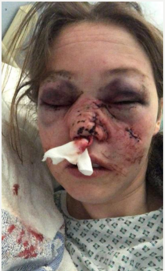
The history should establish whether the patient was hit once or repeatedly, whether one person or a group was involved, and whether a blunt or sharp instrument was used. It should also consider whether the injury may have occurred during domestic violence.
- Reports should use factual wording such as “alleged assault,” documenting what the patient reports and what is found on examination without inventing or exaggerating details.
 |  |
 |
Sports and Work Injuries1213
Sports injury
Work related injury
Lecturer — Sports and Work Reports
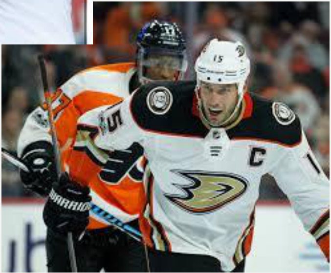
Sports injuries may affect the nose, zygoma, orbit, mandible, or teeth, and boxers may have repeated nasal injuries with a classically bent nose.
- Patients with prominent upper incisors or a Class II appearance who participate in sports may require a mouthguard.
- A dental report should document observed injuries, radiographs, restorability, recommended and alternative treatments, and the discussion with the patient.
- The report must describe findings at examination rather than presumed future findings.
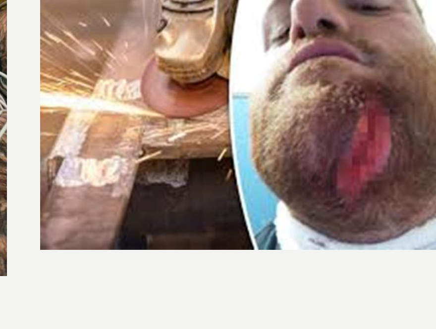
 | 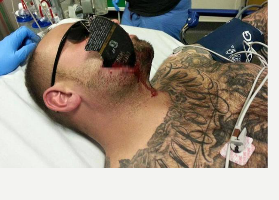 |
 |
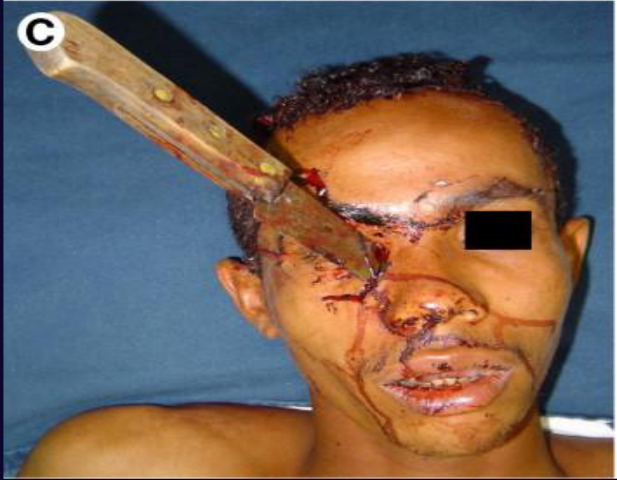
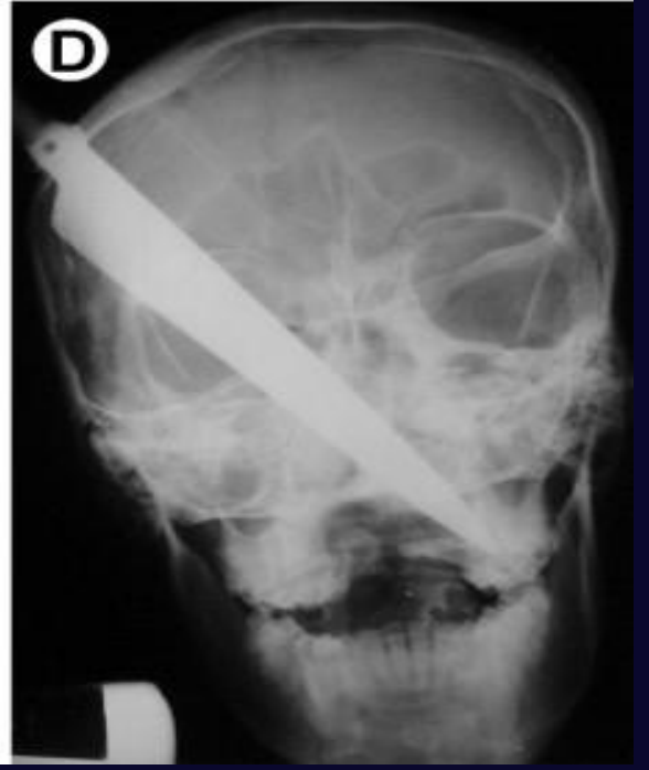
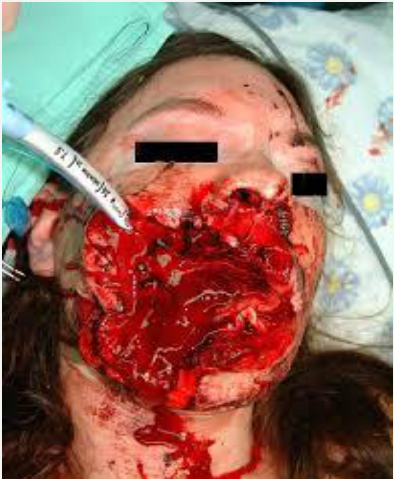
Penetrating Injuries1415
Knife wound
Ballistics
Lecturer — Penetrating Injuries
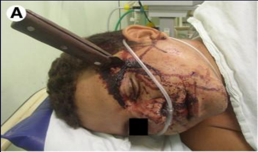
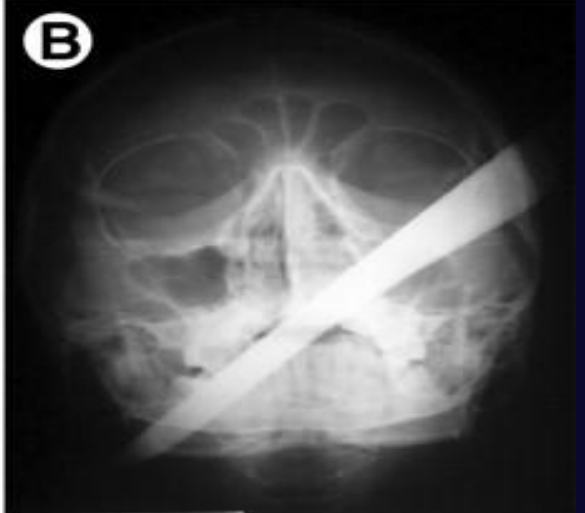
A knife, stick, or other penetrating object should not be removed unless the clinician is prepared to control severe bleeding, because the object may be tamponading an injured artery or another important structure.
- A retained metal object may be unsafe in an MRI scanner because it may move.
- Firearm injuries can cause extensive soft-tissue destruction: the entry wound may be small while the exit wound produces a large defect.
- Airway management is an immediate priority in severe firearm injuries, although this is generally managed in the emergency department.
 |  |  |
 |  |  |
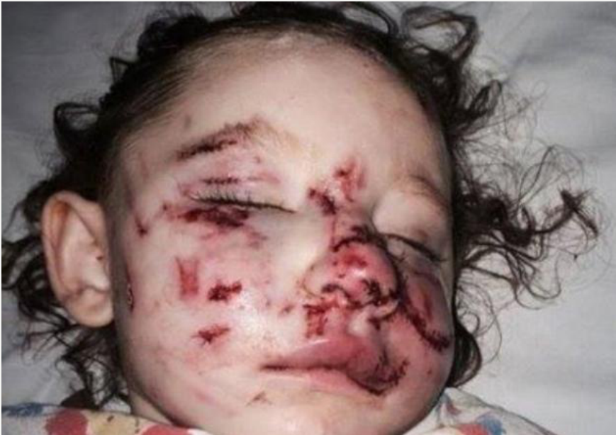
Other Types of Injury16
Lecturer — Other Injuries
Dog-bite reports should be factual, including that the patient alleges being bitten by a particular dog; the RSPCA may request a report. A horse kick may appear relatively benign externally but can cause severe associated injuries.
- A patient struck by a horse should not be moved until the cervical spine has been stabilised if the facial injury is severe.
- In elderly patients, thin skin may shear easily, and bruising or skin loss that appears severe may represent mainly soft-tissue injury once cleaned.
- Blood thinners can make bruising appear worse.
 |  |
Fracture Classification17
- Simple/closed — break does not penetrate skin
- Compound/open — broken bone penetrates skin
- Comminuted — breaks into many fragments
- Compression — bone is crushed, e.g. in osteoporosis
- Depressed — broken bone pressed inward, e.g. skull fracture
- Impacted — bone ends forced into each other
- Spiral — ragged break when excessive twisting forces applied to bone, e.g. sports fracture
- Greenstick — bone breaks incompletely — common in children
- Pathological — fracture in abnormal bone, spontaneous or following minor trauma
Lecturer — Mandibular Fractures
The lecturer added further distinctions and site-specific details about fractures.
- Fractures may also be described as displaced or undisplaced.
- A comminuted fracture is exposed to the air and is more liable to infection.
- A greenstick fracture may occur in children because the bone is malleable.
- Mandibular fractures may involve the condyle, coronoid, symphysis, parasymphysis, body, or angle.
- Approximately 30% of mandibular fractures involve the condyle, and the angle is also a common fracture site.
- Symphysis and parasymphysis fractures may be associated with numbness of the lower lip because of the mental nerve.
- Horizontal mandibular fractures are uncommon because of the direction of pull from the muscles of mastication.
- Mandibular fractures are frequently bilateral because the mandible is an intact, U-shaped bone; the lecturer estimated that approximately 60–70% are bilateral.
Lecturer — Delayed Subdural Haematoma
A slowly developing subdural haematoma may occur in an elderly patient after a fall, particularly when the patient is taking aspirin or warfarin.
- Blood may gradually collect under the dura and compress the brain.
- Head injury severity may be assessed using the Glasgow Coma Scale.
Spot the Fracture1819202122
R L
Lecturer — Panoramic Assessment
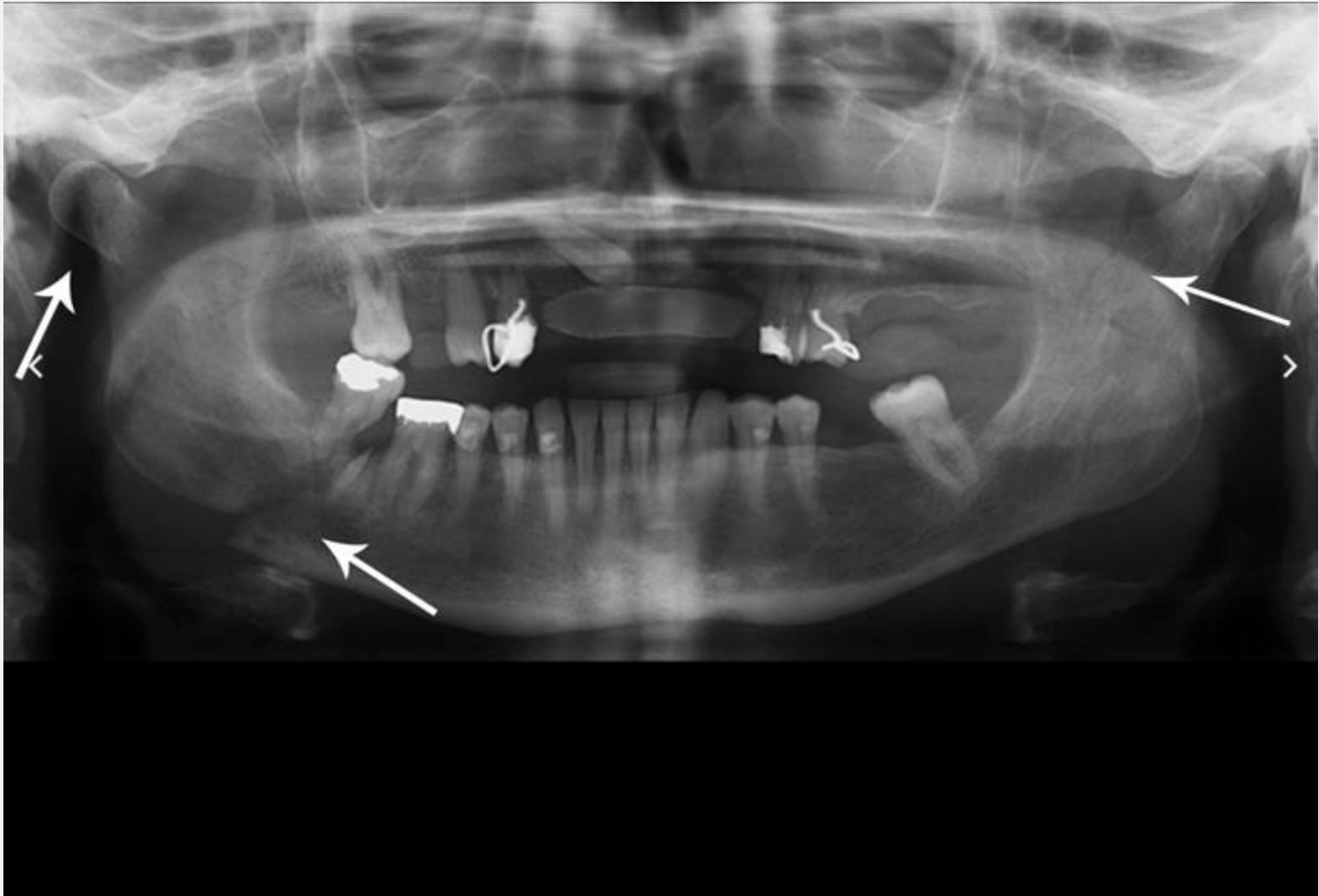
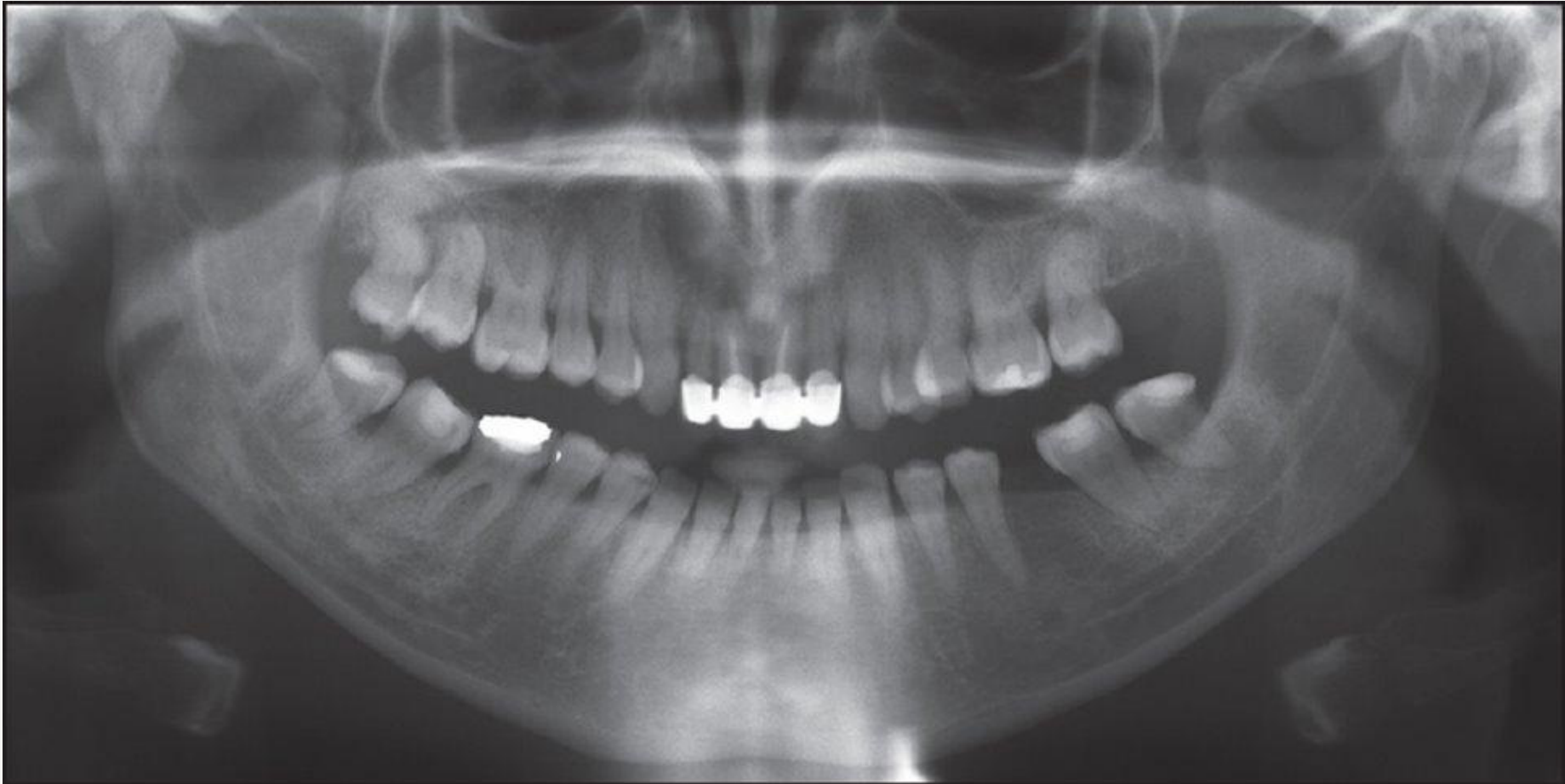
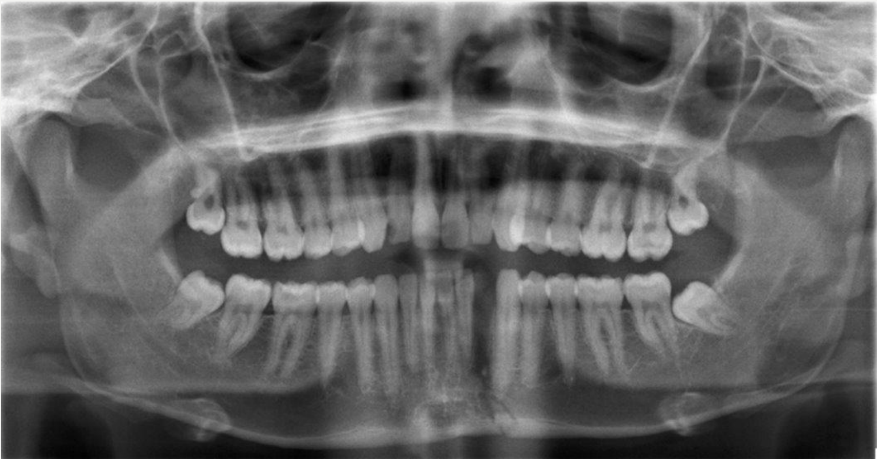
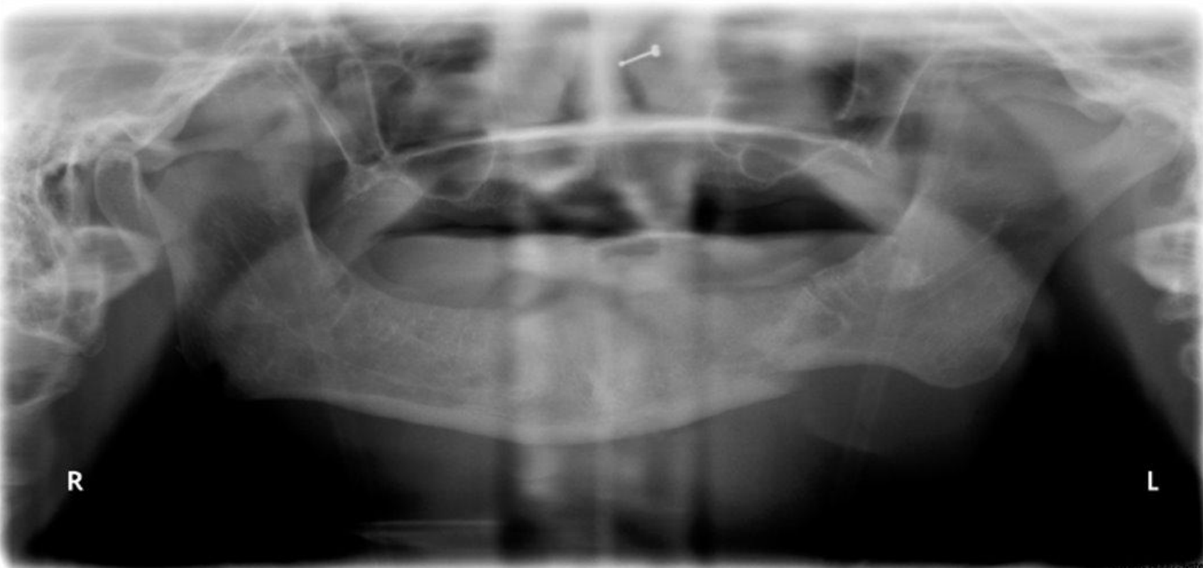
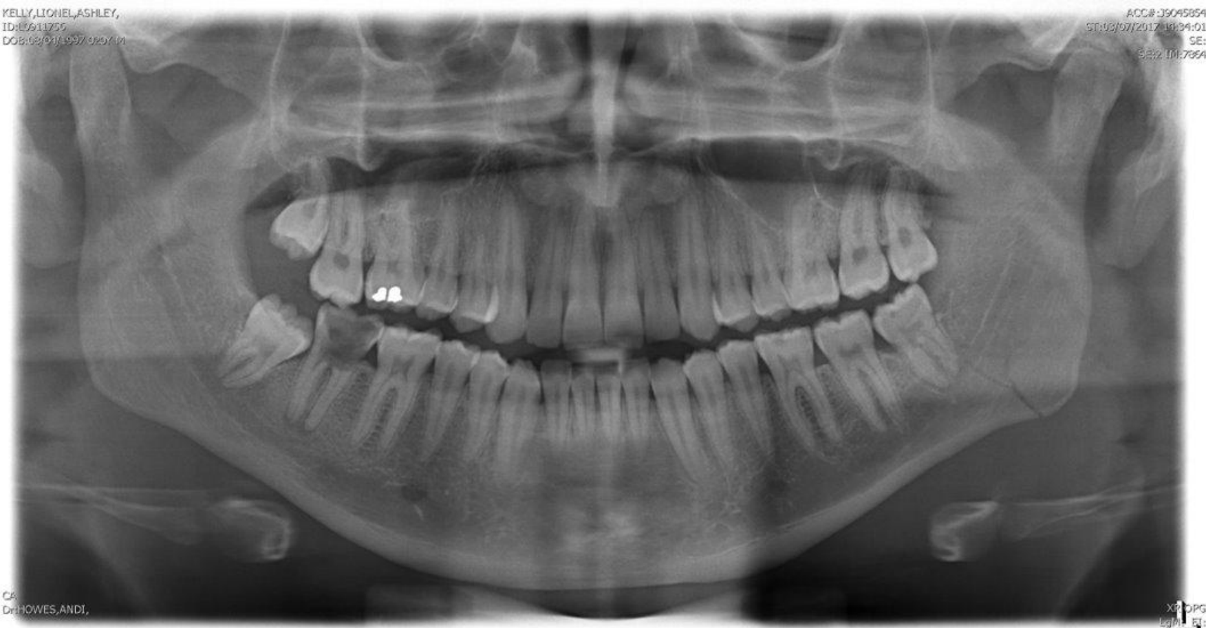
Panoramic radiographs should be interpreted systematically, beginning by confirming the patient and radiograph are correct.
- Trace the mandible from one condyle to the other, assessing the condyles, ramus, angle, body, symphysis, parasymphysis, inferior border, alveolar bone, and teeth.
- Compare both sides because mandibular fractures are commonly bilateral.
- Use two views at approximately 90 degrees, such as an OPG and a posterior-anterior mandibular view, because a fracture may be missed on one view and appear on another.
Relevant Anatomy
Zygomatic Complex2324
ALERT
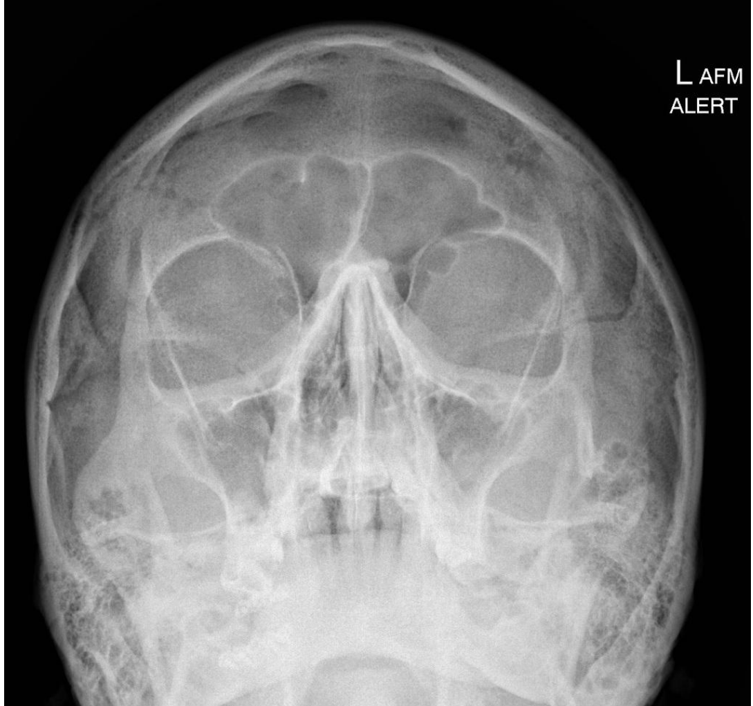
- Isolated zygomatic arch
- A severely depressed isolated zygomatic arch fracture may interfere with mandibular movement and mouth opening. (Image labels: L, AFM)
 |  |
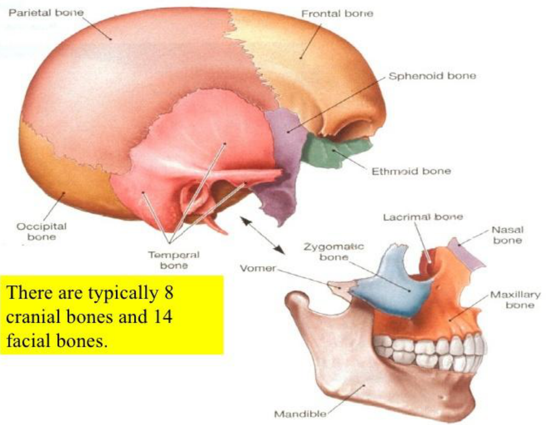
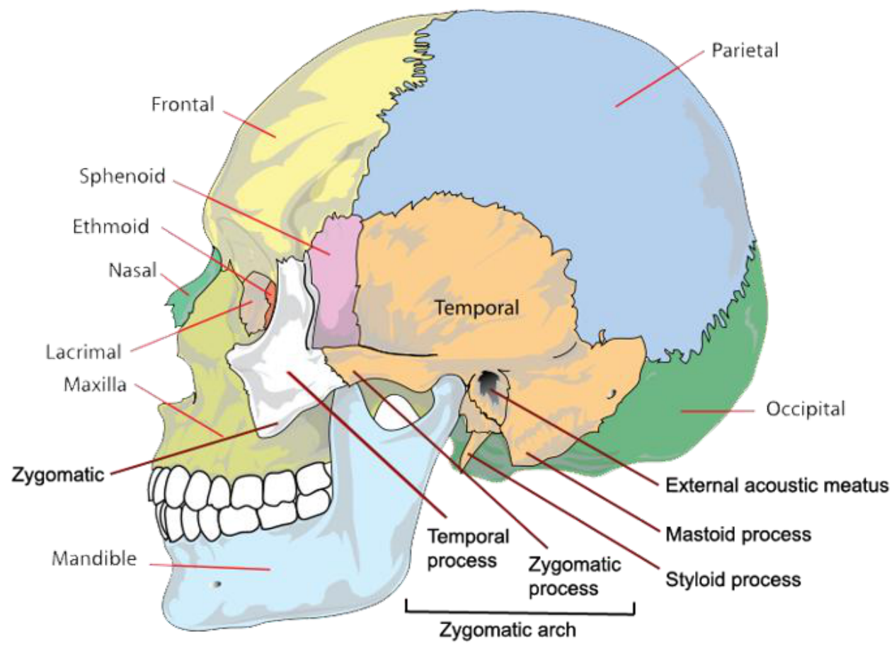
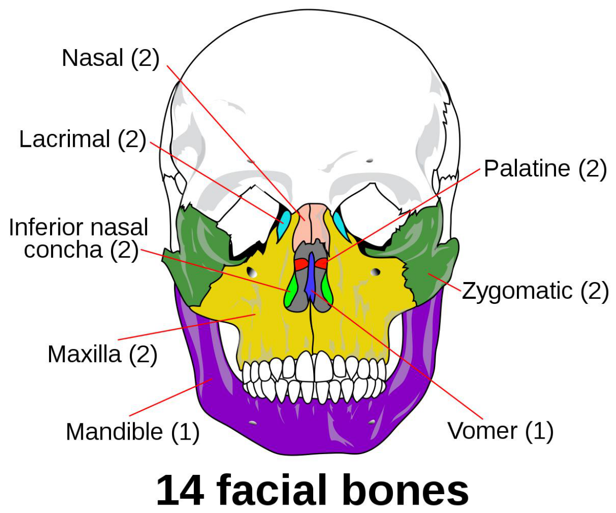
Skull and Facial Bones252627
Skull — vault and base, facial skeleton
There are typically 8 cranial bones and 14 facial bones.
Lecturer — Occlusal Changes
The maxilla and mandible are particularly important to dentistry because they are tooth-bearing bones.
- Even a small movement of either bone can produce a noticeable change in occlusion.
- Patients may present with a changed bite, a new gap, a premature contact, dentures that no longer fit, or teeth close to a fracture becoming non-vital and developing an associated sinus.
Cranial bones:
- Parietal bone
- Frontal bone
- Sphenoid bone
- Ethmoid bone
- Occipital bone
- Temporal bone
Facial bones:
- Lacrimal bone
- Nasal bone
- Zygomatic bone
- Vomer
- Maxillary bone
- Mandible
Cranial bones and facial bones
- Frontal
- Sphenoid
- Ethmoid
- Nasal
- Lacrimal
- Maxilla
- Zygomatic
- Mandible
- Parietal
- Temporal
- Occipital
Temporal bone features:
- External acoustic meatus
- Mastoid process
- Styloid process
- Temporal process
- Zygomatic process
- Zygomatic arch
Lecturer — Zygomatic Injury
The zygomatic bone can be considered a four-sided stool, with extensions to the frontal bone, inferior orbit, lateral maxilla, and temporal bone.
- Any of these extensions may fracture.
- Compare the opposite side for suture widening, steps, loss of smooth bony contours, and displacement.
- Zygomatic injury may be associated with subconjunctival haemorrhage, infraorbital nerve paraesthesia, damage to teeth supplied by that nerve, or altered occlusion.
14 facial bones
- Nasal (2)
- Lacrimal (2)
- Inferior nasal concha (2)
- Maxilla (2)
- Mandible (1)
- Palatine (2)
- Zygomatic (2)
- Vomer (1)
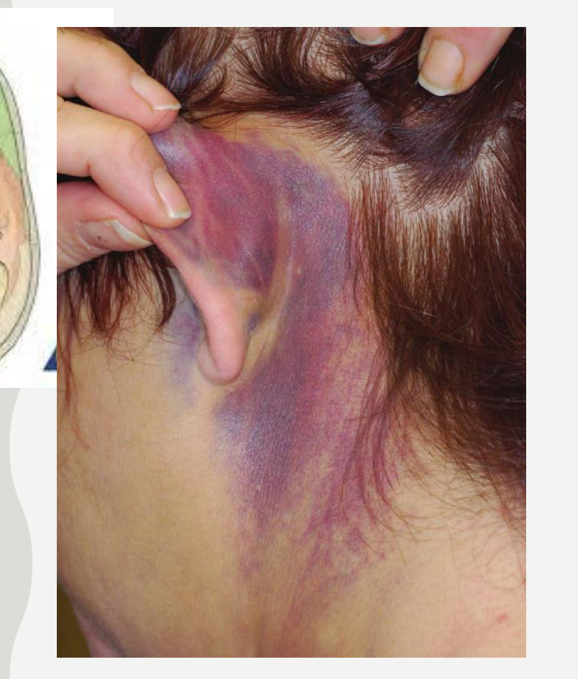
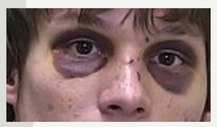
Base of Skull Fractures2829
-
Base of skull fracture — break of a bone in the base of the skull
-
Symptoms include bruising behind the ears, bruising around the eyes, or blood behind the ear drum
-
A cerebrospinal fluid (CSF) leak occurs in about 20% of cases
-
Result in fluid leaking from the nose or ear
-
Stabilize neck / C-spine
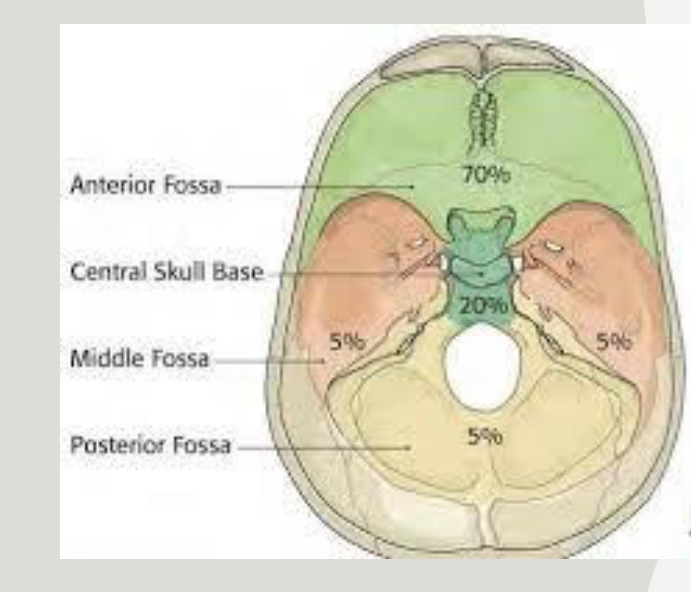
Lecturer — Cranial Fossa Signs
Base of skull fractures may involve the anterior, middle, or posterior cranial fossae.
- Cerebrospinal fluid may leak from the ear, or through the nose when the anterior cranial fossa is involved.
- These injuries should be identified and managed in the emergency department.
 |  |
 |  |
Head Injury
Contact (head strike)30
- Scalp laceration
- Skull fracture
- Coup
- Epidural haematoma
- SDH (subdural haematoma)
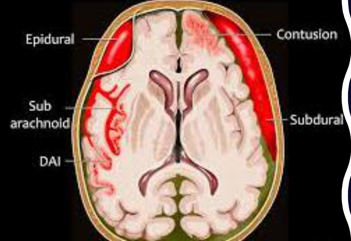 | 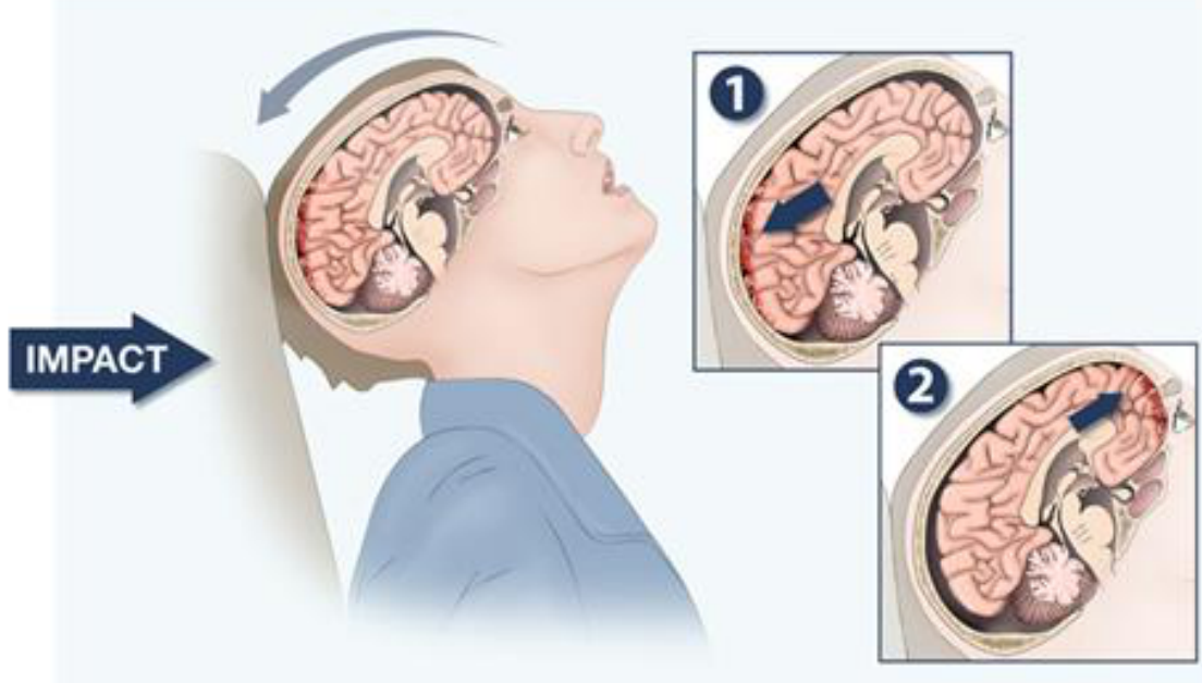 |
Acceleration (violent head motion)
- Concussion
- Contrecoup
- SDH (subdural haematoma)
- DAI (diffuse axonal injury)
Injury types shown: epidural, contusion, subarachnoid, subdural, DAI
(Figure labels: 1 — impact; 2 — impact)
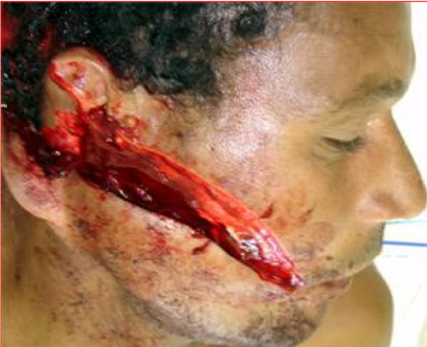
Soft Tissues31
oral cavity and teeth-bearing areas
Lecturer — Facial Nerve Injury
Deep facial lacerations may injure the facial nerve within the parotid region; injury to the main trunk may produce facial drooping.
- Temporal branch injury prevents forehead wrinkling.
- Zygomatic branch injury prevents tight eye closure and eye protection.
- Buccal branch injury causes difficulty puffing out the cheeks, controlling food, or controlling a denture, and food may collect in the sulcus.
- Deep lacerations must be thoroughly cleaned before suturing.
 |  |
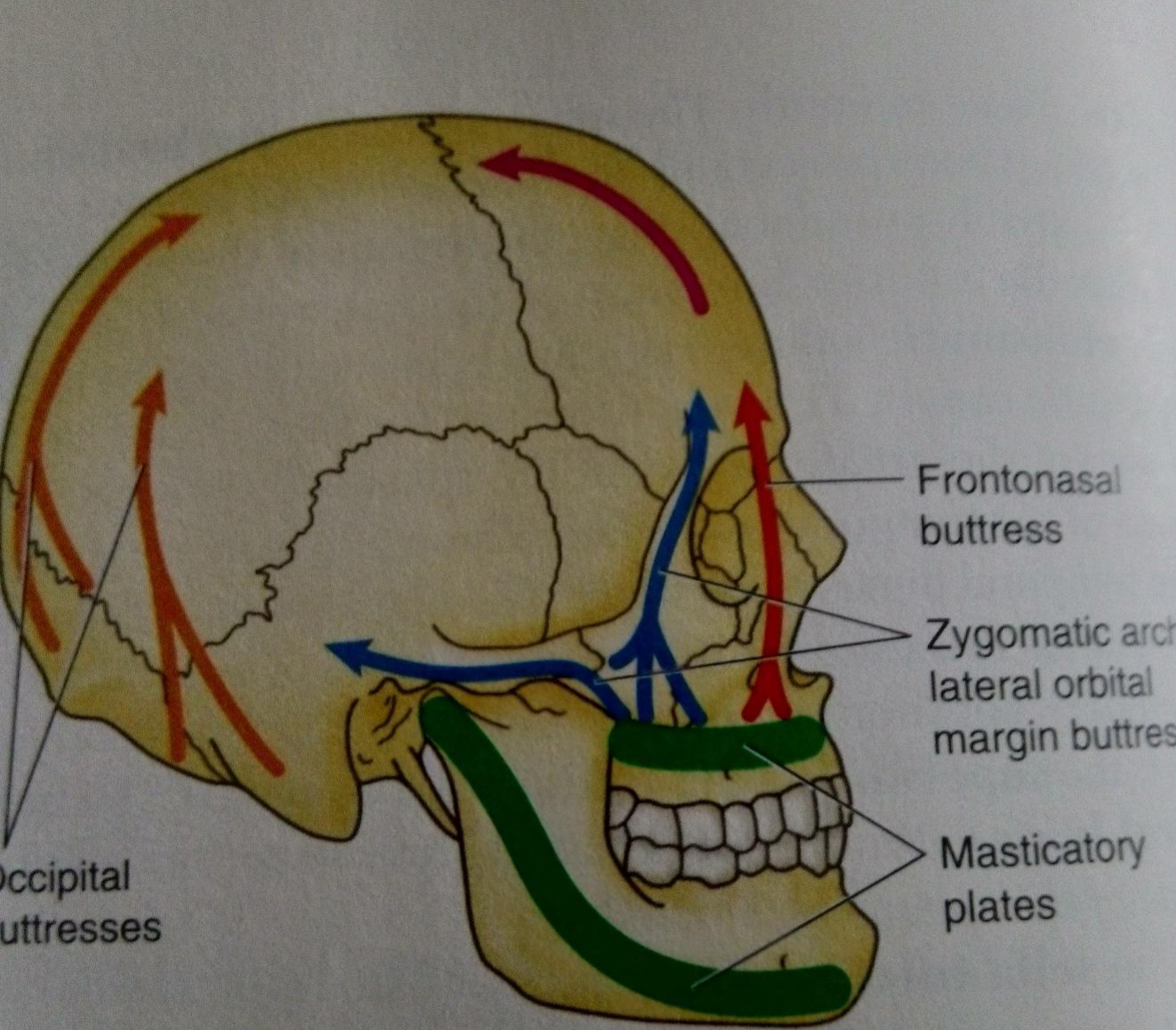
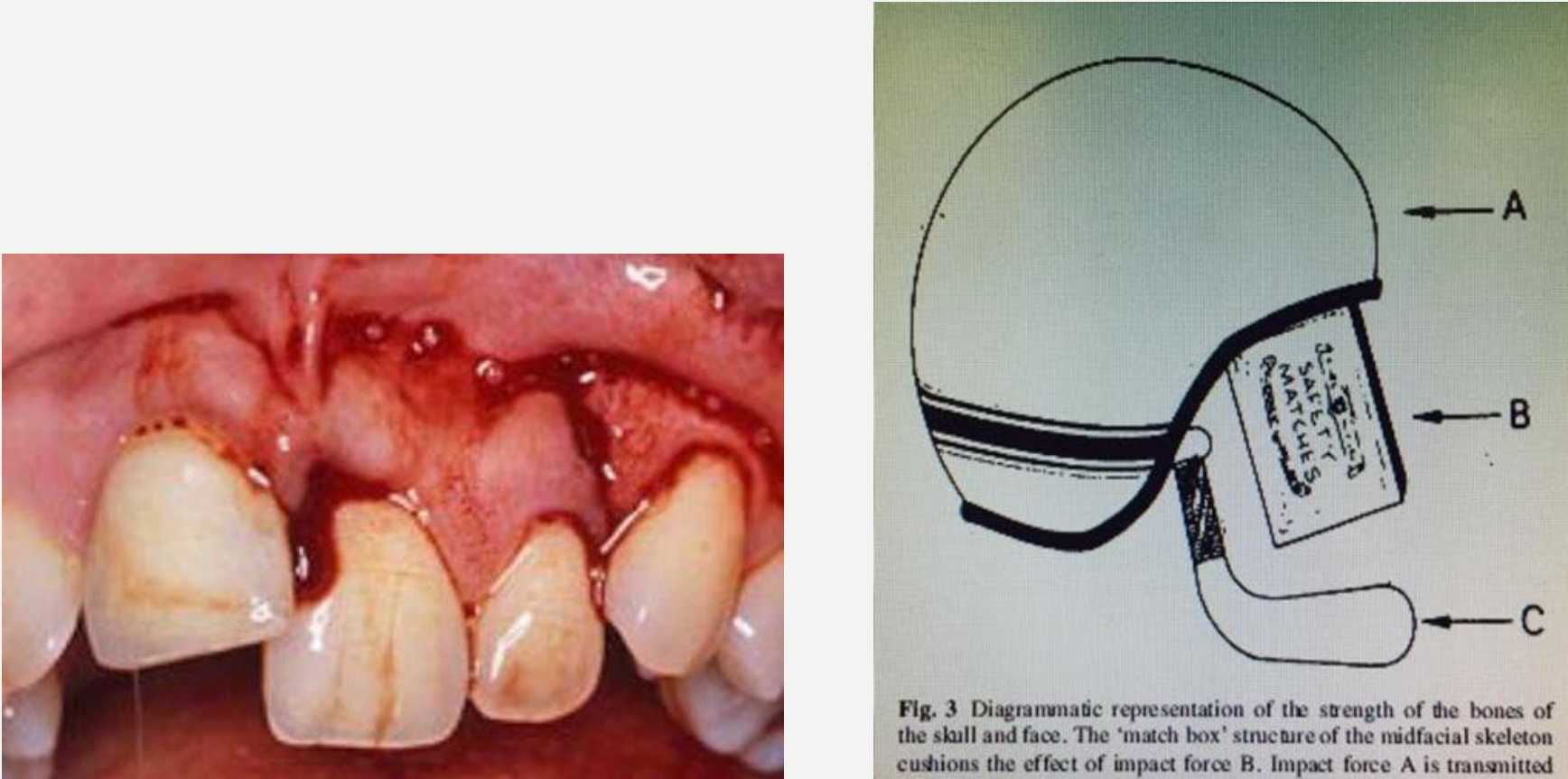
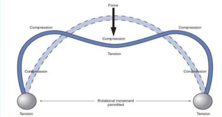
Buttresses and Crumple Zones32
One hypothesis theorizes that the sinuses function as a crumple zone to distribute and absorb energy to protect the brain and other critical structures in the event of facial trauma.
Lecturer — Midface Collapse
Severe midface trauma may cause the face to collapse and produce major changes in facial shape and occlusion.
- The tongue may fall backward and contribute to breathing difficulty.
Buttresses shown in the figure:
- Frontonasal buttress
- Zygomatic arch / lateral orbital margin buttresses
- Masticatory plates
- Occipital buttresses
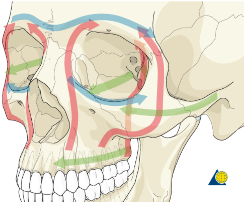
FIGURE 7.13. Buttresses of cranium. The buttresses are thicker portions of cranial bone that transmit forces around weaker regions of the cranium.
Lecturer — Zygomatic Buttresses
The paranasal sinuses are air-filled spaces that may function as crumple zones and help protect the brain by absorbing energy.
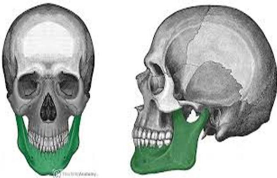
Mandibular Anatomy3334
Anatomical structures
- Lower third of facial skeleton prominent — more commonly fractured c.t. middle third
- Fracture alone or in combination with other facial fracture
- U-shaped bone (more sensitive to lateral impact c.t. frontal impact) — look for contralateral fracture
- Articulates with temporal bone at TMJ (condyle safety mechanism / ‘crumple zone’)
- Craniomandibular articulation — beware, may have head / C-spine injury
- Muscle attachment (e.g. bilateral # — tongue may drop back obstructing airway)
Lecturer — Mandibular Fracture Assessment
The condyle is a growth centre in children, so condylar injury may affect mandibular growth and later cause a deviated or asymmetric mandible.
- The mental nerve exits through the mental foramen and may be affected by symphysis or parasymphysis fractures.
- Inferior alveolar nerve injury may cause numbness of the lower lip.
 |  |
 |
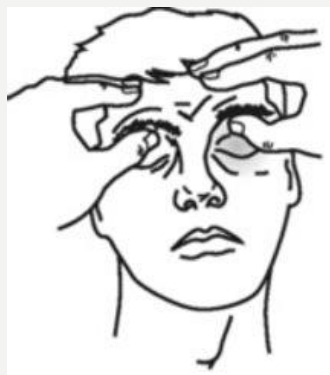
Examination35
 | 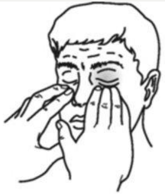 | 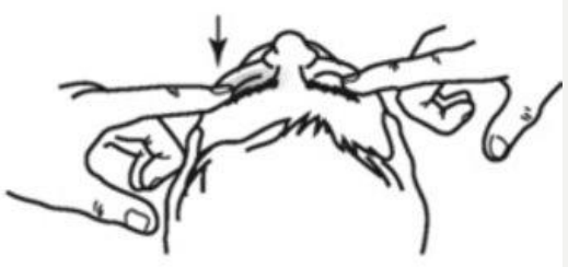 |
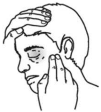 | 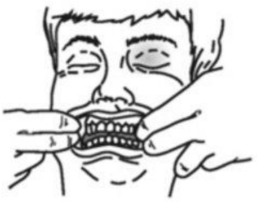 |  |
Specific Fractures
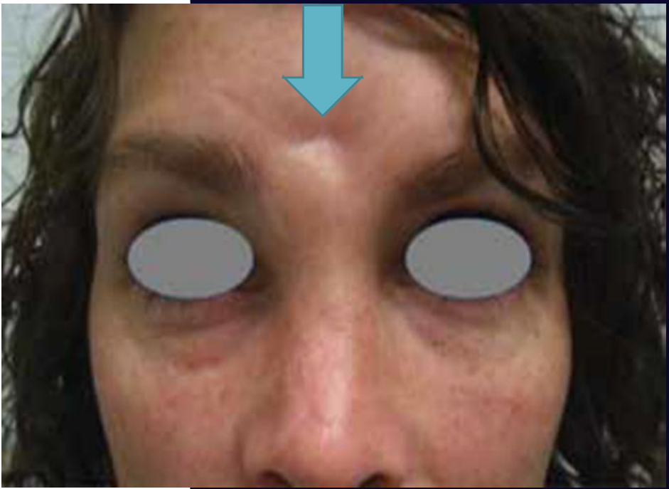
Frontal Bone Fractures3637
-
Frontal fractures — high energy
-
Usually involves the anterior table
-
Produces a cosmetic defect
-
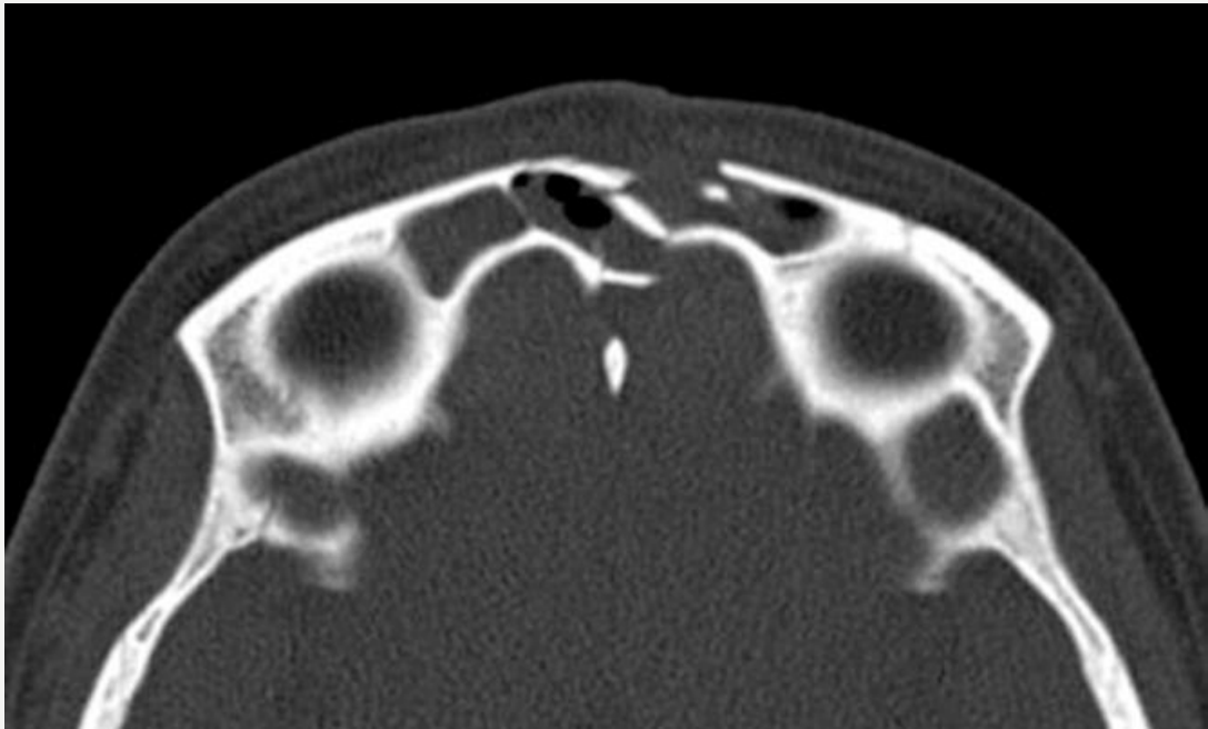
Posterior table fractures are less common but more significant as they involve the cranial cavity
-
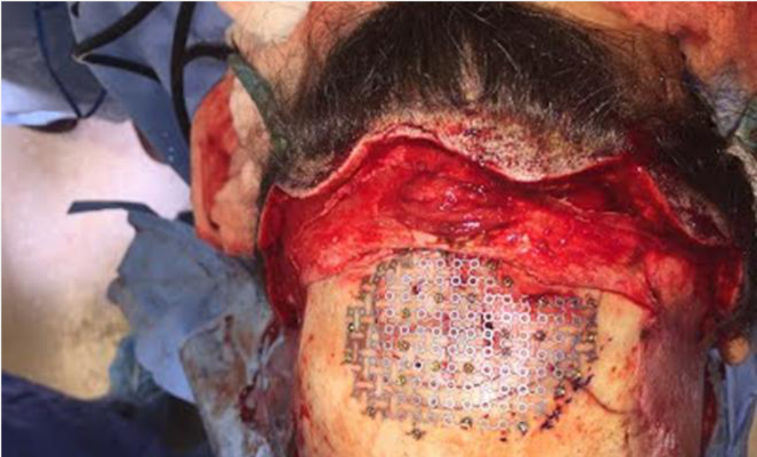
Requires joint management by maxillofacial and neurosurgical teams
Lecturer — Frontal Sinus Injury
The frontal bone contains the frontal sinus and includes anterior and posterior tables.
- Both-table fractures may communicate with the cranial cavity and allow cerebrospinal fluid to leak through the nose.
- These injuries are generally evaluated with CT in the emergency setting.
 |  |
 |
Nasal Bone Fractures38
- Most common facial fracture (> 50%)
- Main cause is interpersonal violence
- Assess for asymmetry and mobility clinically
- Manage epistaxis
- Allow 5-7 days for the oedema to resolve, facilitating precise operative intervention
Lecturer — Nasal Assessment
Nasal trauma may result from boxing, repeated altercations, sports injuries, punches, or vehicle accidents, and repeated trauma can produce a classically bent nose.
- Assessment should include a septal haematoma, cosmetic deformity, and nasal breathing.
- A septal haematoma should be treated promptly to avoid septal perforation.

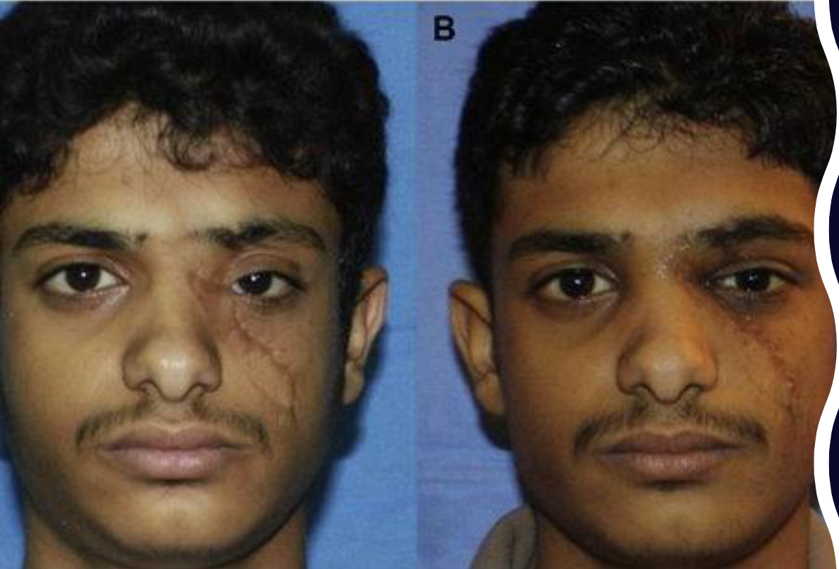
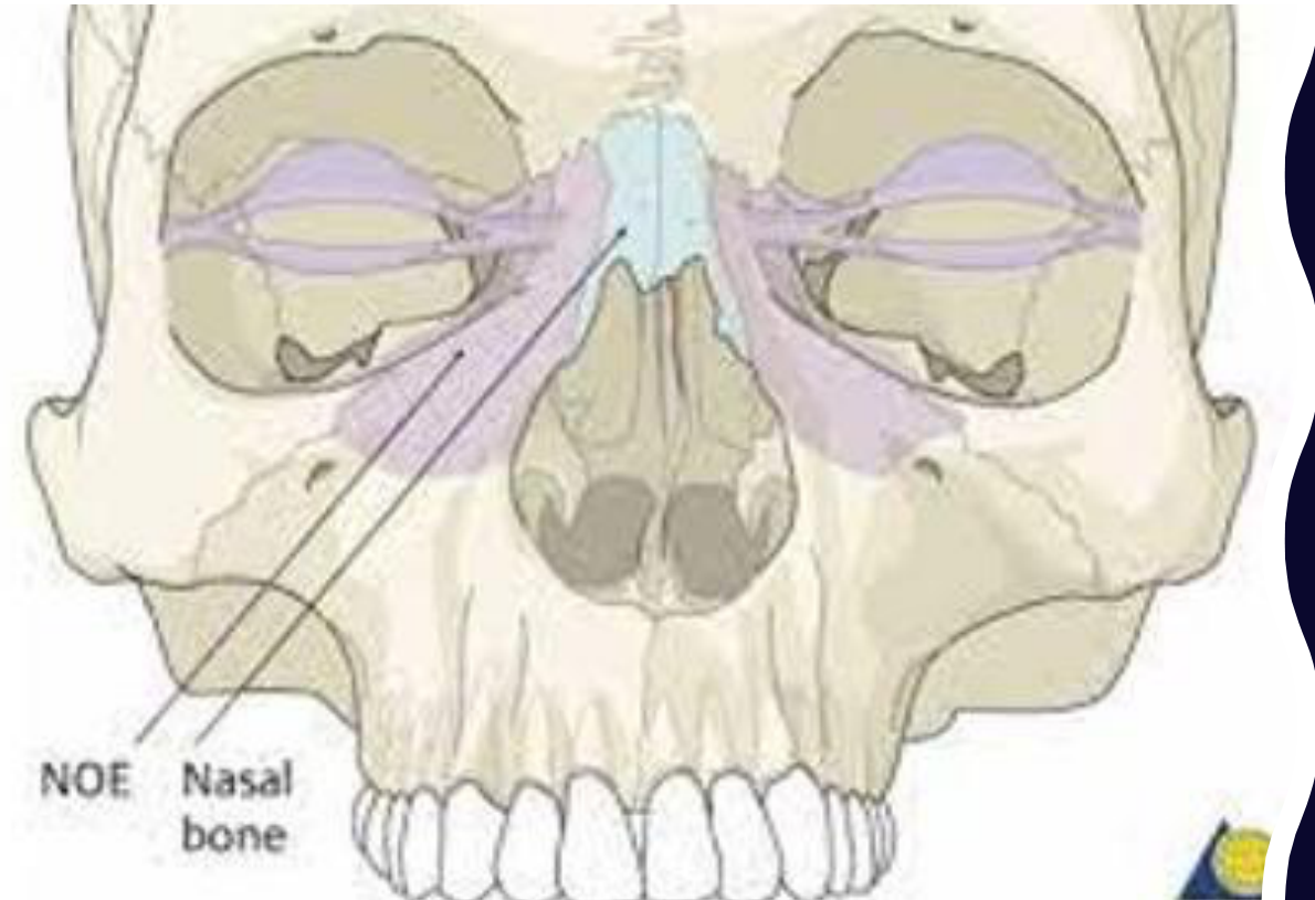
Naso-Orbital-Ethmoidal Fracture39
Panel B: NOE fracture — nasal bone.
Lecturer — NOE Fracture
Naso-orbital-ethmoidal fractures may widen the distance between the eyes if the supporting ligament is severed or displaced.
- Reduction of the bone alone may not correct the eye position.
- The resulting telecanthus and the complexity of treating these injuries make them among the most difficult facial fractures to manage.
 |  |
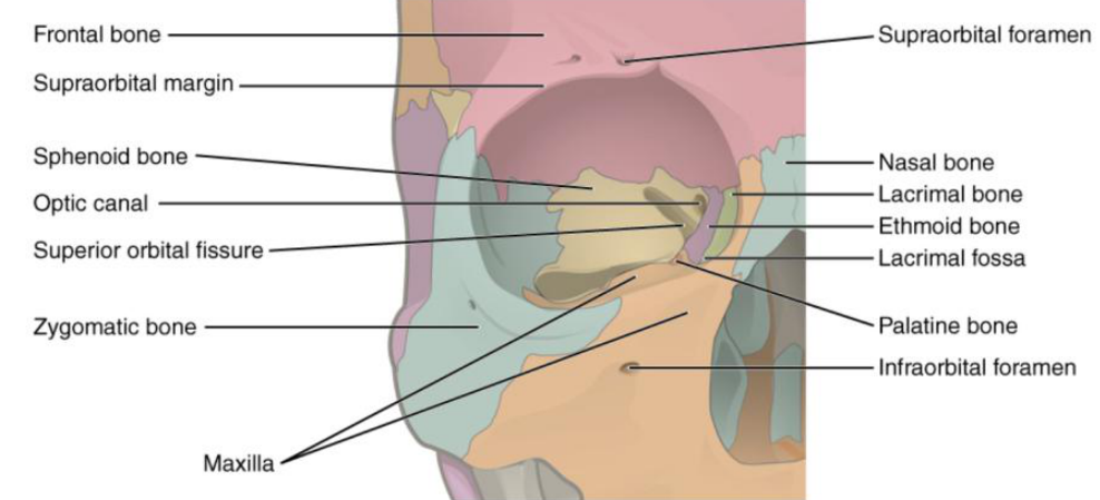
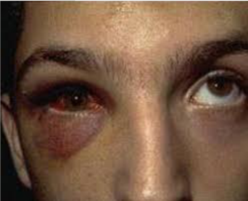
Orbital Fracture40
- Medial and inferior walls of the orbital skeleton are exquisitely thin
- Floor and lateral wall commonly involved in zygomatic complex fractures
- Also occur as an isolated injury due to a direct blow to the orbit or orbital rim
- Assessment of the eye is paramount when these injuries occur
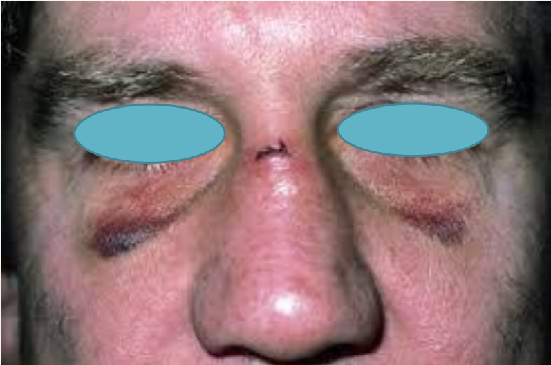
- Subconjunctival and periorbital haematoma are common
- Diplopia occurs when the orbital muscles and soft tissues are trapped
Lecturer — Paediatric Orbital Fractures
In children, the orbital floor may fracture as a greenstick or trapdoor fracture, allowing muscles to become trapped as the bone springs back.
- This can produce an oculocardiac reflex with vomiting and feeling unwell.
- Paediatric injuries require treatment because of the possibility of muscle entrapment.
 |  |
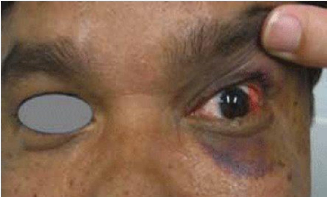
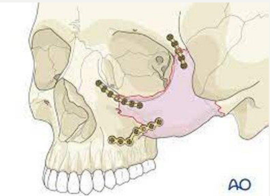
Zygomatic Fractures4142
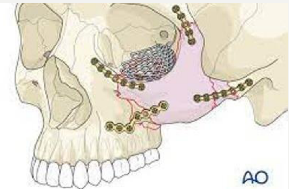
Zygomatic complex fracture — open reduction and internal fixation.
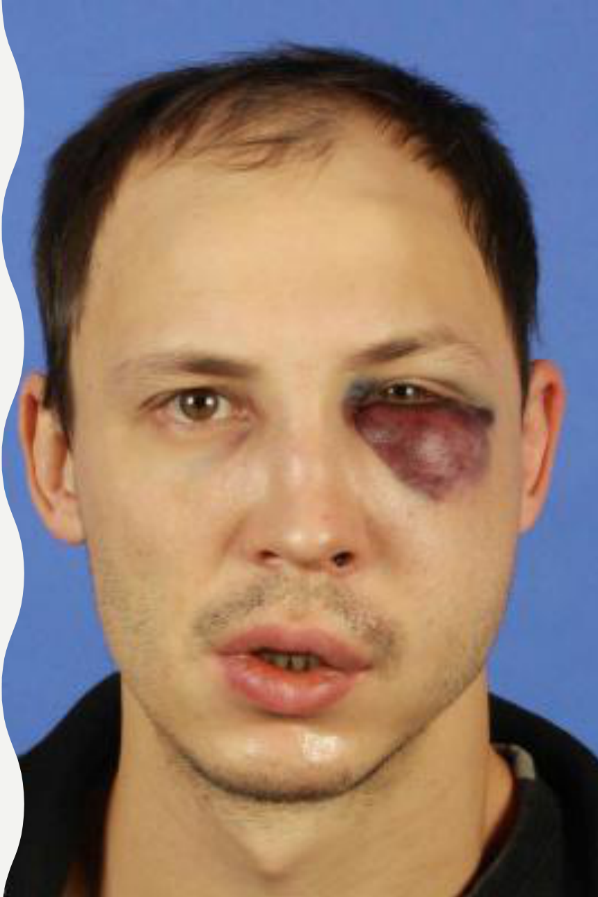
- Periorbital swelling and ecchymoses
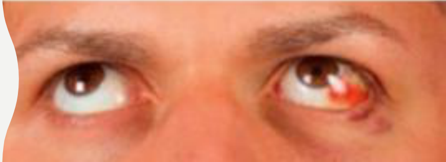
- Subconjunctival haemorrhage
- Palpable step at the infraorbital margin or the lateral brow area
- Infraorbital nerve paraesthesia
- Depression of the malar eminence
- Diplopia is common due to orbital involvement — entrapment of extraocular muscles
Lecturer — Zygomatic Assessment
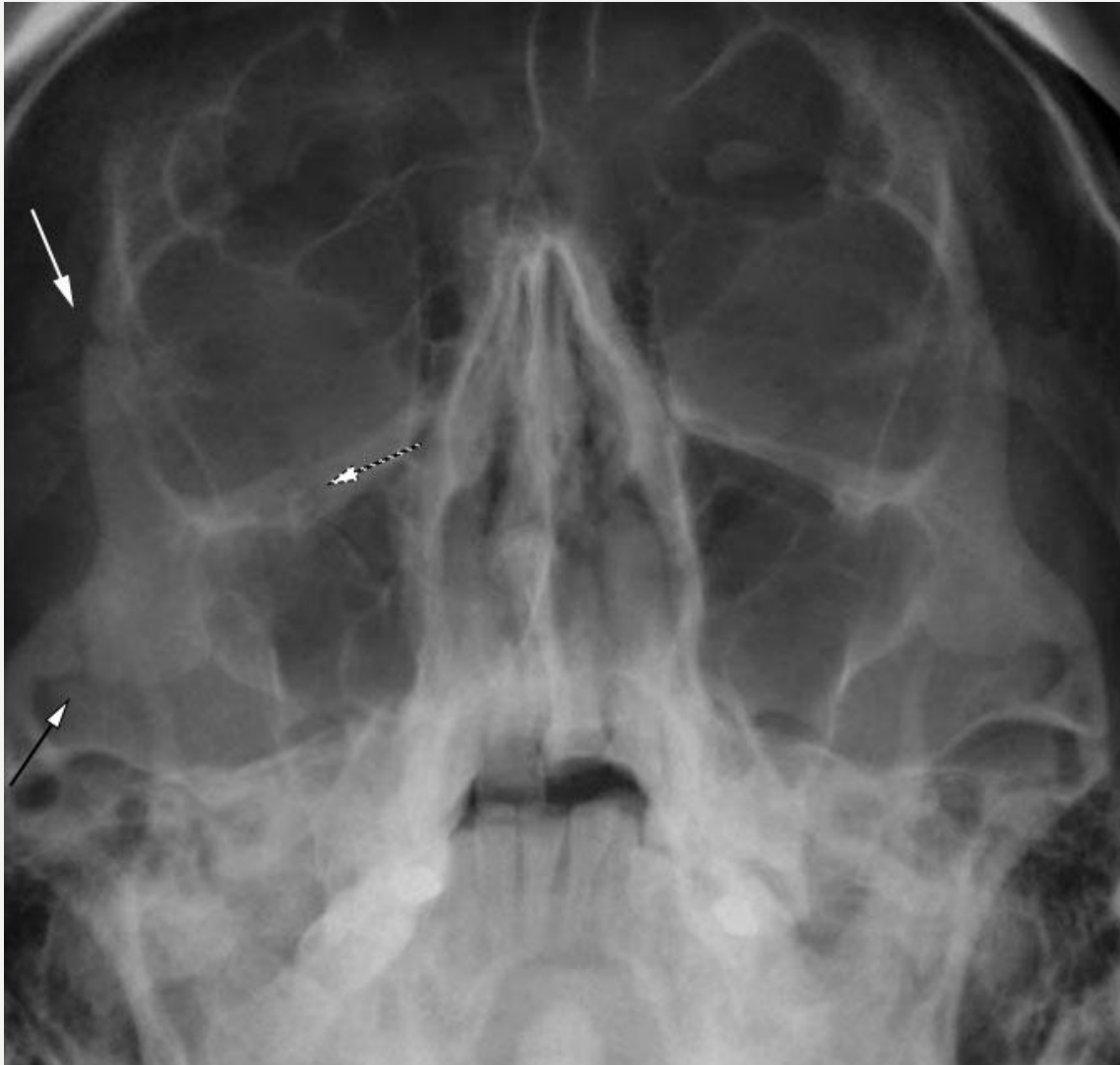
Zygomatic fractures may be identified by comparing both sides on an occipitomental view.
- Findings can include widening of the zygomaticofrontal suture, an irregular lateral maxillary wall, and widening near the temporal articulation.
- An isolated depressed zygomatic arch fracture may restrict mouth opening or interfere with mandibular function.
 |  |
 |  |
Midface Fractures4344
-
Bony pillars
-
Fragility (and prominence)
- Nasal
- Maxilla (anterior wall)
- Zygomatic arch
- Zygomatic complex
- Orbit
-
Compressible air-filled energy-absorbing crumple zone vs frontal bone
-
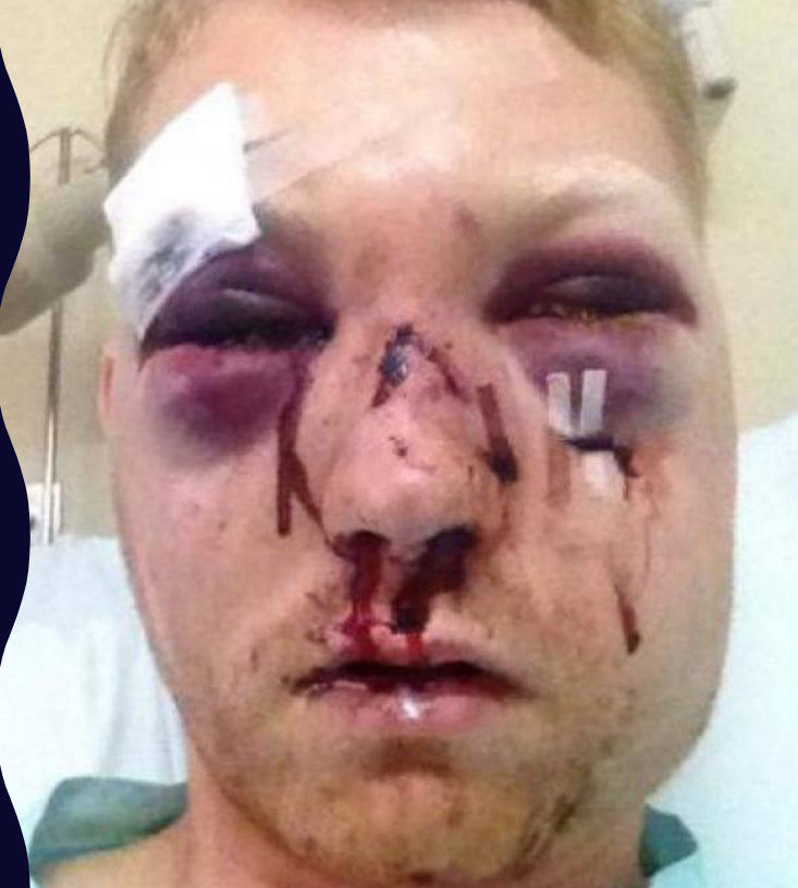
Facial swelling with bilateral periorbital haemorrhages (raccoon signs)
-
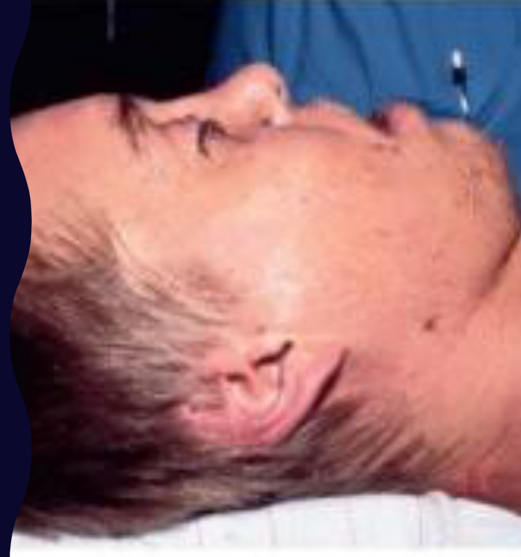
Flattening and elongation of the midface
-
Dish face deformity
-
Test mobility of the maxilla
-
Patients often complain of malocclusion
-
Beware airway obstruction
Lecturer — Midface Displacement
Midface fractures do not necessarily occur in clean, symmetrical patterns; one side may be higher while the other is lower.
- Because the maxilla is tooth-bearing, even a small displacement can affect occlusion.
- Patients may later report a changed bite, a long or altered facial appearance, numbness, or problems with teeth near the fracture.
 |  |
 |
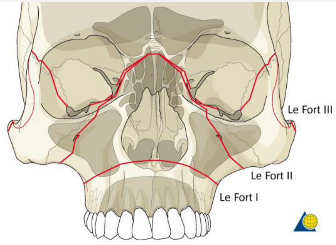
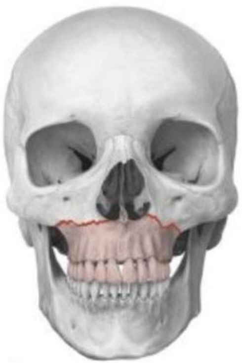
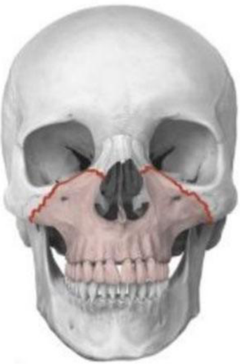
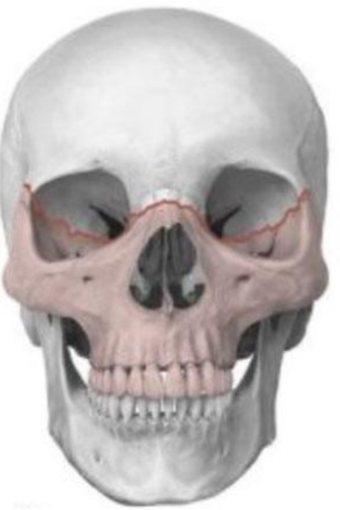
Le Fort Fractures4546
- Le Fort III
- Le Fort II
- Le Fort I
Lecturer — Le Fort Classification
Actual injuries may not fit cleanly into one Le Fort classification, with one side showing a Le Fort I pattern while the other has a higher fracture.
- The important dental implication is the effect on occlusion.
- If a healed fracture leaves only one high tooth causing premature contact, odontoplasty may be considered rather than reopening the entire fracture.
 |  |
 |  |
Mandibular Fractures47484950
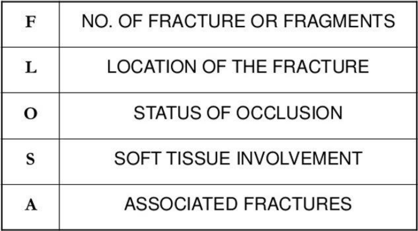
| Letter | Meaning |
|---|---|
| F | No. of fracture or fragments |
| L | Location of the fracture |
| O | Status of occlusion |
| S | Soft tissue involvement |
| A | Associated fractures |
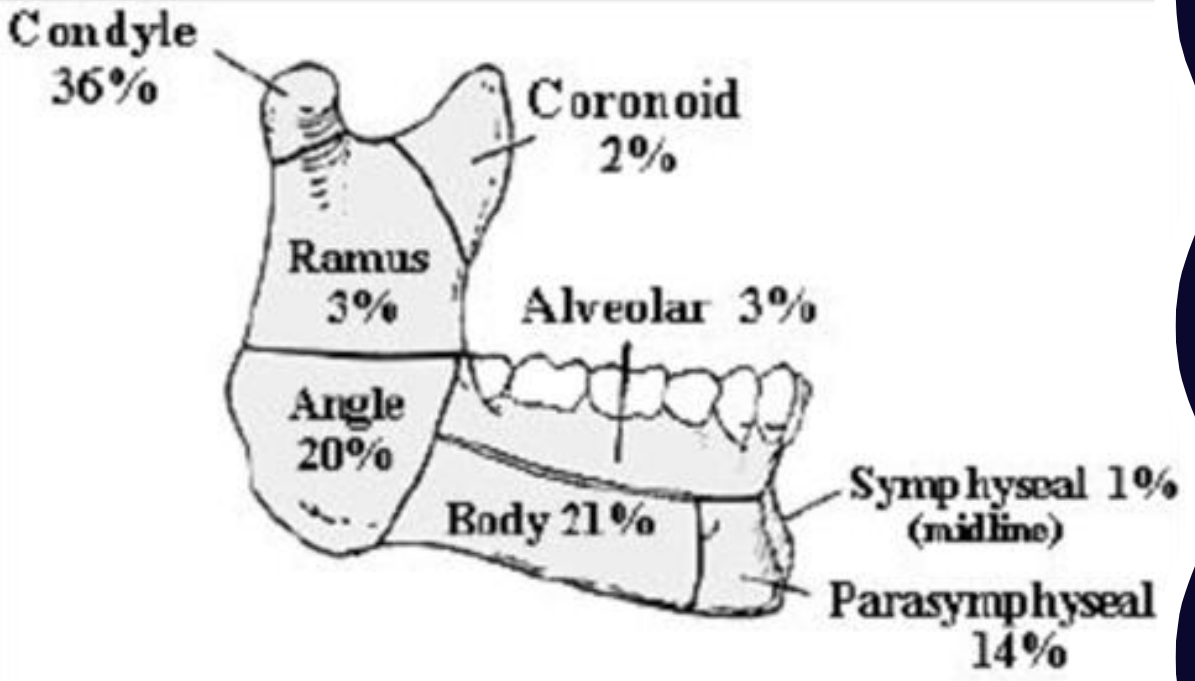
- Condyle (intra/extracapsular, high neck/low condyle)
- Angle
- Body
- Parasymphysis
- Symphysis
- Ramus
- Coronoid
- Alveolus
Distribution of fracture sites: condyle 36%, angle 20%, body 21%, parasymphyseal 14%, ramus 3%, coronoid 2%, alveolar 3%, symphyseal (midline) 1%.
Lecturer — Mandibular Fracture Patterns
A fracture through the parasymphysis may produce a visible step between teeth and a gap between the canine and premolar.
- Adjacent teeth may later become devitalised or develop a buccal sinus from apical infection.
- Condylar fractures in children require prolonged monitoring because of the risk of impaired growth and later mandibular asymmetry.
- Pain
- Swelling
- Limited mouth opening
- Mental paraesthesia
- Deviation of mandible (condyle fracture)
- Malocclusion
- Step deformity
- Mobility at site (+/- missing or mobile teeth)
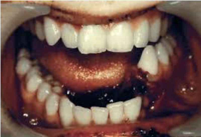
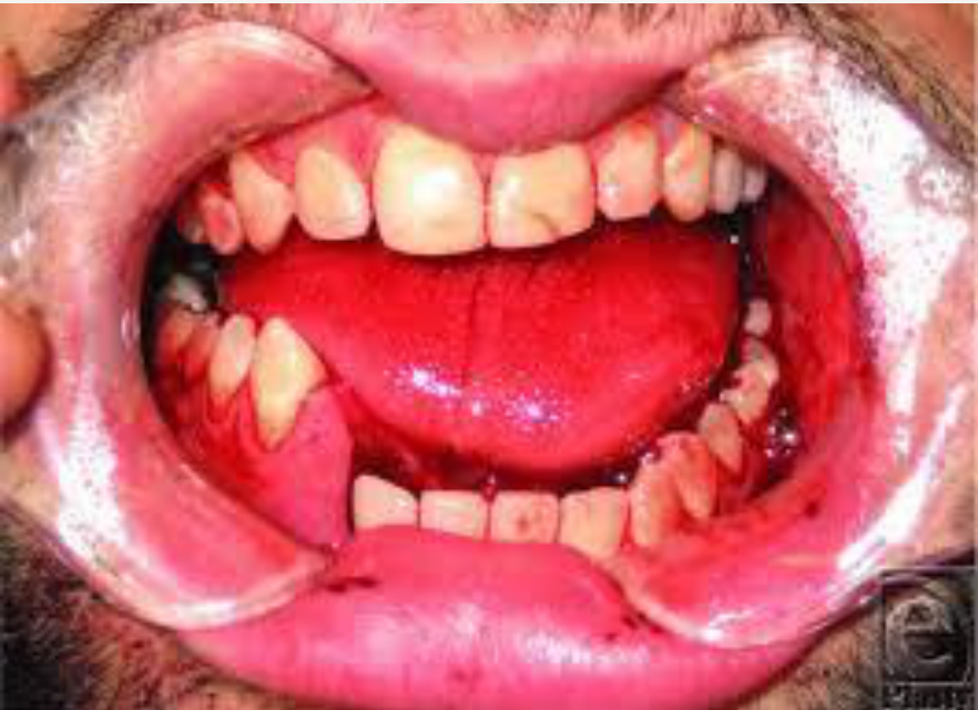
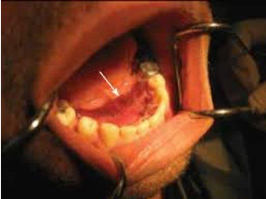
- Haematoma (lingual haematoma pathognomonic)
Lecturer — Mandibular Examination
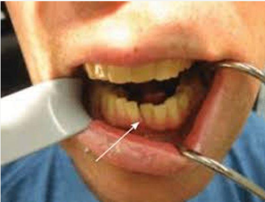
A lingual haematoma should be treated as a mandibular fracture until proven otherwise.
- In an edentulous patient, occlusion cannot be assessed with teeth, but a lingual haematoma or dentures that no longer fit may still indicate injury.
- An unexplained new gap, tooth devitalisation, sinus, or altered bite should be reconciled with the history and raise suspicion of an unrecognised fracture.
Lecturer — Meaning of Reduction
Reduction means returning the fractured bone to its correct or original position. Displaced or overlapping bone ends should be repositioned and aligned before healing.
- The plate holds the bone in the correct position while the body heals the fracture; it does not itself heal the bone.
- Ideally, the plate has an equal number of holes on each side of the fracture and sits flat across the bone.
- Care is required around the inferior alveolar nerve when exposing a mandibular fracture.
- Plates and screws may loosen or become exposed through thin mucosa, sometimes requiring referral for plate removal.
- Titanium plates are inert and generally do not trigger airport alarms.
- Closed reduction does not directly expose the fracture; adjacent anatomy and occlusion are used to estimate the correct alignment.
- Closed reduction may be useful when a condylar fracture is too high to accept a plate.
- It may also be used when there is insufficient bone for adequate plate holes on both sides or when the patient is medically unfit for general anaesthesia.
- Immobilisation may require a liquid diet for approximately four to six weeks.
Treatment
Reduction51
realignment of bone ends
- Open versus closed reduction
- Immobilization – to allow the healing process
- Internal fixation versus external fixation
Lecturer — Zygomatic Arch Reduction
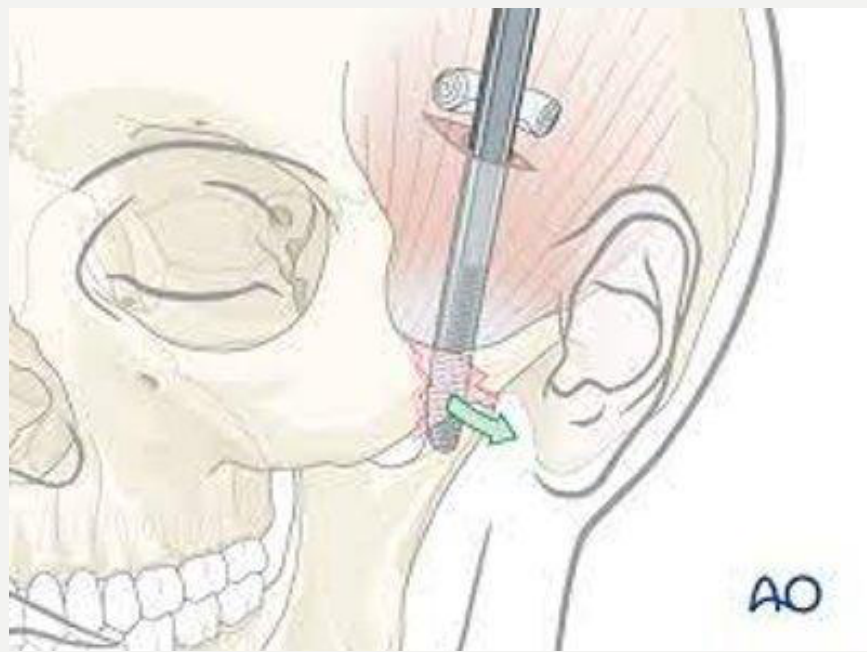
Some zygomatic arch fractures can be reduced without directly exposing the fracture. The surgeon opens the fascia and slides over the temporalis muscle beneath the zygomatic arch, using its anatomy to push the depressed arch outward.
- This is a closed reduction because the fracture is not directly visualised.
- Treatment is particularly important when depression restricts mouth opening or mandibular function.
- The patient should avoid contact sports and further impacts during healing.
- An isolated arch is not a major weight-bearing unit.
Lecturer — External Fixation
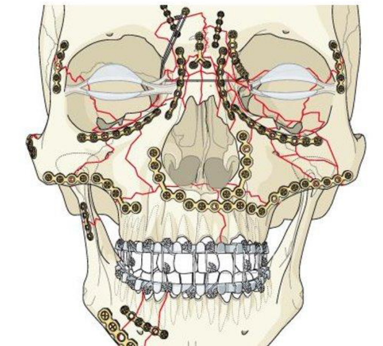
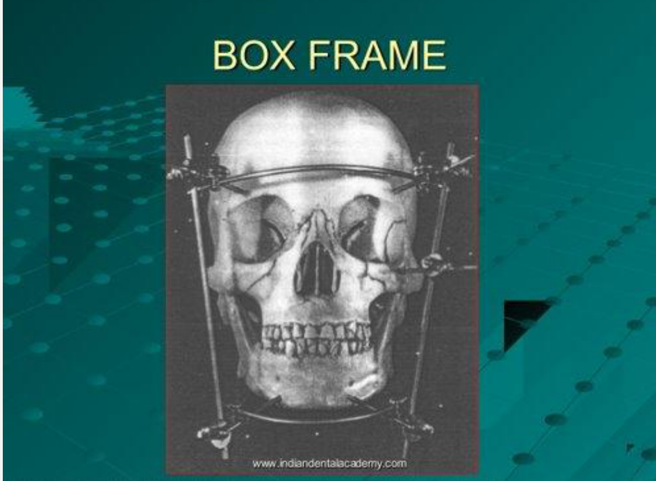
Excessive movement prevents healing and may result in fibrous union or non-union. External fixation may be used when the fracture is highly comminuted, when there are too many fragments to plate effectively, or when the bone or periosteal blood supply should be preserved.
- Historically, severe midface fractures were sometimes managed with a box frame.
- The frame compressed the facial structures and relied on correct occlusion as an indication that the fracture had been reduced.
- External fixation and prolonged wiring are used less often because they restrict mouth opening, complicate cleaning, and delay normal function.
 |  |
Open Reduction and Internal Fixation52
- Mini-plate
- Intra- or extraoral approach
- Direct visualisation and reduction of fracture
- Earlier return to normal function
- Better nutrition
- Better oral hygiene
- More expensive
- Periosteal stripping disturbs vascular envelope
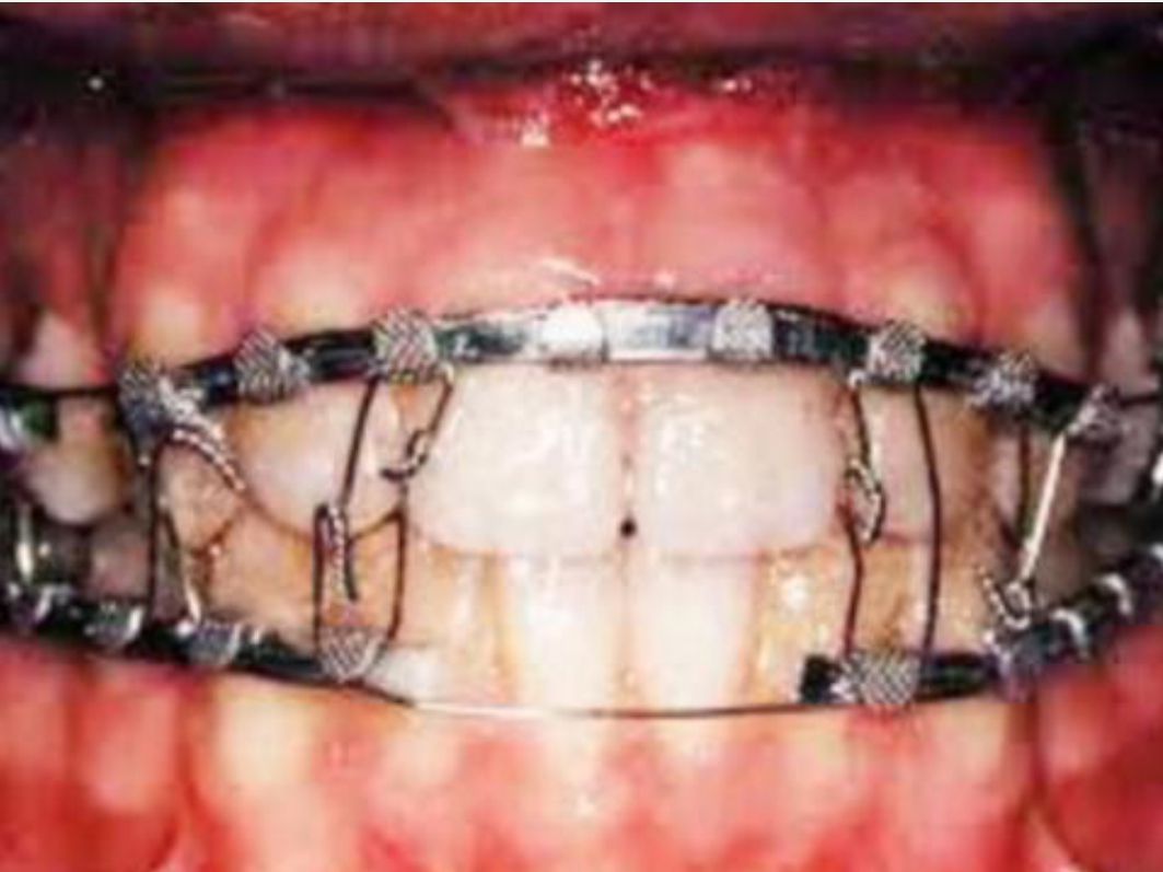
Closed Reduction53
- IMF screws, arch bars, eyelet wires, bonded brackets
- Less expensive
- Does not traumatize vascular envelope
- Period of immobilization (difficulty eating/weight loss)
- Requires intact dentition
- Requires patient compliance
- Risk damage to roots
- Gingivae inflammation / poor oral hygiene
Zygomatic Fracture Reduction54
- Open reduction
- Closed reduction
 |  |
Immobilization55
- Internal fixation
- External fixation
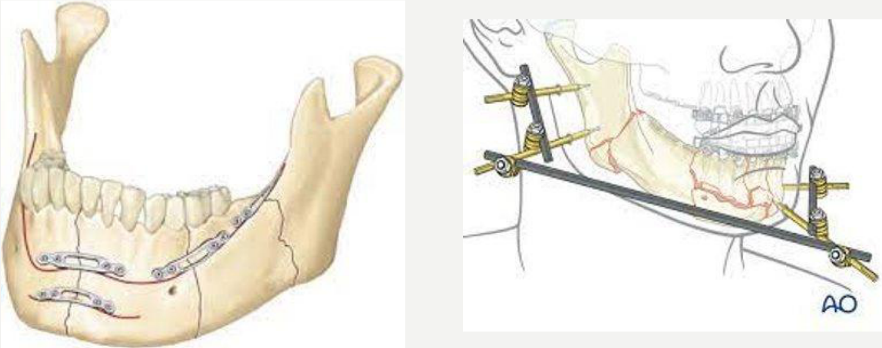
Special Considerations
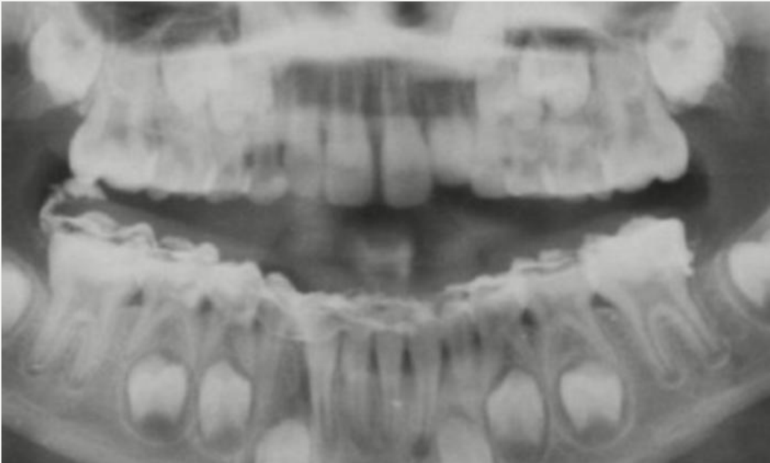
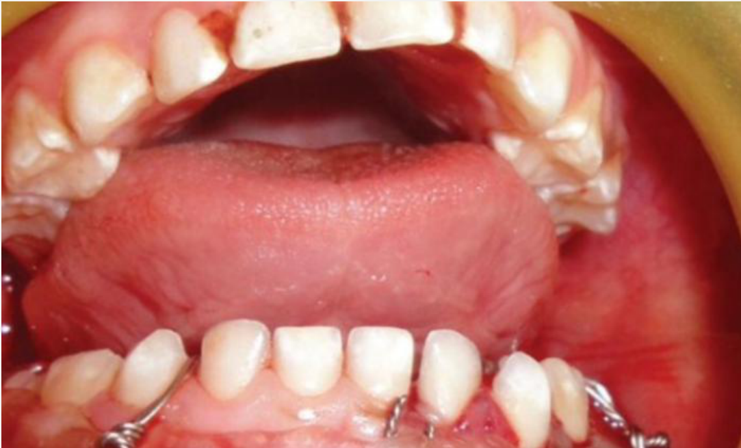
Paediatric Mandibular Fractures56
-
Bony growth centres and developing tooth buds
-
Restoration of mandibular continuity important for function and future craniofacial development
-
Conservative management if minimally displaced
-
TMJ – growth centre in children, so beware of late sequelae (ankylosis)
-
A condylar injury may interfere with mandibular growth.
-
The child may later develop a deviated or asymmetric mandible if growth is affected.
-
Even after treatment, children require monitoring for years while they continue to grow.
-
Conservative treatment may be chosen in some cases, but the child must still be reviewed regularly.
-
Paediatric orbital floor fractures may be trapdoor fractures with muscle entrapment and an oculocardiac reflex.
 |  |
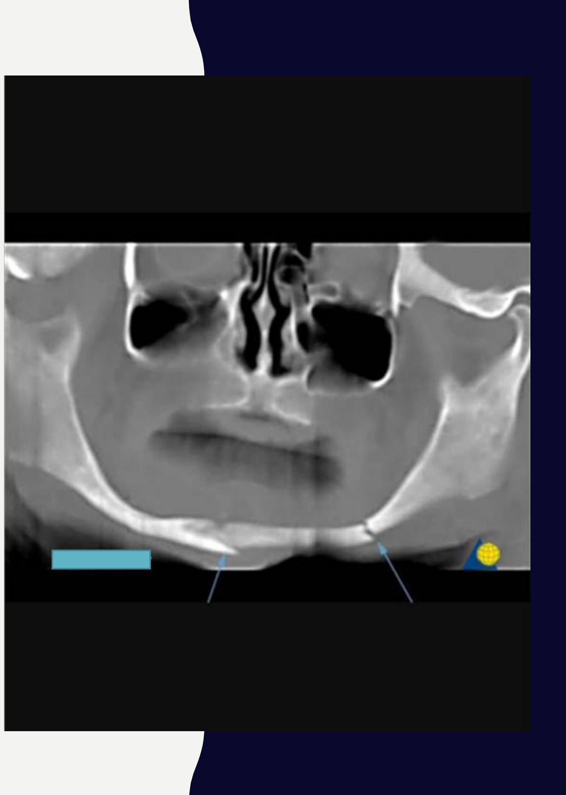
Edentulous Mandible57
-
Atrophy of the mandible, which becomes less resistant to traumatic forces
-
Individuals with systemic co-morbidities
-
Poor healing (fibrous/non-union)
-
Poor bone stock
-
Mandibular fractures are more difficult to identify in edentulous patients because occlusion cannot be checked using teeth.
-
Useful findings include:
- A lingual hematoma.
- Dentures that no longer fit.
- Inability to wear the dentures.
- A history of a fall that may have been forgotten.
-
OPG examination should proceed systematically from one condyle to the other.
-
A fracture may involve one condyle and the opposite body.
-
If the fracture is identified late, it may already have begun healing in a slightly incorrect position.
-
Management depends on:
- Whether the fracture is still mobile.
- Whether it has become solid.
- The patient’s general health.
- Whether surgery and general anaesthesia are appropriate.
-
If the fracture has healed and the patient is not fit for surgery, a new denture may be made despite a residual step.
-
An elderly patient may have poor blood supply or osteoporosis.
-
Avoiding periosteal stripping may be important.
-
External fixation or denture modification may be considered according to the individual situation.
Bone Healing
Healing Timeline58
Healing takes 4–6 weeks, or longer for large bones and elderly patients.
Lecturer — Healing Factors
In a healthy young person, mandibular bone healing takes approximately four to six weeks, but the first four weeks are particularly important because the fracture is still healing.
- Healing may also be delayed by osteoporosis, Prolia treatment, osteogenesis imperfecta, poor nutrition, bone deformity, or excessive movement at the fracture.
- After healing, routine dental treatment can generally be carried out normally; a fracture treated twenty years earlier should not be considered at risk of displacement from routine dental treatment.
Repair of Fractures59
- Formation of haematoma
- Fibrocartilage callus (mass of connective repair tissue) – splinting the break
- Bony callus replaces the fibrocartilage callus
- Bone remodelling in response to mechanical stress – healed fracture
Lecturer — Fracture Stability
Excessive movement interferes with healing because the fracture needs to remain stable while the body forms the callus and repairs the bone.
- If the fracture remains mobile, it may develop a fibrous union or a non-union.
- Splinting or fixation keeps the bone ends together; plates and screws maintain the correct position but do not perform the healing.
- Nutrition and general health also influence the repair process.
Postoperative Care60
- Soft diet important in patients who have undergone open reduction internal fixation
- Patients placed in intermaxillary fixation need to be advised about nutritional issues and oral hygiene instructions
- Postoperative radiographs and clinical assessment to check for:
- Fracture healing
- Fracture segment alignment and status of occlusion
Lecturer — Intermaxillary Fixation
Wired or immobilised patients may have difficulty cleaning their teeth and can develop tartar accumulation, sore gums, redness, and swelling.
- Dental management may include oral hygiene instruction, several hygiene visits, and scaling and polishing.
- During the first four weeks, care should be taken not to disturb the healing fracture; after healing, the patient can generally be treated normally.
Lecturer — Teeth Near Fractures
Teeth near a fracture may later become non-vital, so the dentist should assess their vitality and restorability.
- The assessment should consider the need for root canal treatment, extraction, and replacement options.
Lecturer — Forensic Reports
Reports for courts, health funds, or workers’ compensation should identify the clinician and patient, document the examination date and clinical findings, and include relevant radiographs and treatment recommendations.
- The report should include the clinician’s name, practice, qualifications, and AHPRA number, together with the patient’s name and date of birth.
- The history should be recorded as an allegation, and the report should remain factual without invented symptoms, speculation, or embellished language.
- It should state whether teeth are restorable or unrestorable and justify the recommended treatment. Health funds may request a separate report in addition to the clinical notes, and reports may be charged to the health fund when requested.
- Treatment options, such as an immediate partial or spoon denture followed by a later implant, should be documented together with the discussion with the patient.
Summary61
- Mechanism of injury important in history taking
- Patterns of injury from type of mechanism deployed
- Injury prevention as part of continuum of care — mouthguard, helmets.
Lecturer — Facial Trauma Assessment
Trauma is the transfer of energy, and injury severity depends on the amount and pattern of energy absorbed.
- The commonest facial fractures discussed are nasal and mandibular fractures.
- The history should establish the mechanism, speed, height, vehicle position, seat-belt use, and associated injuries.
- Severe facial injury requires consideration of cervical spine injury and appropriate immobilisation.
- A lingual hematoma is a highly significant sign of mandibular fracture.
- Mandibular fractures should be assessed systematically on radiographs, tracing from one condyle to the other and comparing both sides.
Lecturer — Dental Trauma Presentation
Because the maxilla and mandible are tooth-bearing structures, fractures may present to the dentist through altered occlusion, dentures that no longer fit, tooth mobility or displacement, tooth devitalisation, a sinus or swelling, or nerve paraesthesia.
- Children with condylar fractures require long-term growth monitoring.
- Elderly and edentulous patients may present late and require assessment of denture fit, fracture mobility, general health, bone quality, and treatment suitability.
- Reduction aligns the fracture, while fixation holds it in position as the body heals.
Acknowledgement62
All images are available online.
Clinical Cases
Case: Multifacial injury, clinical photo and 3D CT
Question
Scenario: A patient presents with severe facial trauma following a motor vehicle accident or road traffic accident.
What’s shown: A clinical photograph of a child’s face showing significant injury, alongside a 3D CT reconstruction of the skull.
Consider: Identify the facial injuries visible on the imaging and determine the immediate management step required for the cervical spine.
Answer
Observations:
- Multifacial injury involving the orbits, maxilla, and mandible.
Reasoning: The extent of the facial fractures indicates a high-energy impact. Because of the severity of the trauma, the cervical spine must be assumed to be injured until proven otherwise.
Outcome: The patient must have their cervical spine stabilized with a collar.
Takeaway: In cases of severe maxillofacial trauma, always assume a concurrent cervical spine injury and immobilize it with a collar until it is cleared.
Case: Intraoral photo of the lower jaw
Question
Scenario: An intraoral clinical photograph is presented to demonstrate clinical signs associated with facial trauma.
What’s shown: An intraoral view of the lower jaw showing a displaced injury and a collection of blood on the lingual side.
Consider: What is the key clinical finding visible in this image, and what does it indicate?
Answer
Observations:
- A lingual hematoma is present.
- The mandibular injury is displaced.
Reasoning: The lecturer points out that a lingual hematoma is the classical, pathognomonic picture of a fractured mandible. Even if the displacement is not severe, the presence of a lingual hematoma equals a fractured mandible until proven otherwise. Other signs of a mandible fracture can include deranged occlusion, a large step deformity, inferior dental (ID) nerve numbness, swelling, and discomfort.
Outcome: The exhibit serves to diagnose a fractured mandible based on the pathognomonic sign.
Takeaway: A lingual hematoma is pathognomonic for a mandibular fracture and should be considered diagnostic until proven otherwise.
Case: Work-related facial injury, clinical photo and report
Question
Scenario: A patient presents with a work-related facial injury and requires a report for workers’ compensation.
What’s shown: A clinical photograph of a male patient lying supine, alongside a split image illustrating the mechanism of injury.
Consider: How should the clinician document the findings and prognosis for a medico-legal or workers’ compensation report?
Answer
Observations:
- An associated laceration (which was sutured in the emergency department).
- Fractured lateral incisor and canine.
- Teeth 11, 12, and 13 are non-vital and severely fractured.
Reasoning: When writing a report for workers’ compensation or health funds, the clinician must only document objective findings and radiographic evidence observed at the time of the examination. Symptoms cannot be fabricated, as the reports are subject to audit. The severe fractures render the affected teeth unrestorable.
Outcome: The report should state the clinical findings, note that the teeth are non-vital and likely to be devitalized, and conclude that teeth 11, 12, and 13 require extraction and replacement.
Takeaway: Medico-legal and workers’ compensation reports must strictly reflect objective clinical and radiographic findings at the time of assessment without fabricating symptoms.
Case: Impaled object to the head, clinical photo and skull radiographs
Question
Scenario: A patient presents to the emergency department or a rural clinic with an impaled object in the head following a fall.
What’s shown: A clinical photograph of a patient lying supine, alongside frontal and anteroposterior skull X-rays demonstrating a large radiopaque object (a knife) embedded in the skull.
Consider: What is the critical immediate management principle for an embedded foreign body in this context?
Answer
Observations:
- A knife is embedded in the patient’s head, clearly visible on clinical examination and radiographs.
Reasoning: The impaled object may be tamponading a major blood vessel. Removing it outside of a controlled operating theater, without the ability to immediately control catastrophic bleeding, is highly dangerous. Additionally, the metallic nature of the knife is an absolute contraindication for an MRI, as the magnetic field could move the object.
Outcome: The object must not be removed unless the patient is in a theater prepared to manage severe hemorrhage. The patient cannot undergo an MRI scan.
Takeaway: Never remove an impaled object in the field or emergency department, as it may be tamponading a major vessel; also remember that metallic objects contraindicate MRI.
Case: Ballistic injury to the face, clinical photos
Question
Scenario: A patient presents with severe facial trauma following a ballistic injury (e.g., a suicide attempt with a firearm).
What’s shown: Clinical photographs showing a small entry wound and a massive exit wound with extensive soft tissue destruction and facial defect.
Consider: What are the primary clinical concerns and initial management priorities for severe ballistic facial injuries?
Answer
Observations:
- A small, localized entry wound.
- A large, destructive exit wound causing massive soft tissue loss and facial defect.
Reasoning: Ballistic injuries cause widespread, non-localized tissue destruction that extends far beyond the entry point. The massive soft tissue damage and distortion of anatomy can make it difficult to identify the mouth and airway.
Outcome: The immediate priority is securing the airway (e.g., intubation), which may fall outside the dentist’s immediate purview but is life-saving.
Takeaway: Ballistic injuries to the face cause extensive, non-localized soft tissue destruction, and airway management is the immediate priority due to anatomical distortion.
Case: Panoramic radiograph of facial trauma
Question
Scenario: A patient presents with facial trauma, and a panoramic radiograph (OPG) is taken to evaluate for fractures.
What’s shown: A panoramic dental radiograph showing the maxilla and mandible.
Consider: How should the clinician systematically evaluate the OPG for fractures, and what clinical signs correlate with the radiographic findings?
Answer
Observations:
- The right condyle is overriding the ramus, shortening the ramus height.
- A radiographic step is visible between the right second molar (tooth 7) and first molar (tooth 6).
- A fractured coronoid process is present on the left side.
Reasoning: A systematic review from one condyle to the other is essential. Condylar overriding shortens the ramus, leading to premature posterior contact and an incorrect bite. A step in the dentition indicates an alveolar or body fracture. Because the mandible is a U-shaped ring, 60-70% of fractures are bilateral; finding one fracture necessitates checking the contralateral side. A coronoid fracture does not affect the occlusion. Clinical signs such as a gingival tear, swelling, bleeding, or a numb lip (indicating inferior alveolar nerve damage) correlate with the fracture sites.
Outcome: The condylar fracture and the fracture between teeth 6 and 7 affect the bite and require treatment, whereas the coronoid fracture does not affect the bite and is not treated.
Takeaway: Mandibular fractures are frequently bilateral (60-70%), so always systematically check the entire mandible on an OPG when one fracture is found, and correlate radiographic steps with clinical signs like occlusal discrepancies or nerve damage.
Case: Panoramic radiograph of the mandible
Question
Scenario: A patient presents to the dentist complaining that their bite does not feel quite right, having previously attended the Emergency Department for a facial injury that was initially missed.
What’s shown: A panoramic radiograph of the mandible.
Consider: Identify the radiographic findings and determine the appropriate management, considering the potential impact on occlusion and growth.
Answer
Observations:
- The right condyle (neck/head) is fractured.
- The fractured bone edges are overlapping and not smooth.
- There is shortening of the ramus on the affected side.
Reasoning:
- The overlapping and irregular edges indicate a displaced fracture of the right condyle.
- The shortening of the ramus causes the condyle to sit higher, leading to an incorrect occlusion with a premature bite on the affected side.
- In children, a condylar fracture can damage the growth center, potentially causing a skewed mandible as they grow, which requires years of monitoring.
- In adults, it results in pain or occlusal disharmony.
- Because there is shortening and displacement at the condylar neck, the case requires referral.
Outcome:
- The patient is referred for management due to the displaced fracture with ramus shortening.
Takeaway:
- Mandibular fractures can be missed in the ED, especially if undisplaced or if only a single view is taken; always compare both condyles on an OPG, as condylar fractures alter the occlusion and require careful monitoring in children to prevent growth disturbances.
Case: Parasymphysis fracture on panoramic radiograph
Question
Scenario: A patient presents with a misaligned bite or a gap between the lower front teeth, possibly a month or two after a minor trauma they may have forgotten about.
What’s shown: A panoramic radiograph showing a fracture in the parasymphysis region between teeth 32 and 33, with a visible step.
Consider: What is the diagnosis, and what complications should be suspected?
Answer
Observations:
- Fracture in the parasymphysis between teeth 32 and 33.
- A large step is visible at the fracture site.
Reasoning: The misalignment of the bite and the gap between the teeth correspond to the displaced parasymphysis fracture. A lingual hematoma may also be present. Teeth 32 and 33 are at risk of quiet devitalization, which could later present as a swelling, a buccal sinus, or an apical area trying to discharge an abscess.
Outcome: The patient may present late with devitalized teeth or a sinus if the initial trauma was forgotten or ignored.
Takeaway: Maintain an index of suspicion for forgotten trauma when teeth suddenly devitalize or a step and gap are present without an obvious recent cause.
Case: Bilateral mandibular fractures in an edentulous patient
Question
Scenario: An elderly edentulous patient presents complaining that their dentures no longer fit, possibly a month or more after a fall they may not remember.
What’s shown: A panoramic radiograph of an edentulous mandible displaying overlapping bone ends.
Consider: What is the underlying cause of the ill-fitting dentures, and what does the radiograph reveal upon systematic examination?
Answer
Observations:
- Fracture of the right condyle with overlapping of the condyle and the body of the ramus.
- Fracture of the left body of the mandible with overlapping bone ends.
Reasoning: Mandibular fractures are often bilateral due to the U-shape of the bone. In an edentulous patient, a clinical step cannot be checked, but a lingual hematoma or the sudden inability to wear dentures are key indicators. Systematically tracing the mandible on the OPG from the right condyle to the left reveals the bilateral fractures (right condyle and left body) that have likely started to heal in a slightly wrong position.
Outcome: Management depends on whether the fracture has already healed. If it has healed with a slight step, the lower denture may just need to be remade. If not, the fracture may need to be reduced and plated, though the patient’s fitness for general anesthesia must be considered.
Takeaway: Always take an OPG and systematically trace the mandible in elderly edentulous patients presenting with ill-fitting dentures, as they may have sustained and forgotten bilateral fractures.
Case: Panoramic radiograph showing a left mandibular angle fracture
Question
Scenario: A panoramic radiograph is presented for systematic evaluation to identify a fracture.
What’s shown: A panoramic dental radiograph (OPG).
Consider: Where is the fracture located, and what are its characteristics?
Answer
Observations:
- A fracture is present in the left angle of the mandible.
- It appears as a crack that is not displaced or very minimally displaced.
- The fracture line extends through the left mandibular third molar (tooth 8).
- The patient may or may not have associated symptoms like numbness.
Reasoning: A systematic approach from one condyle to the other reveals the fracture in the left angle. The minimal displacement and its path through the third molar are noted, which raises the question of whether the tooth should be extracted during fracture fixation.
Outcome: The decision to extract the third molar during fracture fixation is deferred to the treating team.
Takeaway: Use a systematic approach (e.g., condyle to condyle) when evaluating panoramic radiographs for fractures, and carefully note the fracture’s relationship to adjacent teeth, such as third molars.
Case: Occipitomental view showing a right zygomatic complex fracture
Question
Scenario: An occipitomental (OM) view of the skull is presented to evaluate the facial bones, prompted by a scenario of a patient with a history of a black eye and an altered bite following sports trauma.
What’s shown: A frontal X-ray view of the skull (OM view) showing the frontal bone, maxilla, and zygomatic bones.
Consider: What abnormalities are visible on the right side of the facial skeleton, and what is the diagnosis?
Answer
Observations:
- The zygomaticofrontal suture on the right is wider than on the left.
- There is a step in the intraoral buccal vestibule on the right.
- The lateral wall of the maxilla on the right is not smooth.
- The temporozygomatic suture on the right is wider and displaced compared to the left.
Reasoning: By comparing the right side to the smooth, intact left side, asymmetry and discontinuities are identified. Conceptualizing the cheekbone as a four-sided stool helps identify that fractures or displacements occur at the sutures (the “legs”) or the body. The widening of the sutures and irregular lateral wall indicate displacement of the zygoma.
Outcome: The diagnosis is a fractured right zygomatic complex. The lecturer notes that a dropped zygoma can affect the bite in the first quadrant, potentially causing devitalization of teeth (e.g., 13 and 14), which requires documentation for legal or insurance purposes.
Takeaway: When evaluating an OM view for a suspected zygomatic fracture, systematically compare the sutures and bony contours of both sides. Isolated zygomatic fractures are easier to identify this way, whereas pan-facial fractures are more complex. A displaced zygoma can affect maxillary occlusion and tooth vitality, which must be documented.
Case: Frontal skull X-ray (OMV) evaluating the facial skeleton
Question
Scenario: An occipitomental (OMV) frontal X-ray of the skull is presented for evaluation following facial trauma.
What’s shown: A frontal radiograph of the skull showing the facial bones and sutures.
Consider: Systematically compare the left and right sides of the facial skeleton to identify any abnormalities and determine the underlying injury.
Answer
Observations:
- The left side of the facial skeleton appears normal, with smooth contours of the maxillary wall and zygomatic bone.
- On the right side, there is widening of the sutures and an irregularity in the maxillary wall.
- The temporal zygomatic suture on the right is located in a different position compared to the left.
Reasoning: By systematically comparing both sides of the OMV X-ray (checking the frontal bone sutures, maxillary walls, and zygomatic bones), the asymmetry and displacement on the right side become evident. The altered location of the temporal zygomatic suture and the widened sutures indicate that the zygoma is displaced or has fractured just in front of the suture at a weaker point.
Outcome: In a clinical scenario following such a fracture, the patient may present with an altered bite and devitalization of adjacent teeth (e.g., teeth 13 and 14). This would require a factual, contemporaneous medico-legal report for health funds or court, detailing the patient’s allegations, the clinical findings on the day, and the recommended treatment (e.g., extraction or restoration), without confabulation.
Takeaway: When evaluating facial trauma radiographs like the OMV, systematically compare the left and right sides of the facial skeleton to easily identify asymmetries and fractures.
Case: Frontal skull X-ray evaluating the zygomatic arches
Question
Scenario: A frontal (AP) skull X-ray is presented to evaluate a suspected facial fracture.
What’s shown: A frontal radiograph of the skull focusing on the zygomatic bones and arches.
Consider: Evaluate the zygomatic complex and arches to determine the extent of the fracture and its potential clinical implications.
Answer
Observations:
- The left zygomatic arch is depressed.
- The rest of the left zygoma is intact.
- The frontal zygomatic suture, inferior orbit, and lateral maxillary wall are all fine and show no signs of fracture.
Reasoning: By comparing the sides and tracing the zygomatic bone, the depression is isolated to the left arch. Because the other components of the zygomatic complex (frontal zygomatic suture, inferior orbit, lateral maxillary wall) are intact, this is diagnosed as an isolated zygomatic arch fracture rather than a full zygomatic complex fracture.
Outcome: If the arch is severely depressed, it can interfere with mandibular function, preventing the patient from opening their mouth properly. A patient might present with this complaint if the fracture was missed or untreated, and they may also have a visible dimple over the depressed arch.
Takeaway: An isolated zygomatic arch fracture can be identified on a frontal X-ray when the arch is depressed but the rest of the zygomatic complex remains intact; clinicians must maintain a high index of suspicion for this injury if a patient presents with restricted mouth opening and an altered bite.
Case: Deep facial laceration involving the parotid
Question
Scenario: A patient presents with a severe, deep soft tissue injury to the face following an assault.
What’s shown: A clinical photograph showing a severe, deep laceration on the face.
Consider: What underlying structures are at risk in this region, and what are the clinical consequences of injury to these structures?
Answer
Observations:
- The laceration is deep and involves the parotid region.
Reasoning: The parotid gland carries the facial nerve. A deep laceration here can injure the entire trunk of the facial nerve, causing the face to droop, or individual branches. Injury to the temporal branch prevents wrinkling the forehead. Injury to the zygomatic branch impairs the ability to protect the eye. Injury to the buccal branch prevents puffing out the cheeks, leading to food accumulation in the sulcus and difficulty controlling a denture. It also causes the corner of the mouth to paradoxically go up due to the unopposed action of the depressors.
Outcome: Not explicitly stated, but emphasizes the anatomical risks and functional deficits.
Takeaway: Deep lacerations to the parotid region risk injury to the facial nerve and its branches, which can cause specific functional deficits depending on the branch affected.
Case: Healed facial laceration with buccal branch deficit
Question
Scenario: A patient presents to the dental clinic a month after having a facial laceration sutured in the emergency department, complaining of difficulty controlling their denture and cheeks.
What’s shown: An intraoral clinical photograph of a healed laceration.
Consider: What is the cause of the patient’s functional deficit, and is it related to recent dental treatment?
Answer
Observations:
- The laceration has healed following suturing in the emergency department a month prior.
- The patient cannot control their buccal branch (cannot puff out cheeks, trouble with denture control and pressure).
Reasoning: The buccal branch of the facial nerve was likely damaged during the initial injury, not by a recent dental block. The deficit was present before the patient sat in the dental chair.
Outcome: The patient has a persistent buccal branch paralysis affecting denture control and cheek puffing.
Takeaway: Dentists must recognize that functional deficits like buccal branch paralysis may be pre-existing sequelae of past facial trauma, rather than complications of recent dental procedures.
Case: Frontal bone injury with suspected CSF leak
Question
Scenario: A patient presents following a high-energy injury to the frontal bone.
What’s shown: A clinical photograph of the patient’s face, an axial CT scan, and an intraoperative photograph.
Consider: What structures are involved in the fracture, and how does this dictate the management?
Answer
Observations:
- Brain fluid (CSF) leaking from the nose.
- Fractures of both the anterior and posterior tables of the frontal bone on the CT scan.
Reasoning:
- The frontal sinus is an air-filled space, and the posterior table separates it from the cranial cavity where the brain sits.
- A fracture involving the posterior table creates a direct entry to the brain, leading to a CSF leak and a high risk of intracranial infection.
- This is distinguished from an isolated anterior table fracture, which typically presents as a cosmetic “little dent.”
Outcome:
- Because both tables are fractured and there is a direct entry to the brain, the injury requires a joint surgical approach to fix it and prevent infection.
Takeaway:
- When evaluating frontal bone fractures, it is critical to determine if the posterior table is involved; a posterior table fracture creates a direct pathway to the brain and requires urgent joint surgical intervention, unlike isolated anterior table fractures which are primarily cosmetic.
Case: Facial trauma, clinical photographs of the face and eyes
Question
Scenario: A male patient presents following facial trauma.
What’s shown: Clinical photographs of the patient’s face and a close-up of the eyes.
Consider: Identify the clinical signs visible in the images and determine what they suggest about the underlying injury and potential nerve involvement.
Answer
Observations:
- Bruising and swelling on one side of the face.
- Subconjunctival hemorrhage (bleeding under the conjunctiva).
- The hemorrhage extends posteriorly with no visible posterior limit.
Reasoning:
- A simple hematoma from blunt trauma (such as a punch or a ball) would typically have a posterior extent limited to the subconjunctiva.
- When the bleeding or bruising extends and the posterior limit cannot be seen, it indicates the hemorrhage is tracking back and is most likely connected to an underlying fracture, such as a zygomatic fracture.
- The infraorbital nerve exits at the infraorbital foramen in this region. Damage or extensive bruising around this area from the fracture can cause infraorbital nerve paresthesia, leading to numbness in the cheek and maxillary teeth on the affected side. Patients often do not notice this numbness initially because they are distracted by pain, swelling, and eye signs, only realizing it weeks or months later.
Outcome:
- The patient may present months after the initial injury complaining of delayed numbness in the cheek or teeth. The clinician must take a thorough history to link these delayed symptoms to the previous fracture and anatomical damage to the infraorbital nerve.
Takeaway:
- A subconjunctival hemorrhage with no visible posterior limit strongly suggests an underlying facial fracture, and clinicians must always consider anatomical relationships—such as the proximity of the infraorbital nerve to the zygomatic complex—when evaluating delayed symptoms like paresthesia.
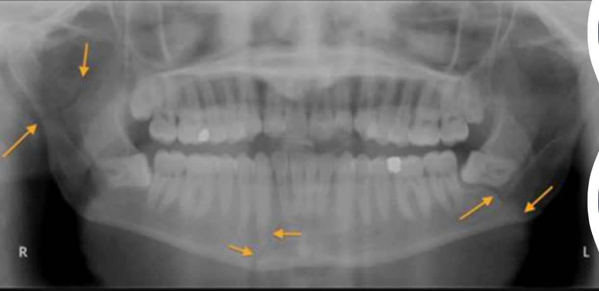
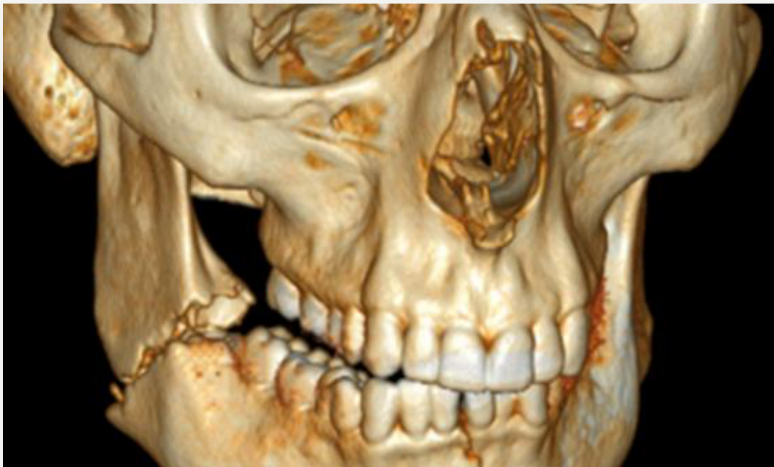
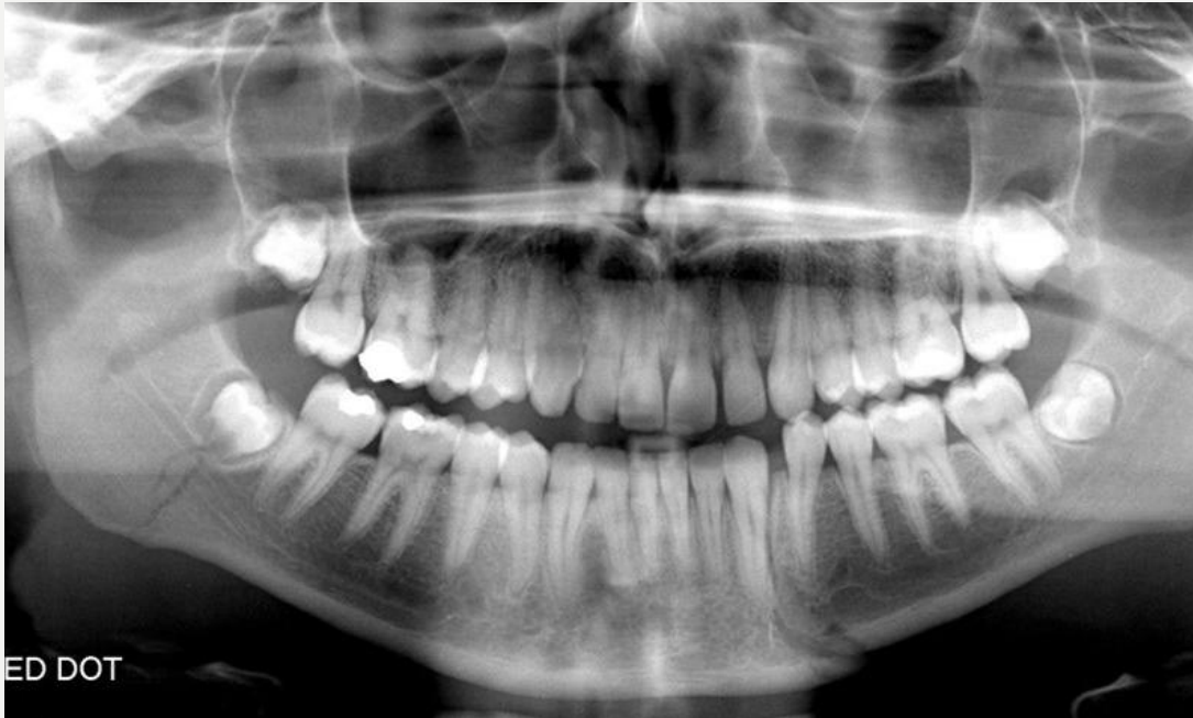
Case: Fractured mandible, radiographs and clinical photos
Question
Scenario: A patient presents following facial trauma with potential signs of a jaw fracture.
What’s shown: A 3D volume-rendered CT reconstruction of the facial skeleton, a panoramic radiograph of the dentition, an intraoral clinical photo of the floor of the mouth, and a close-up clinical photo of the patient’s teeth.
Consider: What are the key clinical and radiographic signs of a fractured mandible, and how do you determine the appropriate management?
Answer
Observations:
- A 3D CT reconstruction and panoramic radiograph showing a fractured mandible.
- An intraoral photo showing a lingual hematoma (bruising under the tongue).
- A clinical photo showing malocclusion, specifically a gap or step in the occlusion.
Reasoning: The lecturer explains that while pain, swelling, paresthesia, and mobility are signs of trauma, a lingual hematoma is a pathognomonic sign of bleeding that definitively indicates a mandibular fracture. A step or gap in the occlusion also points to a fracture. When a patient presents with a gingival tear and an occlusal gap, an OPG is required to determine whether the fracture is confined to the alveolus (the bone around the teeth) or involves the mandible.
Outcome: If the fracture is confined to the alveolus, it can be treated directly. If it is a mandibular fracture, the patient requires referral.
Takeaway: A lingual hematoma is a pathognomonic sign of a mandibular fracture, and radiographic evaluation is essential to differentiate between an alveolar fracture and a mandibular fracture to guide appropriate management.
Case: Mandible fracture with internal and external fixation
Question
Scenario: A patient with a mandible fracture and other severe injuries is presented.
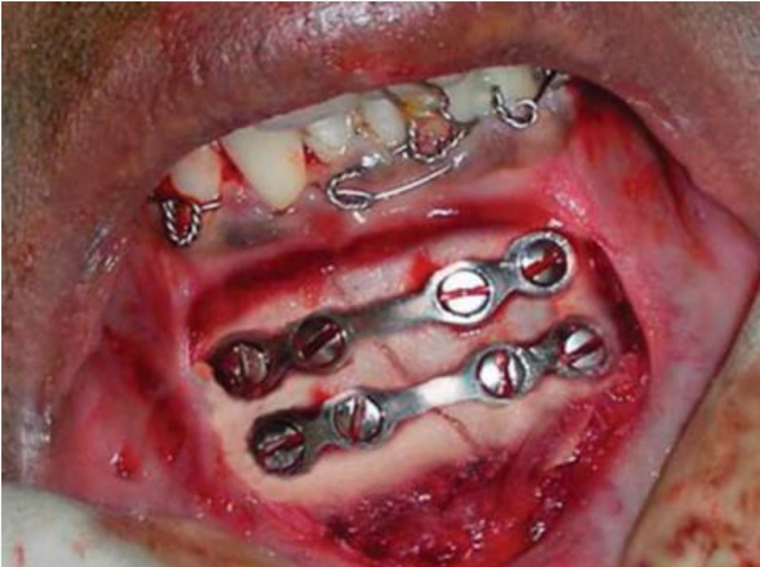
What’s shown: An intraoral clinical photograph showing a mandible fracture treated with plates and screws, as well as wires around the teeth.
Consider: What types of reduction and fixation are being used, and why might both be employed in this patient?
Answer
Observations:
- The mandible fracture is treated with plates and screws.
- There are wires around the patient’s teeth.
Reasoning: The plates and screws represent open reduction and internal fixation, where the gum is lifted to visualize the fracture directly and fix it internally. The wires around the teeth represent external fixation, which relies on adjacent anatomy (occlusion) to guide the reduction. This external fixation is likely used because the patient has other severe injuries, making it too difficult to hold the fracture together with internal fixation alone; it allows the team to reduce and fix the mandible and then concentrate on other areas, such as the maxilla.
Outcome: The patient undergoes open reduction and internal fixation of the mandible, supplemented by external fixation via wires around the teeth to accommodate and manage other severe injuries.
Takeaway: External fixation may be used alongside internal fixation in patients with multiple severe injuries to stabilize a fracture while allowing the surgical team to focus on other critical areas.
Footnotes
-
Original PDF page 1: W1 Maxillofacial Trauma - Mechanisms, Fractures and Bone Healing - Foo, p.1 ↩
-
Original PDF page 2: W1 Maxillofacial Trauma - Mechanisms, Fractures and Bone Healing - Foo, p.2 ↩
-
Original PDF page 3: W1 Maxillofacial Trauma - Mechanisms, Fractures and Bone Healing - Foo, p.3 ↩
-
Original PDF page 4: W1 Maxillofacial Trauma - Mechanisms, Fractures and Bone Healing - Foo, p.4 ↩
-
Original PDF page 5: W1 Maxillofacial Trauma - Mechanisms, Fractures and Bone Healing - Foo, p.5 ↩
-
Original PDF page 6: W1 Maxillofacial Trauma - Mechanisms, Fractures and Bone Healing - Foo, p.6 ↩
-
Original PDF page 7: W1 Maxillofacial Trauma - Mechanisms, Fractures and Bone Healing - Foo, p.7 ↩
-
Original PDF page 8: W1 Maxillofacial Trauma - Mechanisms, Fractures and Bone Healing - Foo, p.8 ↩
-
Original PDF page 9: W1 Maxillofacial Trauma - Mechanisms, Fractures and Bone Healing - Foo, p.9 ↩
-
Original PDF page 10: W1 Maxillofacial Trauma - Mechanisms, Fractures and Bone Healing - Foo, p.10 ↩
-
Original PDF page 11: W1 Maxillofacial Trauma - Mechanisms, Fractures and Bone Healing - Foo, p.11 ↩
-
Original PDF page 12: W1 Maxillofacial Trauma - Mechanisms, Fractures and Bone Healing - Foo, p.12 ↩
-
Original PDF page 13: W1 Maxillofacial Trauma - Mechanisms, Fractures and Bone Healing - Foo, p.13 ↩
-
Original PDF page 14: W1 Maxillofacial Trauma - Mechanisms, Fractures and Bone Healing - Foo, p.14 ↩
-
Original PDF page 15: W1 Maxillofacial Trauma - Mechanisms, Fractures and Bone Healing - Foo, p.15 ↩
-
Original PDF page 16: W1 Maxillofacial Trauma - Mechanisms, Fractures and Bone Healing - Foo, p.16 ↩
-
Original PDF page 17: W1 Maxillofacial Trauma - Mechanisms, Fractures and Bone Healing - Foo, p.17 ↩
-
Original PDF page 18: W1 Maxillofacial Trauma - Mechanisms, Fractures and Bone Healing - Foo, p.18 ↩
-
Original PDF page 19: W1 Maxillofacial Trauma - Mechanisms, Fractures and Bone Healing - Foo, p.19 ↩
-
Original PDF page 20: W1 Maxillofacial Trauma - Mechanisms, Fractures and Bone Healing - Foo, p.20 ↩
-
Original PDF page 21: W1 Maxillofacial Trauma - Mechanisms, Fractures and Bone Healing - Foo, p.21 ↩
-
Original PDF page 22: W1 Maxillofacial Trauma - Mechanisms, Fractures and Bone Healing - Foo, p.22 ↩
-
Original PDF page 23: W1 Maxillofacial Trauma - Mechanisms, Fractures and Bone Healing - Foo, p.23 ↩
-
Original PDF page 24: W1 Maxillofacial Trauma - Mechanisms, Fractures and Bone Healing - Foo, p.24 ↩
-
Original PDF page 25: W1 Maxillofacial Trauma - Mechanisms, Fractures and Bone Healing - Foo, p.25 ↩
-
Original PDF page 26: W1 Maxillofacial Trauma - Mechanisms, Fractures and Bone Healing - Foo, p.26 ↩
-
Original PDF page 27: W1 Maxillofacial Trauma - Mechanisms, Fractures and Bone Healing - Foo, p.27 ↩
-
Original PDF page 28: W1 Maxillofacial Trauma - Mechanisms, Fractures and Bone Healing - Foo, p.28 ↩
-
Original PDF page 29: W1 Maxillofacial Trauma - Mechanisms, Fractures and Bone Healing - Foo, p.29 ↩
-
Original PDF page 30: W1 Maxillofacial Trauma - Mechanisms, Fractures and Bone Healing - Foo, p.30 ↩
-
Original PDF page 31: W1 Maxillofacial Trauma - Mechanisms, Fractures and Bone Healing - Foo, p.31 ↩
-
Original PDF page 32: W1 Maxillofacial Trauma - Mechanisms, Fractures and Bone Healing - Foo, p.32 ↩
-
Original PDF page 33: W1 Maxillofacial Trauma - Mechanisms, Fractures and Bone Healing - Foo, p.33 ↩
-
Original PDF page 34: W1 Maxillofacial Trauma - Mechanisms, Fractures and Bone Healing - Foo, p.34 ↩
-
Original PDF page 35: W1 Maxillofacial Trauma - Mechanisms, Fractures and Bone Healing - Foo, p.35 ↩
-
Original PDF page 36: W1 Maxillofacial Trauma - Mechanisms, Fractures and Bone Healing - Foo, p.36 ↩
-
Original PDF page 37: W1 Maxillofacial Trauma - Mechanisms, Fractures and Bone Healing - Foo, p.37 ↩
-
Original PDF page 38: W1 Maxillofacial Trauma - Mechanisms, Fractures and Bone Healing - Foo, p.38 ↩
-
Original PDF page 39: W1 Maxillofacial Trauma - Mechanisms, Fractures and Bone Healing - Foo, p.39 ↩
-
Original PDF page 40: W1 Maxillofacial Trauma - Mechanisms, Fractures and Bone Healing - Foo, p.40 ↩
-
Original PDF page 41: W1 Maxillofacial Trauma - Mechanisms, Fractures and Bone Healing - Foo, p.41 ↩
-
Original PDF page 42: W1 Maxillofacial Trauma - Mechanisms, Fractures and Bone Healing - Foo, p.42 ↩
-
Original PDF page 43: W1 Maxillofacial Trauma - Mechanisms, Fractures and Bone Healing - Foo, p.43 ↩
-
Original PDF page 44: W1 Maxillofacial Trauma - Mechanisms, Fractures and Bone Healing - Foo, p.44 ↩
-
Original PDF page 45: W1 Maxillofacial Trauma - Mechanisms, Fractures and Bone Healing - Foo, p.45 ↩
-
Original PDF page 46: W1 Maxillofacial Trauma - Mechanisms, Fractures and Bone Healing - Foo, p.46 ↩
-
Original PDF page 47: W1 Maxillofacial Trauma - Mechanisms, Fractures and Bone Healing - Foo, p.47 ↩
-
Original PDF page 48: W1 Maxillofacial Trauma - Mechanisms, Fractures and Bone Healing - Foo, p.48 ↩
-
Original PDF page 49: W1 Maxillofacial Trauma - Mechanisms, Fractures and Bone Healing - Foo, p.49 ↩
-
Original PDF page 50: W1 Maxillofacial Trauma - Mechanisms, Fractures and Bone Healing - Foo, p.50 ↩
-
Original PDF page 51: W1 Maxillofacial Trauma - Mechanisms, Fractures and Bone Healing - Foo, p.51 ↩
-
Original PDF page 52: W1 Maxillofacial Trauma - Mechanisms, Fractures and Bone Healing - Foo, p.52 ↩
-
Original PDF page 53: W1 Maxillofacial Trauma - Mechanisms, Fractures and Bone Healing - Foo, p.53 ↩
-
Original PDF page 54: W1 Maxillofacial Trauma - Mechanisms, Fractures and Bone Healing - Foo, p.54 ↩
-
Original PDF page 55: W1 Maxillofacial Trauma - Mechanisms, Fractures and Bone Healing - Foo, p.55 ↩
-
Original PDF page 56: W1 Maxillofacial Trauma - Mechanisms, Fractures and Bone Healing - Foo, p.56 ↩
-
Original PDF page 57: W1 Maxillofacial Trauma - Mechanisms, Fractures and Bone Healing - Foo, p.57 ↩
-
Original PDF page 58: W1 Maxillofacial Trauma - Mechanisms, Fractures and Bone Healing - Foo, p.58 ↩
-
Original PDF page 59: W1 Maxillofacial Trauma - Mechanisms, Fractures and Bone Healing - Foo, p.59 ↩
-
Original PDF page 60: W1 Maxillofacial Trauma - Mechanisms, Fractures and Bone Healing - Foo, p.60 ↩
-
Original PDF page 61: W1 Maxillofacial Trauma - Mechanisms, Fractures and Bone Healing - Foo, p.61 ↩
-
Original PDF page 62: W1 Maxillofacial Trauma - Mechanisms, Fractures and Bone Healing - Foo, p.62 ↩