Oral Cancer Resection and Reconstruction1
Magdalen Foo, Oral Maxillofacial Surgeon
Learning Objectives2
- Describe the clinical aspect, diagnosis and management of oral cancer
- Understand the role of Multidisciplinary Team in head and neck cancer management
- Appreciate the contribution of the dentist pre, during and post head and neck cancer treatment
Head And Neck Cancer Overview
Epidemiology In Australia3
- 7th most commonly diagnosed cancer in Australia
-
5000 new cases of head and neck cancer diagnosed in 2020
-
1000 deaths from head and neck cancer in 2020
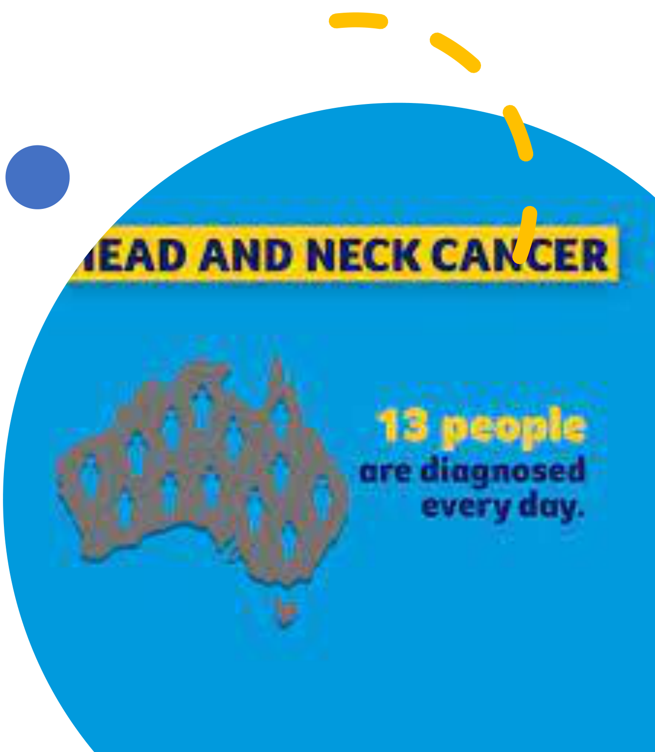
13 people are diagnosed every day.
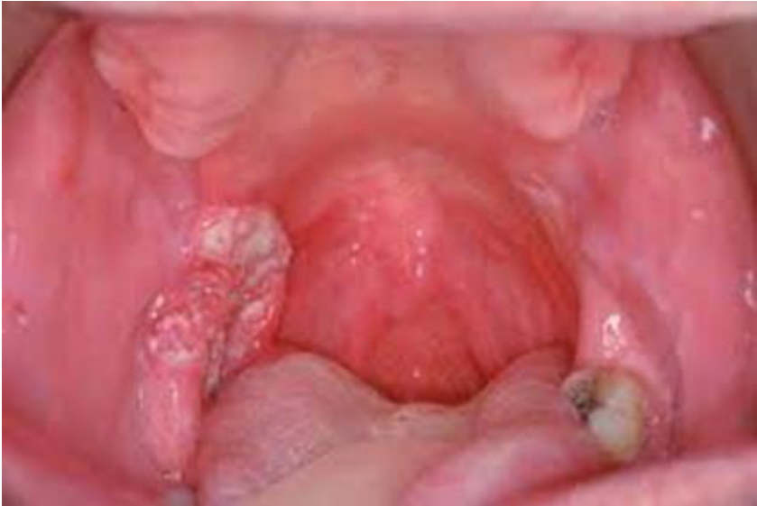
Early Diagnosis And Oral Cancer Screening4
Early diagnosis saves lives
 |  |  |
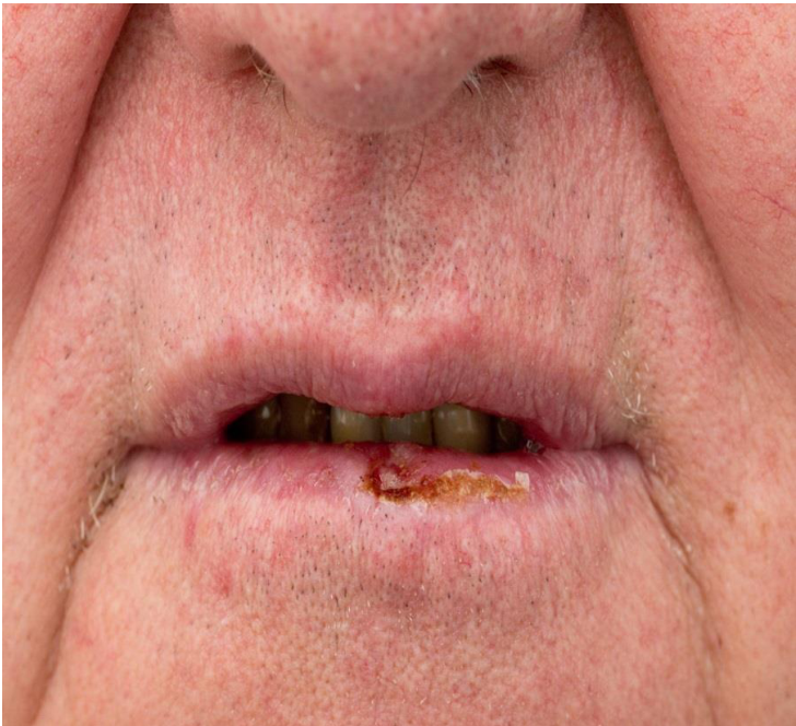 | 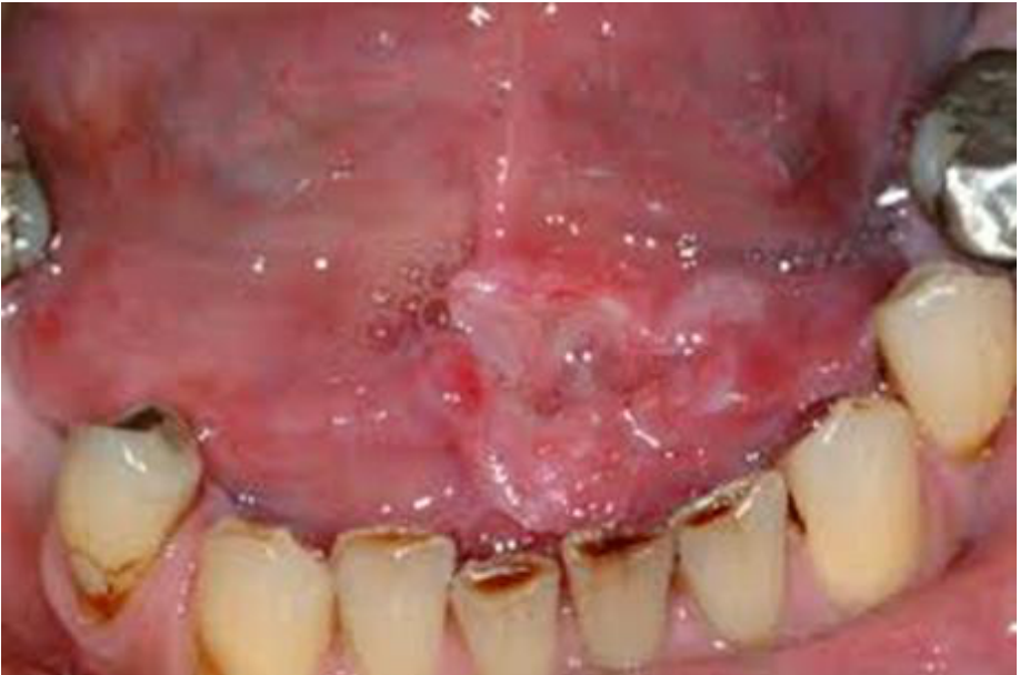 | 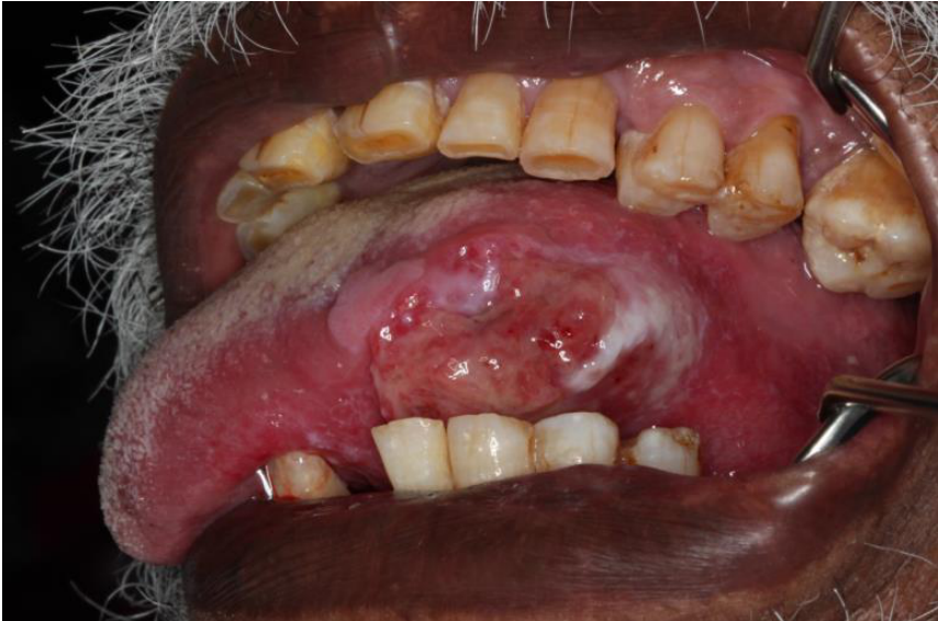 |
The 8-Step Oral Cancer Screening5
- A — Gingiva, mucosa
- B — Lip
- C — Buccal mucosa, posterior gingiva
- D — Vestibule, anterior gingiva, mucosa
- E — Hard palate
- F — Tonsillar area, soft palate
- G — Ventral surface of tongue, floor of mouth
- H — Lateral border of tongue
Signs And Symptoms6
-
Lump in the neck
-
Mobile teeth
-
Swelling or sore on the lip that won’t heal
-
Difficulty or painful swallowing
-
Changes in speech
-
Bleeding or numbness in the mouth
-
White or red patches on the mouth, tongue or gums
-
Unexplained weight loss
-
Changes in speech may be associated with posterior tongue cancers and an oral lump.
-
Teeth-related findings or symptoms may initially appear to be associated with another cause.
Investigations7
-
Biopsy — removal of a small amount of tissue which is sent to a pathologist to determine if cancer cells are present
-
Radiographs — OPG and chest x-ray
-
Imaging — PET, CT and MRI scans to determine the location, stage and spread of cancer
-
A flexible nasal endoscope is used to examine the nose, sinuses, larynx and pharynx
-
A biopsy is performed first to confirm the diagnosis.
-
CT and MRI may be used to assess possible bony involvement.
Clinical Staging TNM8
-
Used to designate the extent of disease and to determine the most appropriate treatment for patient
-
Uniform staging system provides comparisons of results of types of treatment
-
Helps predict treatment response and outcomes
-
T refers to the size of the tumour.
-
N refers to lymph-node involvement.
-
M refers to whether the tumour has metastasised.
-
Staging provides an indication of prognosis.
-
Staging may determine whether a patient qualifies for clinical trials.
-
Staging guides treatment planning.
Multidisciplinary Management
Multidisciplinary Team Meeting9
-
Every patient with oral cancer should be presented at a Multidisciplinary Team (MDT) Meeting
-
Integrated team approach to healthcare whereby medical and allied health professionals consider all relevant treatment options and collaboratively develop an individual care plan for each patient
-
Patients need to be evaluated before, during and after treatment
-
The team considers nutritional status and the effects of surgery on occlusion and swallowing
-
The possible need for radiotherapy or chemotherapy is considered
-
Quality of life and functional recovery are considered
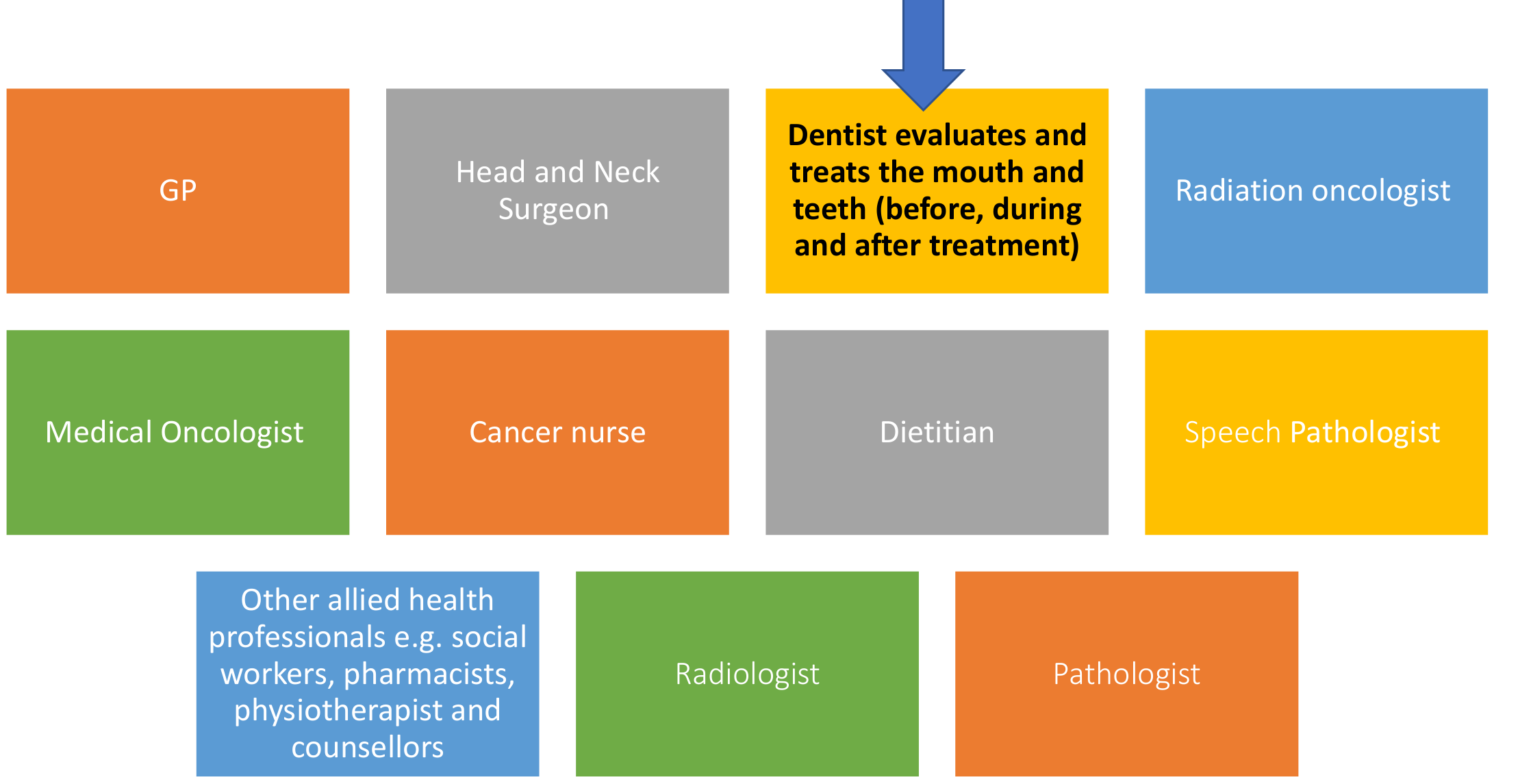
Team Members And Roles10
- GP
- Head and Neck Surgeon
- Dentist — evaluates and treats the mouth and teeth (before, during and after treatment)
- Dental professionals maintain remaining teeth, manage caries and infection, and support oral hygiene
- They address changes in occlusion and denture function and help patients regain oral function where possible
- Dental professionals maintain remaining teeth, manage caries and infection, and support oral hygiene
- Radiation Oncologist
- Medical Oncologist
- Cancer Nurse
- Dietitian
- Nutritional support may be required for severely malnourished patients, substantial weight loss, neglected tumours, or catabolic and anabolic phases during treatment
- A dietitian may assist with dietary modification
- Nutritional support may be required for severely malnourished patients, substantial weight loss, neglected tumours, or catabolic and anabolic phases during treatment
- Speech Pathologist
- Radiologist
- Pathologist
- Other allied health professionals, e.g. social workers, pharmacists, physiotherapists and counsellors
Treatment Modalities11
-
Curative versus palliative
-
Surgery — primary modality of treatment for oral cancer; removes tumour and lymph nodes
-
Adjuvant radiotherapy depends on pathological staging, surgical margins and nodal status
-
Radiotherapy used on its own to treat when organ preservation is a goal, e.g. oropharyngeal cancers; used concurrently with chemotherapy
-
Chemotherapy — standard of care in locally advanced head and neck SCC
-
Surgical treatment includes reconstructing the resulting defect
-
Relevant neck nodes are removed to assess for metastasis
Posterior and Anterior Tongue Tumours
A large posterior tongue tumour may be treated with radiotherapy to avoid removing substantial tissue and creating a debilitating defect.
- Surgery is generally used for anterior tongue tumours when the patient is fit enough.
Lecturer — Treatment Effects
Chemotherapy may be combined with radiotherapy or used when surgery is not possible. If disease is too advanced for surgery, treatment may be palliative and may involve chemotherapy.
- During chemotherapy, decreased red-cell counts may cause anaemia, decreased white-cell counts may increase susceptibility to infection, and decreased platelet counts may increase susceptibility to bleeding.
- During radiotherapy and chemotherapy, patients may experience altered taste, mucositis, opportunistic infections, and dry mouth.
Dental Management Of The Cancer Patient
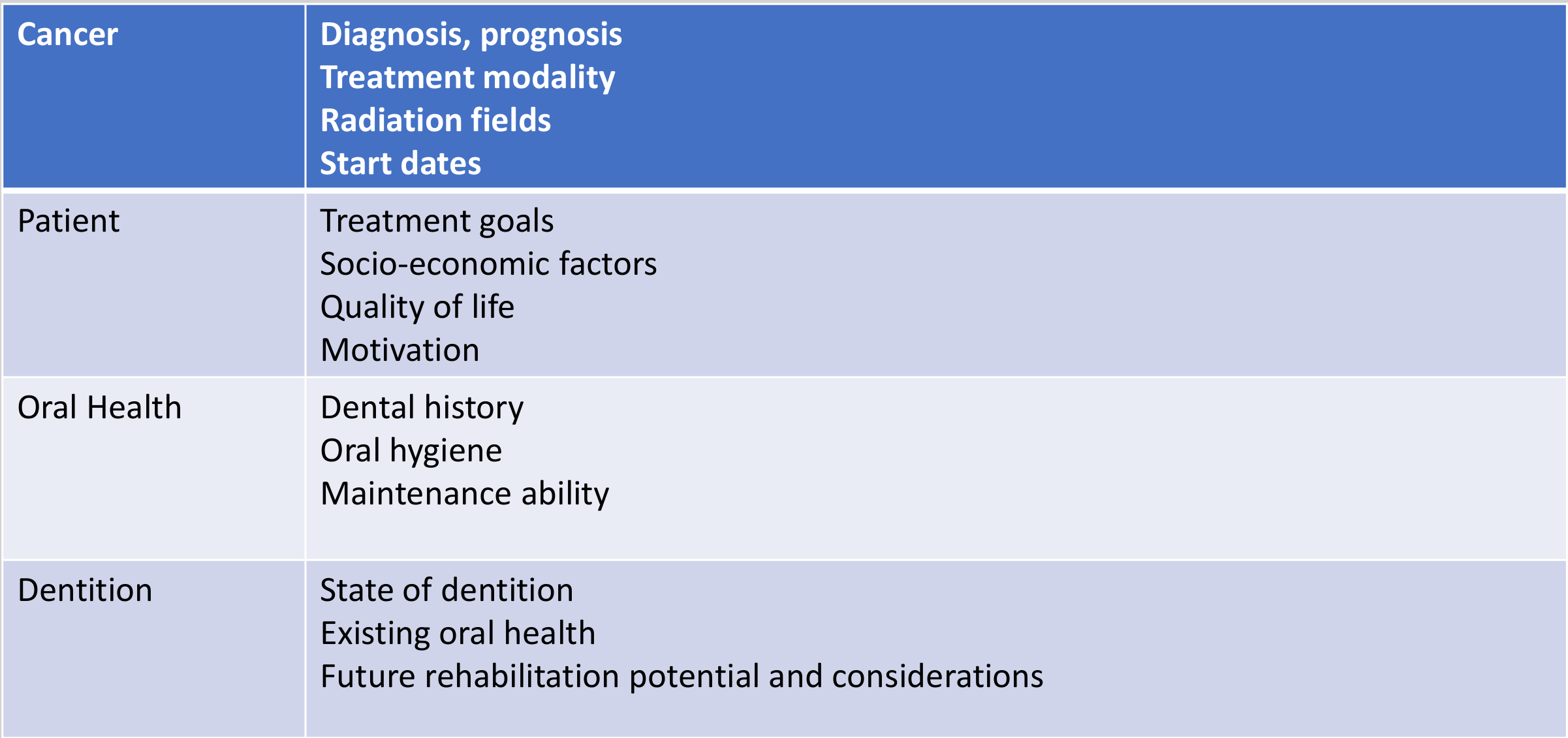
Pre-Treatment Dental Assessment12
| Domain | Assessment factors |
|---|---|
| Cancer | Diagnosis, prognosis Treatment modality Radiation fields Start dates |
| Patient | Treatment goals Socio-economic factors Quality of life Motivation |
| Oral Health | Dental history Oral hygiene Maintenance ability |
| Dentition | State of dentition Existing oral health Future rehabilitation potential and considerations |
Lecturer — Pre-Treatment Factors
- Assess the patient’s nutritional and general condition before treatment.
- Surgery may alter the anatomy, intraoral structure, bite, occlusion, swallowing, and ability to wear a denture.
- Patients may need nutritional support such as supplemental drinks or a soft diet.
- Teeth within the radiation field require particular consideration.
- Some patients require appointments more frequently than the usual six-month interval.
Care During Radiotherapy And Chemotherapy13
- Changes in taste
- Oral hygiene maintenance – mucositis
- Systemic considerations – pancytopenia, opportunistic infections
Lecturer — During Treatment
- Regular review, motivation and maintenance remain important during treatment.
- Patients may require review every three months or every two months.
- Supportive measures include high-fluoride toothpaste, saliva substitutes for dry mouth, management of altered occlusion, and early identification and management of caries.
- Dental care must be adapted to the patient’s changing anatomy and function.
Post-Treatment Dental Management14
- Regular review and maintenance
- Motivation
Lecturer — Post-Treatment Management
- Surgery may mean that a patient is no longer able to wear a denture.
- Treatment should focus on what is practical and beneficial for the individual patient rather than on an ideal treatment plan.
- Oral rehabilitation may include a modified denture, maintenance of remaining teeth, management of caries and infection, and implants where appropriate.
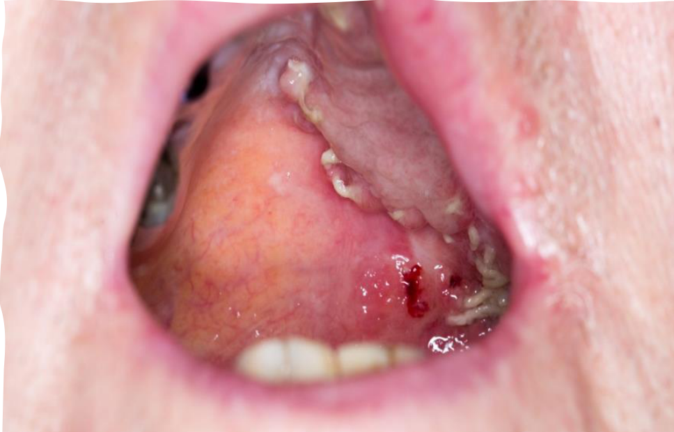
- Patients who have received radiotherapy require particular caution if extraction is being considered.
- Alternatives may include pulp extirpation for pain relief, more frequent maintenance, early management of opportunistic infections, and symptomatic water or other measures for severe dry mouth.
Recommendations For Dental Follow-Up1516
- Initially 3 monthly review for caries and periodontal management
- Once oral condition stabilize, 6 monthly recalls – look out for mucosal changes, xerostomia, candidiasis, changes in occlusion
- Fluoride at each recall – Neutrafluor 5000 toothpaste and fluoride varnish
Lecturer — Follow-Up Planning
- Follow-up should be more frequent for patients with dry mouth, increased caries risk, difficulty chewing, altered occlusion, recurrent infection, or a soft or semisoft diet.
- Dental professionals should continue supporting patients rather than referring them away solely because their care is complex.
- Follow-up should address oral function, including chewing, speaking, eating with family, avoiding drooling, and returning to work where possible.
Assessment And Outcomes
Imaging Assessment Of Mandibular Involvement17
Andreea Nae¹, Gerard O’Leary¹, Linda Feeley², Cassie Fives², Brendan Fitzgerald², Elena Chiriac³, Patrick Sheahan¹
World J Otorhinolaryngol Head Neck Surg 2019 Mar 22;5(2):71-75
The results suggest that combined CT and MRI have diagnostic utility in detecting mandibular invasion by oral cancer, but with significant false positive rate
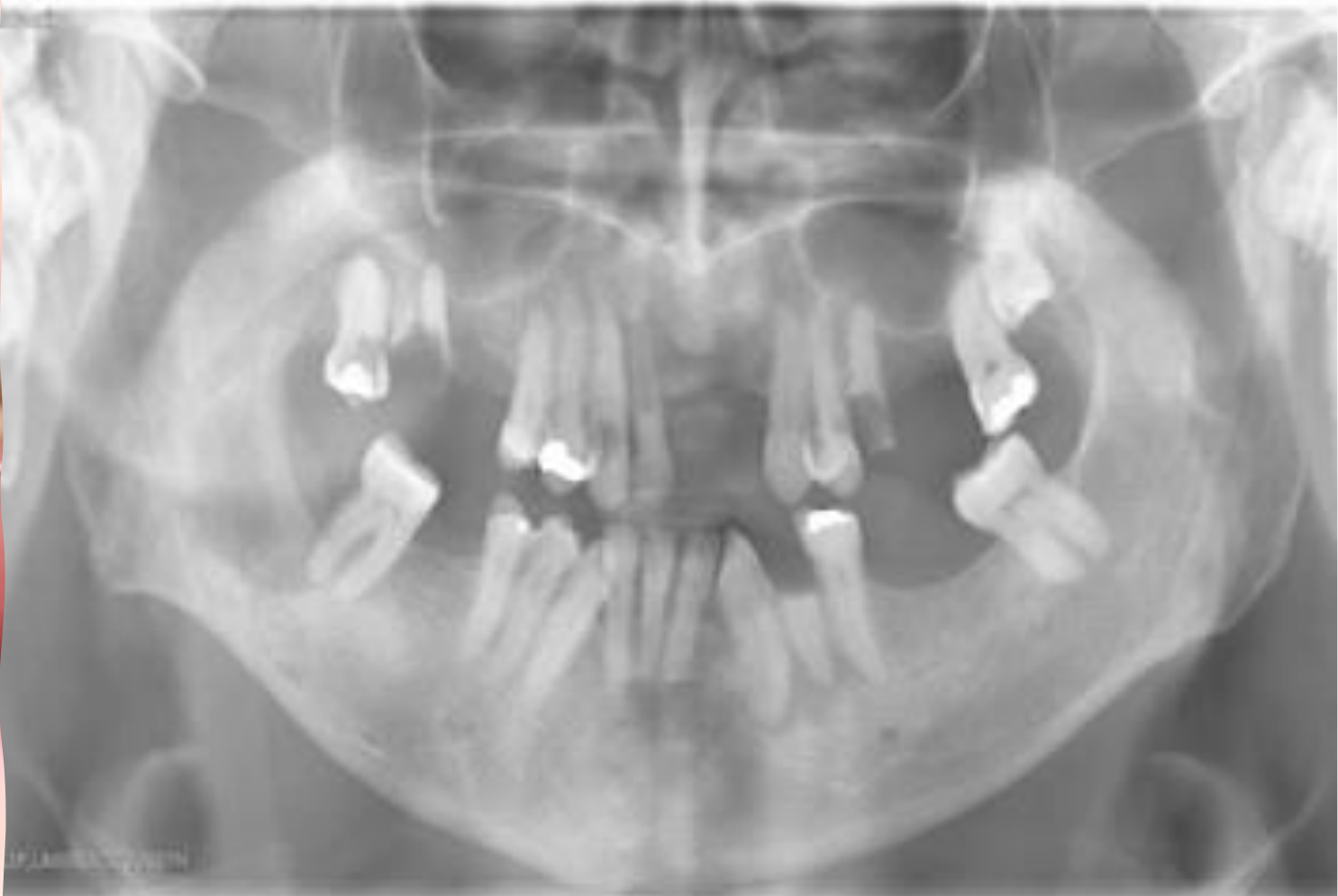
- An OPG may demonstrate invasion of the mandible.
- When bone is involved, reconstruction may require transfer of both bone and soft tissue.
Prognostic Outcomes18
-
Histological grading is a significant predictor for treatment failure and recurrence
-
Tumour size and lymph node status are also prognostic factors
-
Staging helps estimate prognosis and likely outcome.
-
Leaving tumour behind affects survival and may expose the patient to additional radiotherapy.
-
Radiotherapy cannot fully compensate for a close or positive surgical margin.
-
Functional outcomes are also important.
- Patients value the ability to chew.
- Patients value the ability to speak with family.
- Patients value the ability to eat socially.
- Patients value the ability to avoid drooling.
- Patients value the ability to return to employment.
Surgical Margins And Neck Dissection
Impact Of Surgical Margins On Outcome19
The Effect of the Surgical Margins on the Outcome of Patients with Head and Neck Squamous Cell Carcinoma
-
Consider margins of 5 mm and > as clear, 1–5 mm as close, and < 1 mm as involved
-
There is significant impact of positive surgical margins on the outcome in recurrence rate and overall survival
-
Adjuvant radiotherapy/chemotherapy cannot fully compensate for the close and positive margins
-
The surgeon generally removes what can be felt together with approximately an additional 5–10 millimetres.
-
A positive margin means that tumour remains at the resection margin.
Adequate Margin Of Resection20
Richard W Nason, Abdulaziz Binahmed, Kumar A Pathak, Ahmed A Abdooh, George K B Sándor. Oral Surg Oral Med Oral Pathol Oral Radiol Endod 2009 May;107(5):625-9.
Survival improves with each additional millimeter of clear surgical margin. This systematic evaluation of surgical margins suggests that an adequate resection in oral cancer should provide a margin of greater than 3 mm on permanent pathology section.
- An adequate margin was described as approximately 5 millimetres, aiming to clear the tumour while avoiding unnecessary removal of tissue.
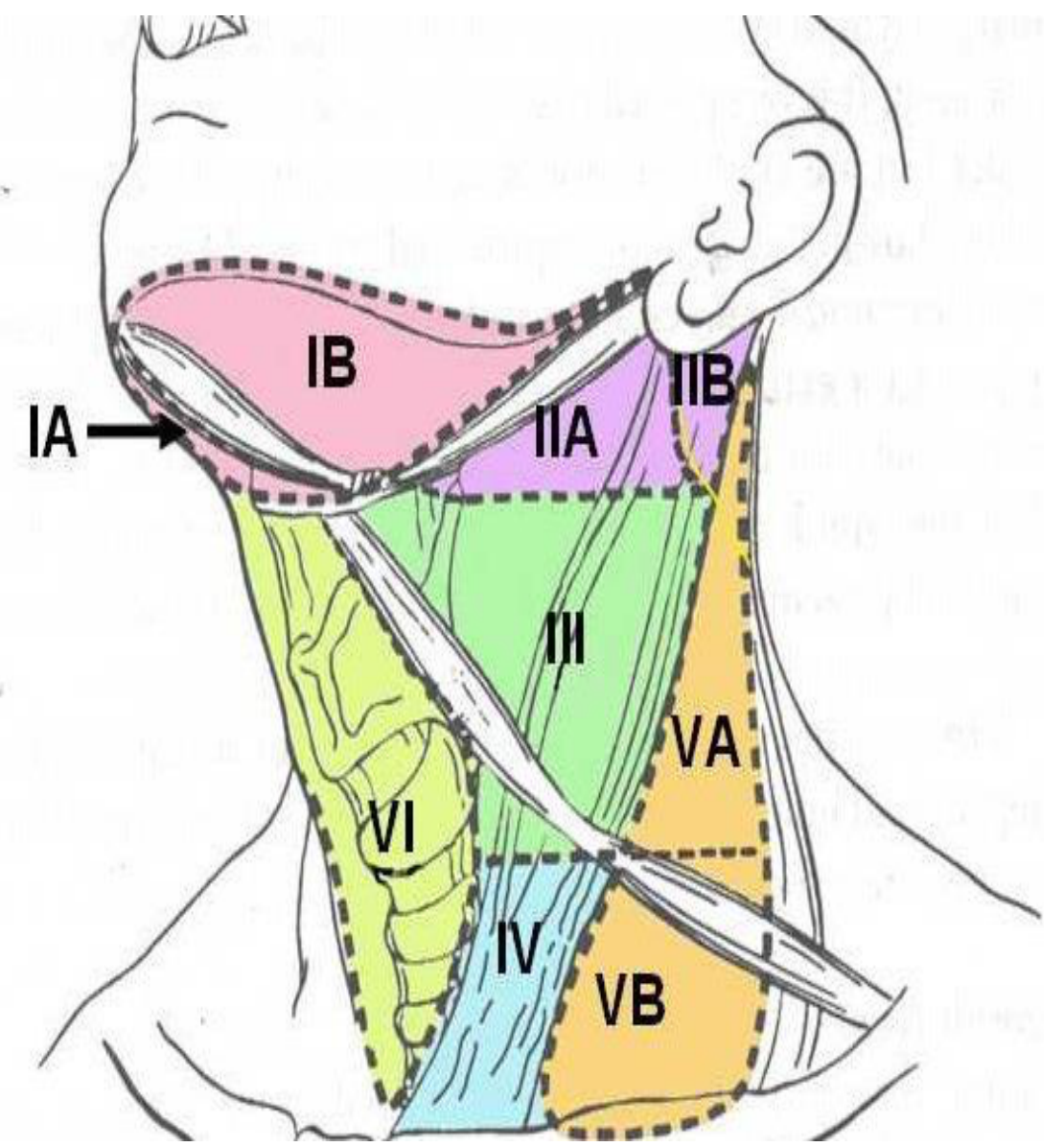
Neck Dissection Principles21
-
Radical neck first described by Crile in 1906, involved removal of all lymphatic structures between mandible and clavicles
-
Shah stratified cervical lymph nodes into 6 group levels
-
Currently, selective neck dissection with the preservation of non-lymphatic structures
-
Level 1 lies beneath the mandible, including the submandibular region.
-
Levels 2, 3 and 4 lie along the jugular digastric chain.
-
Level 1 should be palpated first because metastatic spread usually proceeds to level 1 before levels 2, 3 and 4 and only rarely skips levels.
-
Levels 5 and 6 were described as more relevant to ENT territory.
Ablation And Neck Dissection22
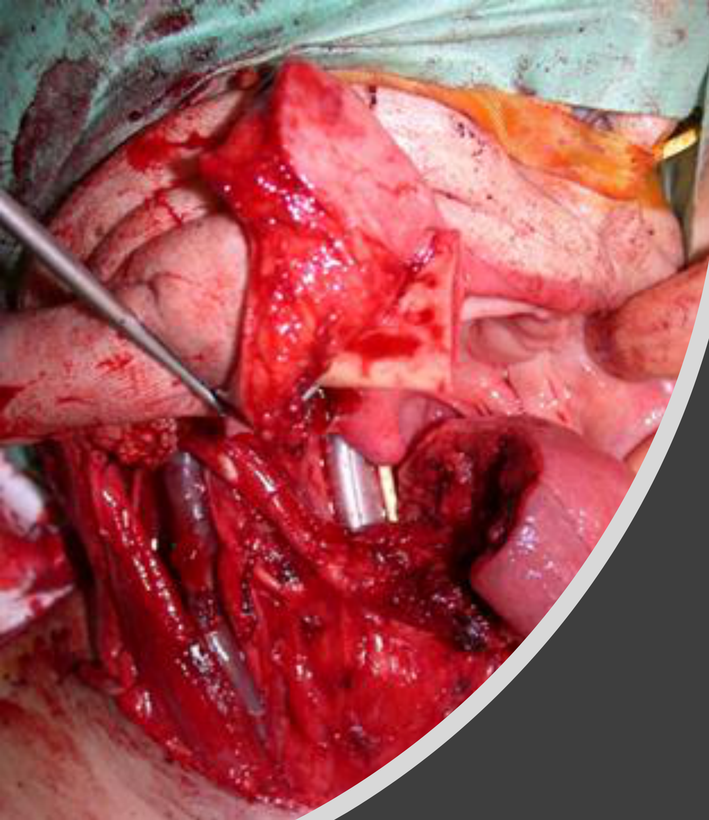
Surgery – ablation and neck dissection
Lecturer — Ablation and Reconstruction
Ablation removes the tumour, while neck dissection removes neck nodes to assess for metastasis.
- After tumour and nodal removal, the defect must be reconstructed with soft tissue or with bone and soft tissue together.
- The reconstructive choice depends partly on whether the tumour has invaded the maxilla or mandible.
Reconstruction Principles
Reconstruction Of Resection Defects23
Head and neck cancer
After resection, more than 50% of the resections require reconstruction of the defect.
This may involve soft tissue reconstruction or involve a bony reconstruction.
Maxillomandibular resection may involve a rim (marginal) or complete segment (segmental).
A marginal mandibulectomy can be reconstructed with local flaps or skin graft, whereas a segmental mandibulectomy requires an osteocutaneous flap.
Lecturer — Reconstruction Goals
Reconstruction is required after removal of the tumour and neck nodes, and may range from allowing the defect to heal to using a free flap or an osseocutaneous graft containing bone and soft tissue. The aim is to restore as much function as possible, not merely to close the defect.
Reconstruction Ladder
- COMPLEX
- Free Flaps
- Regional Flaps
- SIMPLE
- Local Flaps
- Skin Graft
- Primary Closure
- Secondary Healing
Lecturer — Reconstruction Consequences
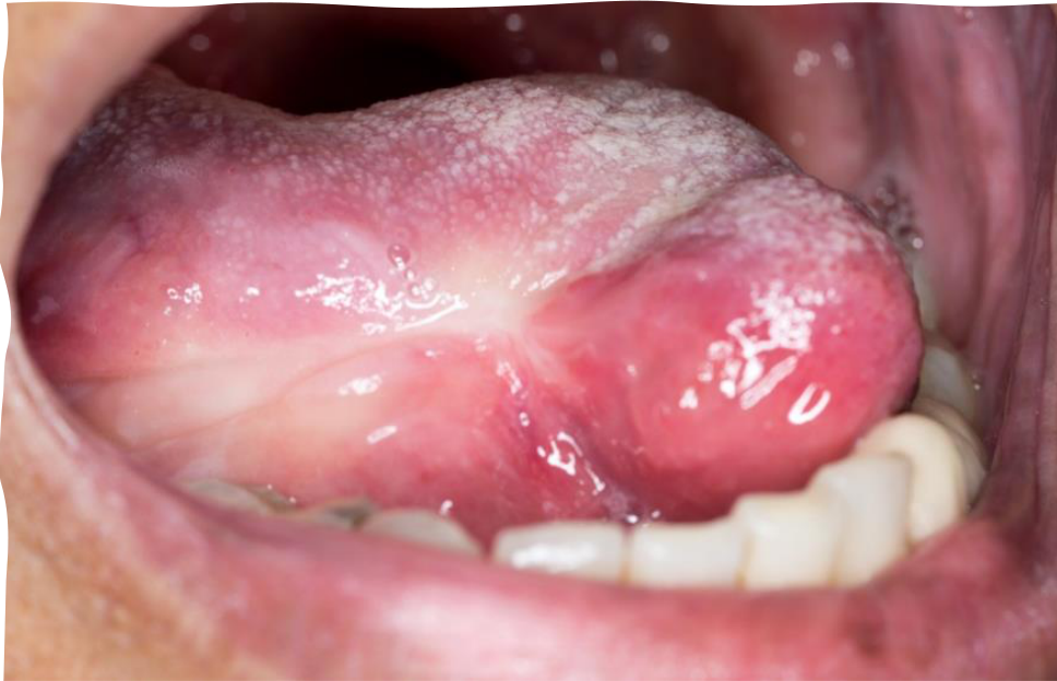
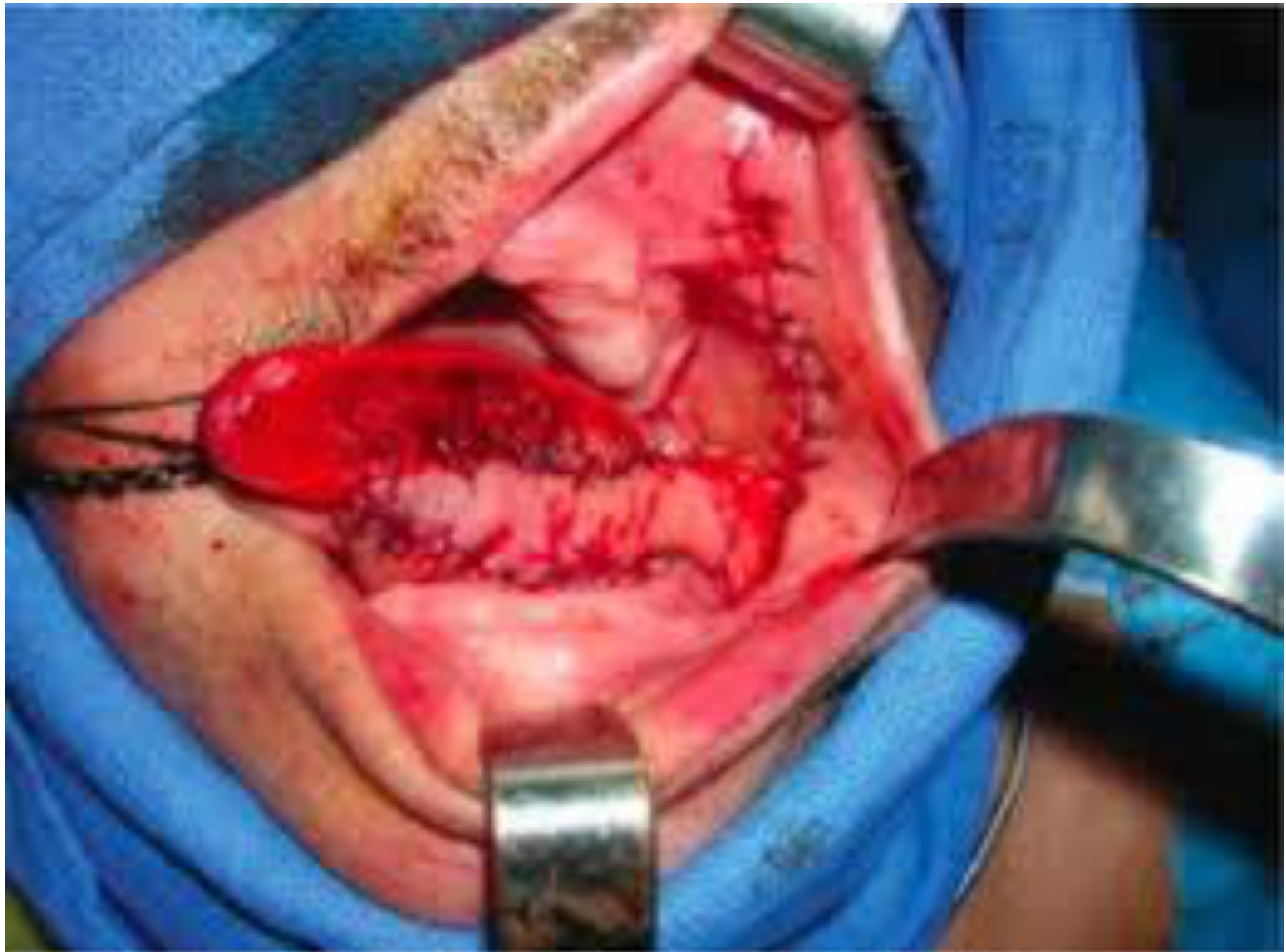
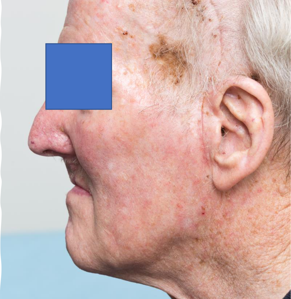
Even a small T1 tumour, described as less than 2 centimetres, may distort the tongue if the edges are pulled together, while larger defects usually cannot be closed simply by bringing the edges together. A local flap may be based on the facial artery, whereas a regional flap may use temporal muscle and skin tunnelled into the defect. These procedures may alter facial appearance, tissue colour, function and dental rehabilitation requirements.
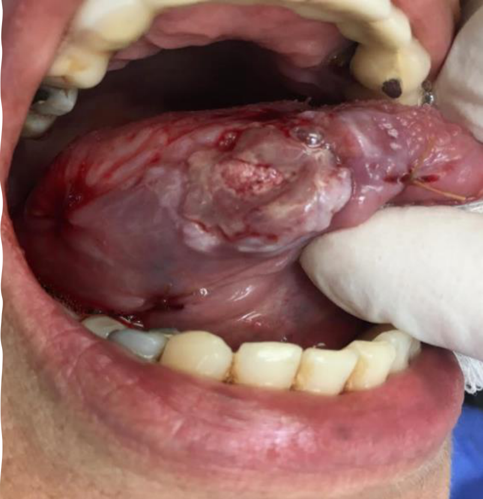
Reconstructive Options By Defect242526
T1 SCC lateral tongue
Primary closure
Local flap
Facial artery musculomucosal (FAMM) flap
Regional flap
Temporalis muscle flap
Lecturer — Defect Planning
Soft-tissue defects may be reconstructed with a soft-tissue graft or flap, whereas defects involving bone require transfer of bone as well as soft tissue. A scapular flap may be useful when there is soft-tissue involvement externally and a greater amount of soft tissue is required. The plan must consider the defect’s size and location, bone involvement, soft-tissue requirements, and the patient’s future ability to chew, speak and use a denture.
History Of Reconstruction
- Before the 1950s, resected cancer was left without reconstruction.
- In 1965, Bakamjian first described the deltopectoral flap.
- In 1973, Daniel and Taylor reported the first free flap, which involved removal of tissue from the donor site and transplantation to another site of the body with anastomosis of the vessels.
- In 1976, Panje and Harashina simultaneously described the use of free flaps to reconstruct defects of the oral cavity.
- In 1979, Ariyan described the pedicled pectoralis major myocutaneous flap.
- In the 1990s, the free flap was the dominant technique for cancer defect reconstruction. The use of osteocutaneous free flaps to reconstruct mandibular defects was advanced.
Poor Reconstruction With Functional And Aesthetic Problems
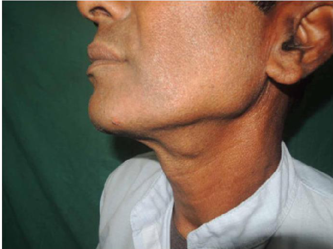
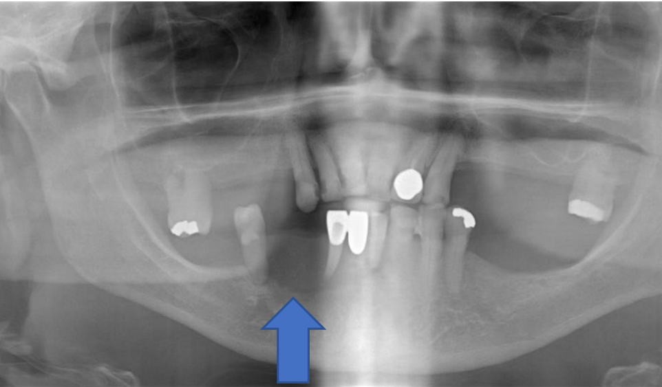
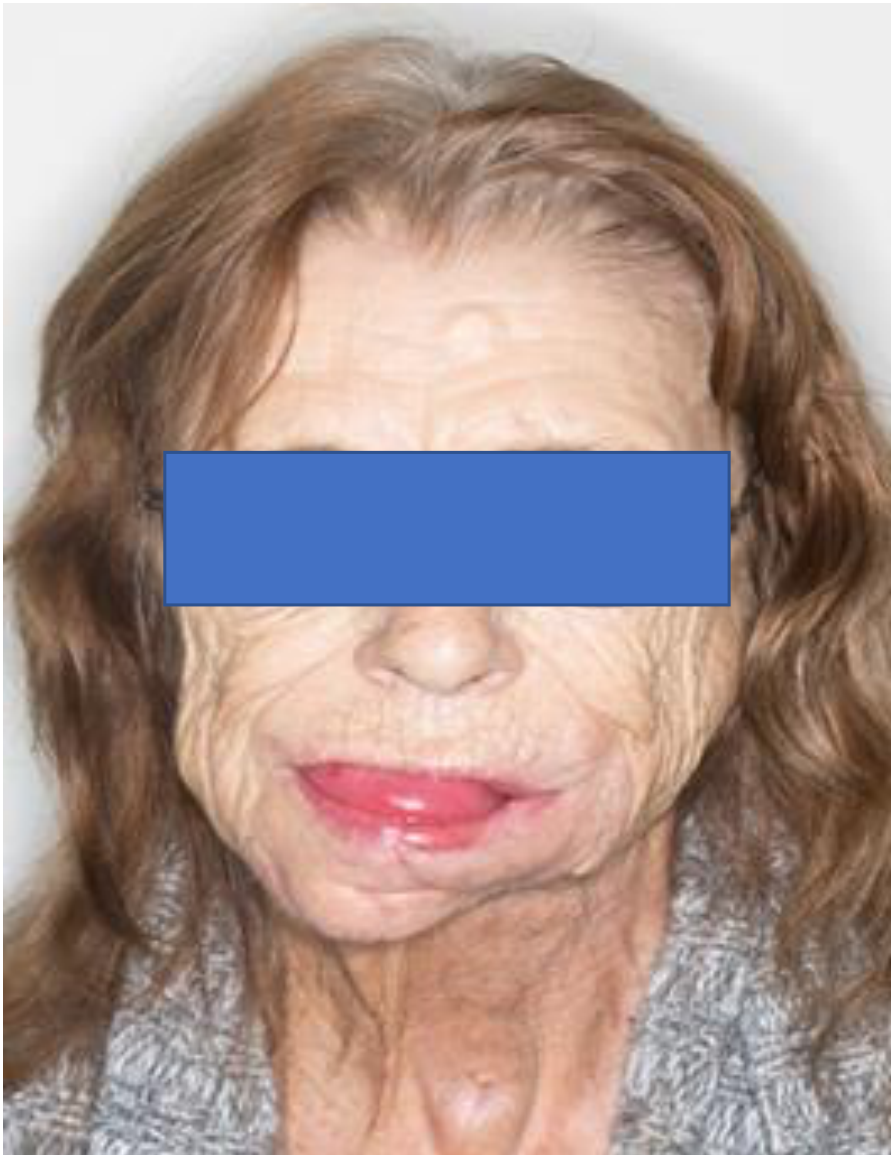
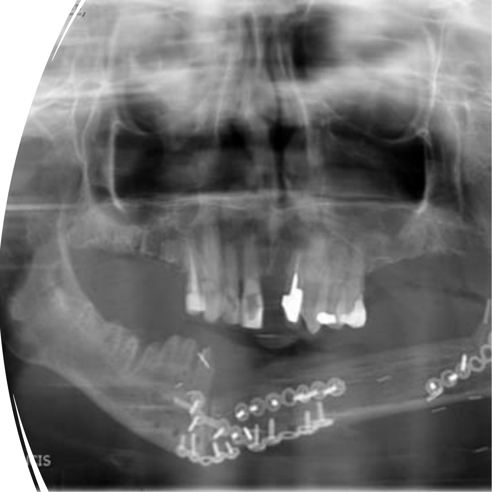
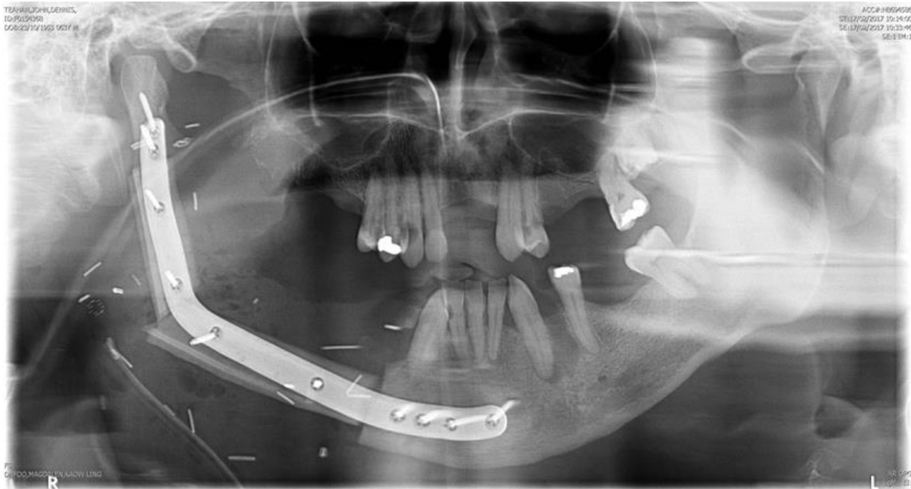
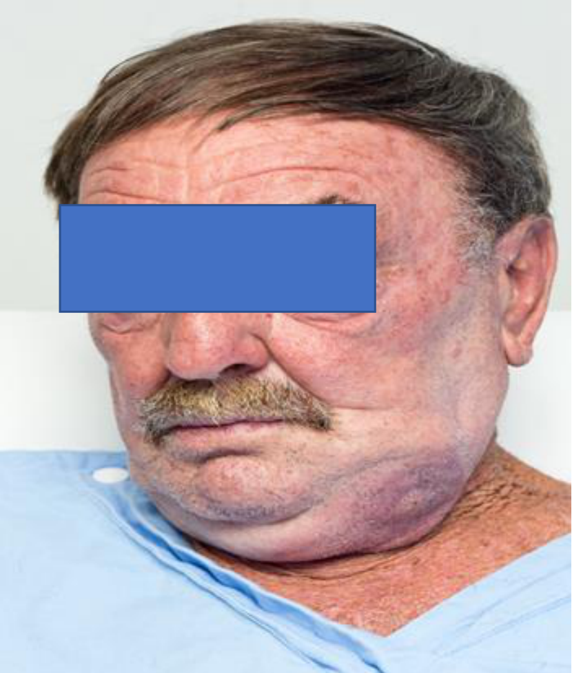
One patient had a poor reconstruction performed elsewhere: the face and chin point were displaced to one side, and the patient could not bring the teeth together. She reported that she would rather be dead than live through the consequences.
- A fibular flap had not been properly osteotomised.
- Multiple miniplates were used, but the reconstruction did not provide an effective result.
- The occlusion was severely compromised.
- A lower denture could not be placed effectively or function properly.
- The case demonstrated that cancer treatment involves more than removing the tumour: reconstruction must be planned carefully, function restored as far as possible, and dental rehabilitation considered from the beginning.
Jaw Reconstruction And Surgical Planning
Challenges In Jaw Reconstruction27
Malposition of the bone graft can negatively affect:
- facial symmetry
- appearance
- support
- occlusal function
- masticatory movement
- dental rehabilitation
Lecturer — Functional Reconstruction
Jaw reconstruction must address both tissue replacement and functional restoration.
- The position of the bone affects the chin point, occlusion, denture placement, chewing, and overall facial appearance.
- Patients may have to live with a semisoft diet if chewing cannot be fully restored.
- Reconstruction should be planned with the patient’s future oral rehabilitation in mind.
Inferior Jaw Reconstruction Pitfalls28
Pitfalls in inferior jaw reconstruction:
- Incorrect positioning of the fibular free flap
- Inadequate fixation of the bone graft
Lecturer — Reconstruction Positioning
Poorly positioned or inadequately osteotomised bone can displace the chin point and compromise occlusion. The condyle must also be positioned correctly, and the fixation plate must hold the reconstruction together effectively.
Three-Dimensional Surgical Planning29
3D printing was first described by Hideo Kodama in 1981, when he started manufacturing 3D plastic models.
Surgical planning was improved, which facilitated resection, flap harvesting, and graft positioning.
Lecturer — Simulation Goals
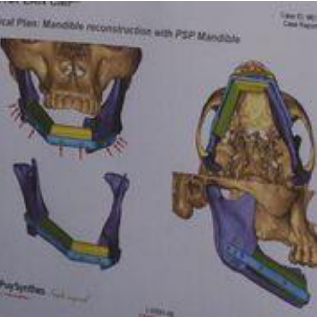
Surgical simulation and planning determine where to make the resection cuts, where to cut the harvested graft, where to perform osteotomies, and how to position the graft accurately. The aim is to make the reconstructed bone fit the defect appropriately.
Benefits Of Digital Planning30
- Increased accuracy
- Improved bone-to-bone contact
- Decreased operating room time
- Lower surgeon stress
- Reduced ischemia time in the microvascular free flap reconstruction
- Enhanced treatment outcome
Lecturer — Ischaemic Time
Ischaemic time refers to the period when the flap is detached from its artery and blood supply before blood flow is restored. The flap should be reconnected and reperfused as quickly as possible.
Case Planning Mandibular SCC31
Mandibular SCC
Pre-Surgical Assessment And Imaging32
Mandibular Invasion and Reconstruction
Imaging showed that the tumour had invaded bone, so reconstruction required bone as well as soft tissue.
- The plan addressed resection of the involved mandible and harvesting and positioning of replacement bone.
- In another case, a lesion extending from approximately tooth 2 to tooth 2 required resection of part of the mandible.
- Future restoration had to be considered, including a soft-tissue graft alone, denture rehabilitation, or possible implant-supported function.
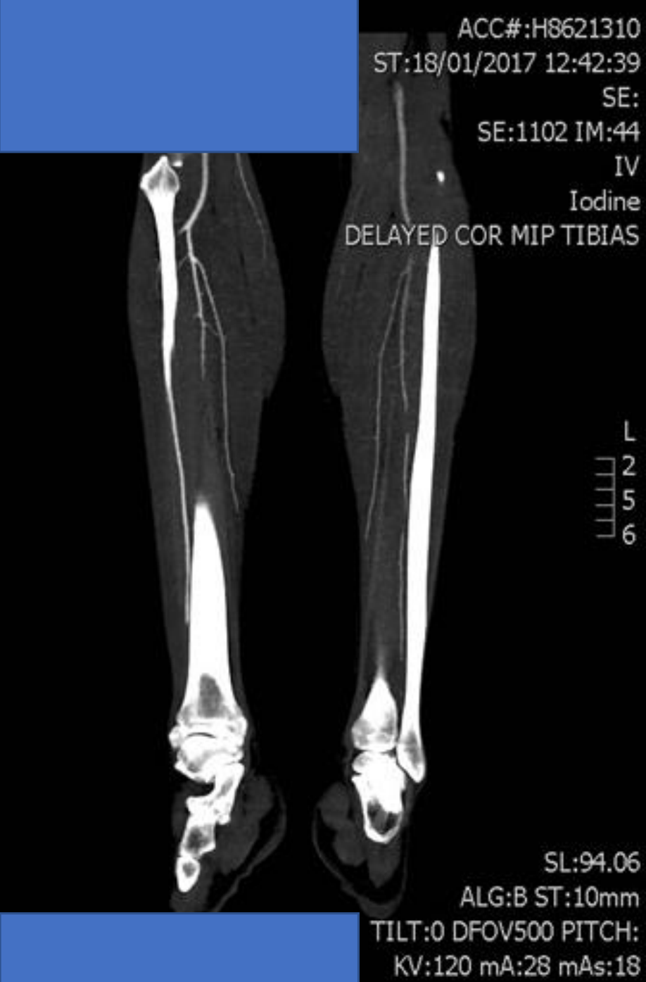
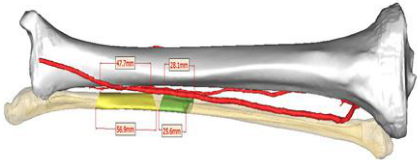
Pre-surgical assessment with angiography.
Lecturer — Pre-Surgical Assessment
Assessment should confirm that the blood supply to the selected flap is adequate. Imaging is also used to evaluate bony invasion and guide the extent of resection, so reconstruction is planned before surgery rather than improvised after tumour removal.
Planning The Resection33
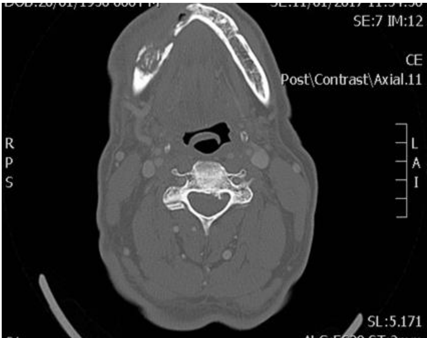
Planning the resection is based on preoperative high-resolution computed tomography (CT).
Lecturer — Resection Planning
Resection planning includes the tumour margins, the mandibular segment to be removed, the required bone graft, the amount and position of soft tissue, and the future position of the chin point and condyle. The aim is to achieve tumour clearance while preserving the possibility of oral rehabilitation.
Surgical Simulation And Templates343536
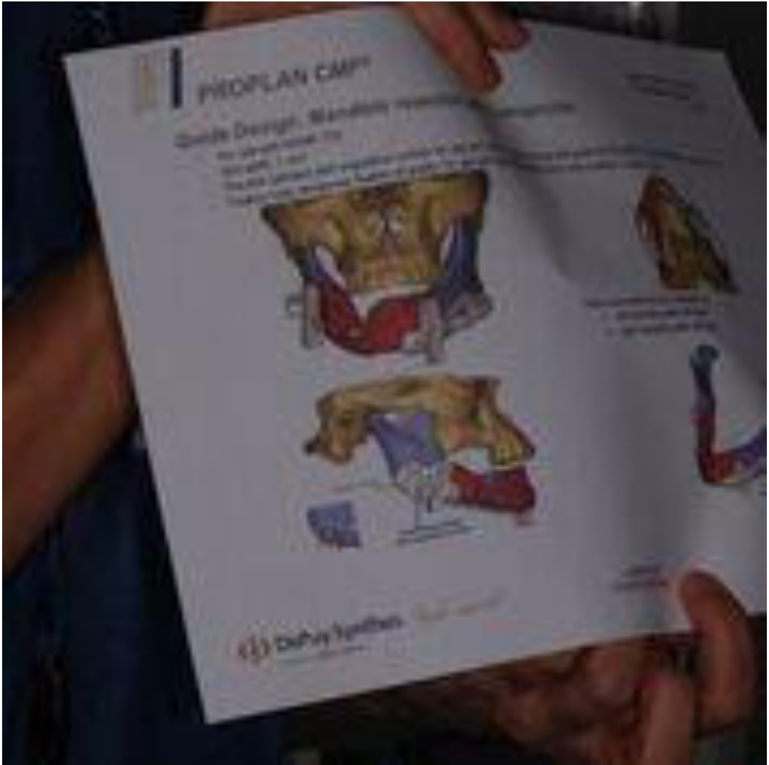
Surgical simulation and planning
3D printed surgical template fabricated to guide the procedure of tumour resection.
Surgical planning
 |  |
 |
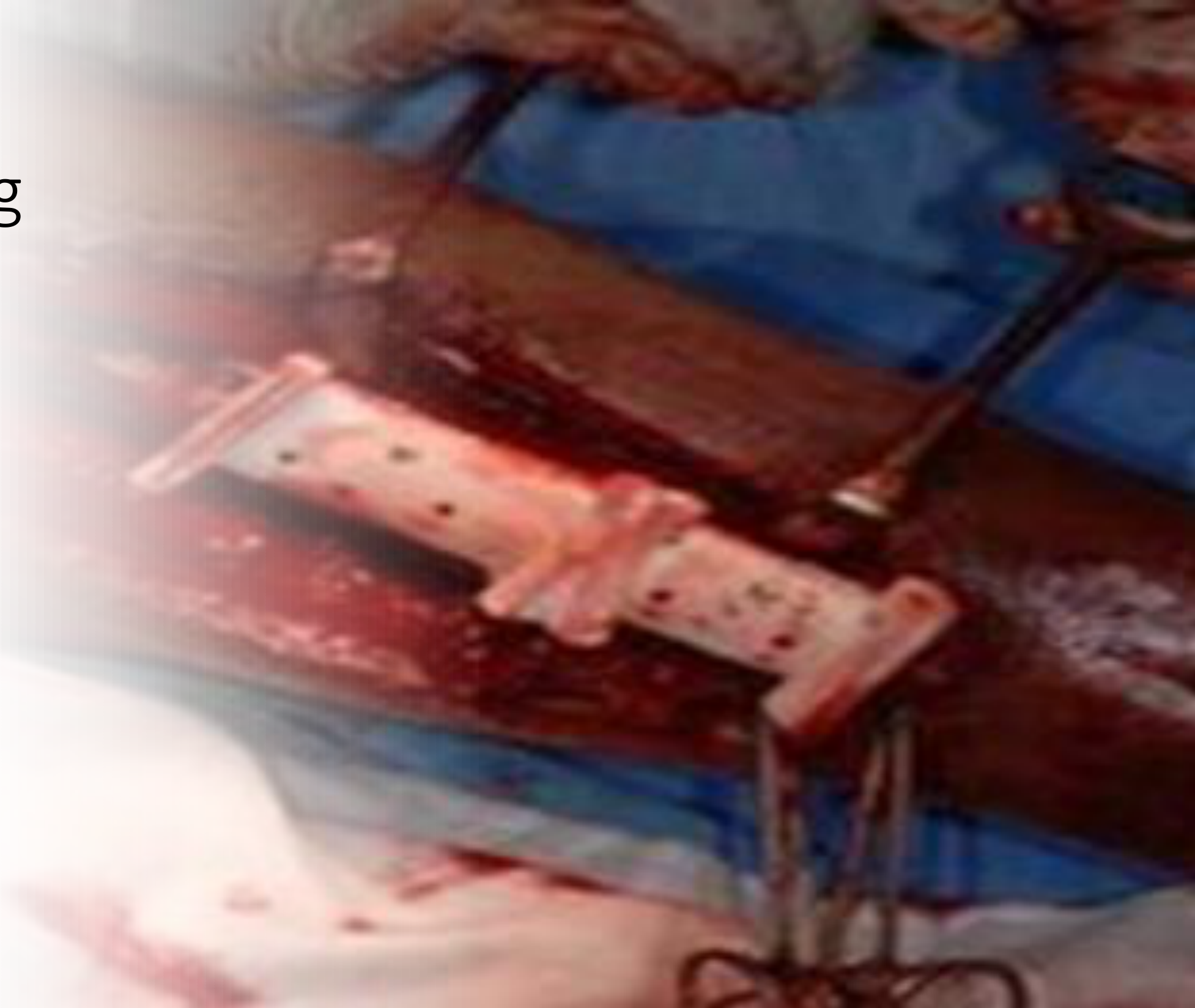
Graft Harvesting3738
Graft harvesting with a surgical template for osteotomies.
Lecturer — Soft-Tissue Requirements
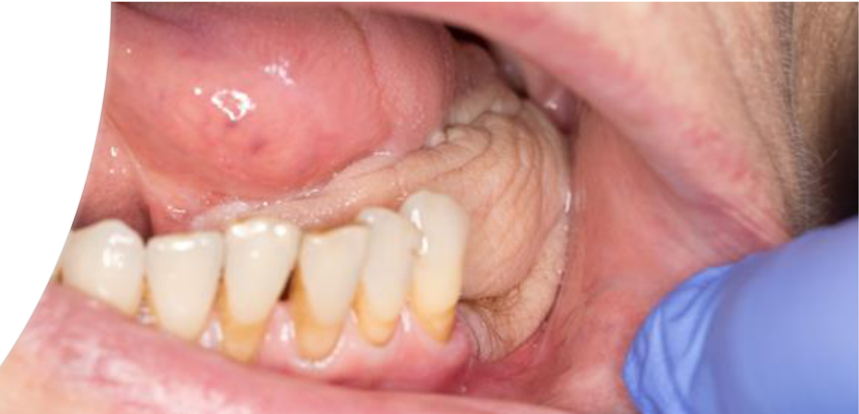
Grafts may contain soft tissue alone or bone and soft tissue, and the selected graft must provide enough tissue for the defect. In a ventral tongue reconstruction, sufficient excess soft tissue is required because tissue should not be pulled tightly across the defect; a donor-site scar may result, and a thigh graft may be considered when the donor site is a particular concern.
 |  |
Case Examples And Flap Techniques
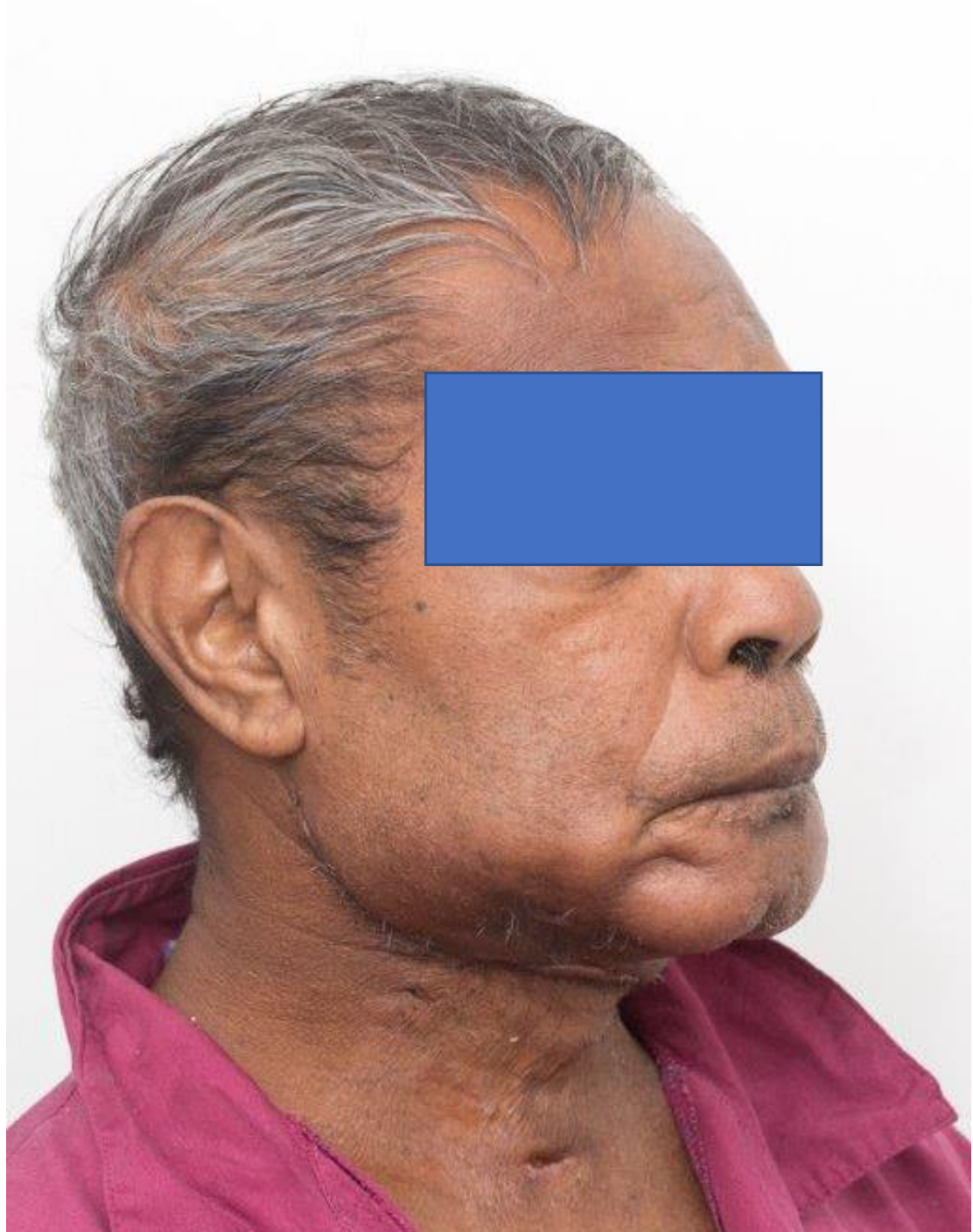
Post-Operative Results39
Post-op photo
Improved Mandibular Reconstruction
A better reconstruction demonstrated correct positioning of the chin point, bone osteotomised in the appropriate locations, a reconstruction plate holding the bone in position, and a correctly located condyle.
- The patient could chew on some of the remaining teeth and retain some oral function.
- This contrasted with the earlier reconstruction, in which the chin point, occlusion, and condyle were incorrectly positioned.
Scapular Flap40
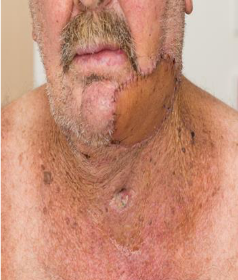 |  |
Radial Forearm Free Flap Reconstruction414243
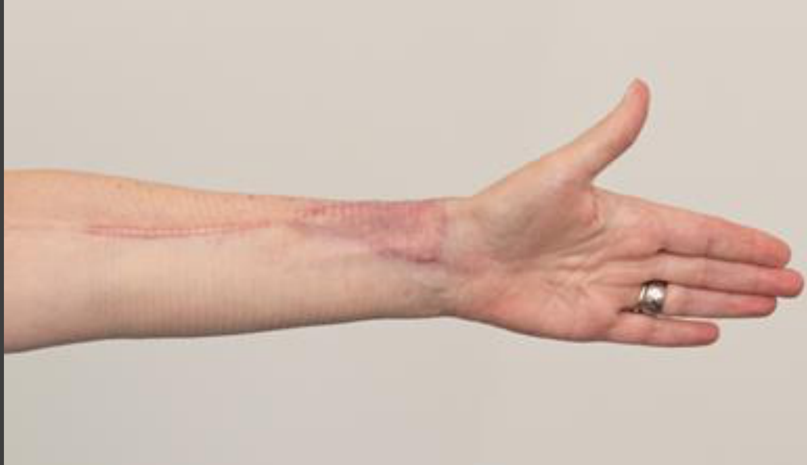
Pre-op assessment — Allen’s test
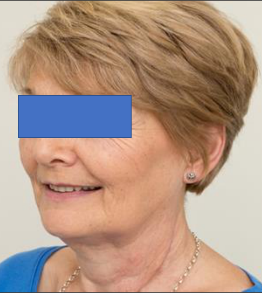
Marginal mandibulectomy
Radial forearm free flap reconstruction
Lecturer — Free Flap Tension
When soft tissue is transferred, it must be positioned with adequate excess so that it is not under excessive tension. The functional result should be considered alongside the appearance of the donor-site scar.
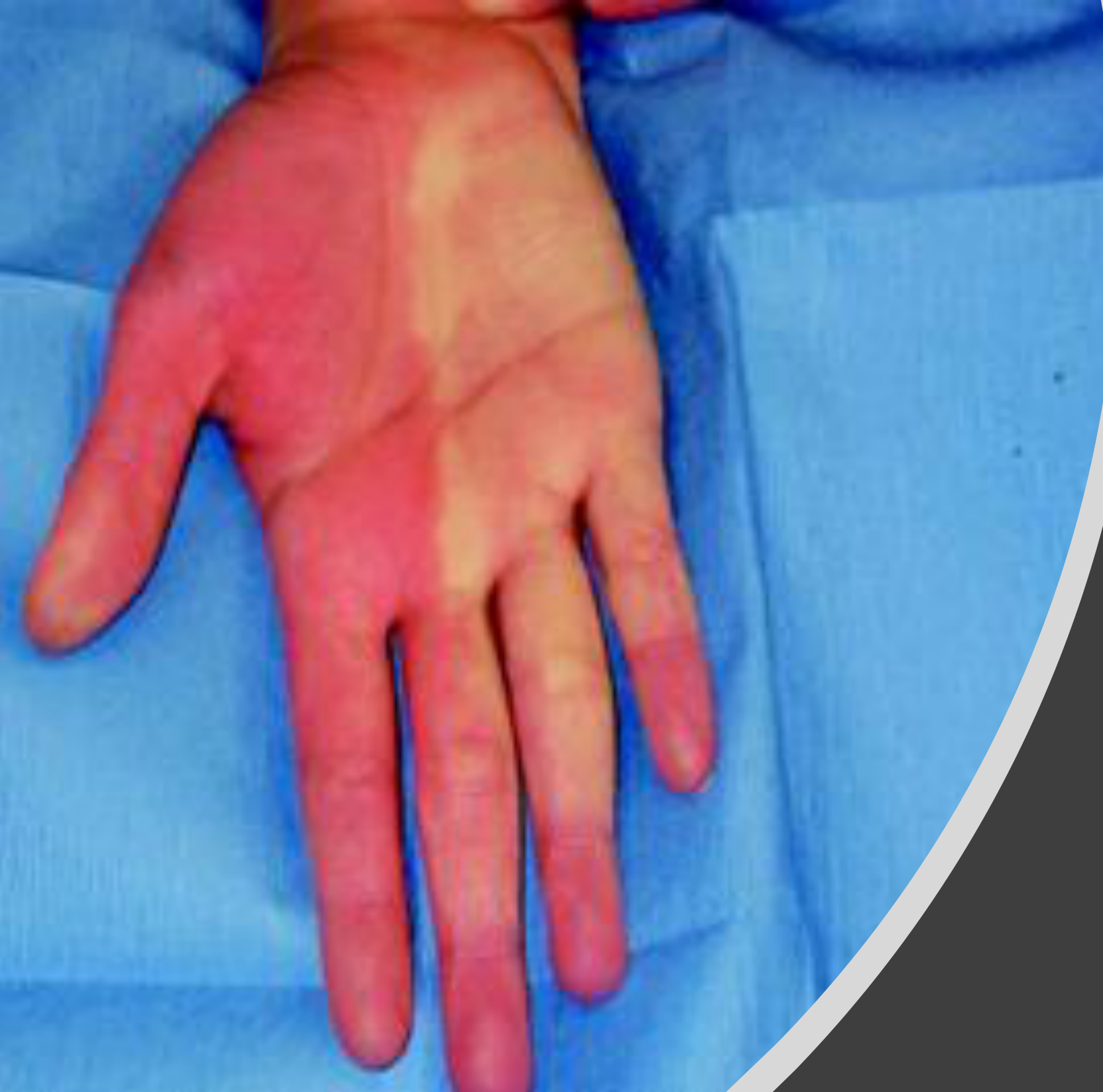
Reconstruction And Oral Rehabilitation44
Lecturer — Oral Rehabilitation
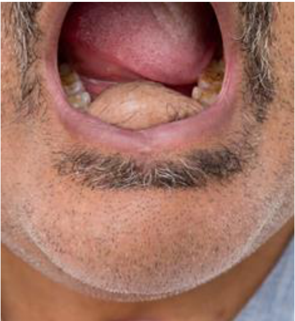
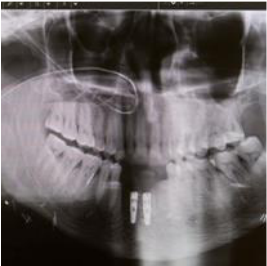
Reconstruction should facilitate chewing, speaking, swallowing, denture use where possible, and social eating. Bone implants may improve chewing function, but a denture may not fit after surgery and some patients may never be able to use one again.
- Treatment differs between patients and may include a modified denture, maintenance of remaining teeth, soft-tissue or bone reconstruction, implants, a modified or semisoft diet, and more frequent oral hygiene and maintenance care.
 |  |
Quality Of Life
Definition Of Quality Of Life4546
- Individual’s perception of their position in life in the context of the culture and value systems in which they live and in relation to their goals, expectations, standards and concerns
Lecturer — Functional Quality of Life
Quality of life is strongly linked to function after treatment.
- Important concerns include whether patients can chew, speak, eat with their family, avoid drooling and return to work.
- Patients who cannot speak or eat, or who drool continuously, may be unable to return to their jobs and may need social-service assistance, including financial help.
- Quality of life must be considered throughout cancer treatment and reconstruction.
QOL
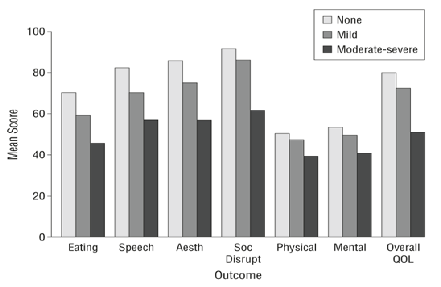
University Of Washington Quality Of Life Questionnaire47
- Pain
- Appearance
- Activity
- Recreation
- Swallowing
- Chewing
- General questions
- a) Health Related QoL month before developing cancer
- b) Health Related QoL during past 7 days
- c) Overall QoL
Quality Of Life By Treatment Modality48
- The 2 issues most important to patients were activity and mood
- A greater proportion of those who received adjuvant radiotherapy rated QoL as worse than at diagnosis compared to those who received primary surgery only — this group experienced more pain and poorer swallowing
Lecturer — Treatment Complications
Treatment may reduce quality of life through changes in anatomy and oral function.
- Potential problems include altered anatomy, impaired occlusion, difficulty chewing or speaking, dry mouth, increased caries, mucositis and opportunistic infections.
- Patients may be unable to wear a denture or may require a semisoft or modified diet.
- Technically unsuccessful reconstruction may severely affect facial appearance and function, while radiotherapy and chemotherapy may add oral complications requiring ongoing dental care.
Applying Quality Of Life Data49
- Health related QoL in treatment planning, and more personalised follow-up support
- Results can be invaluable when counselling patients with a new diagnosis
Lecturer — Individualised Support
Treatment should be individualised, with the dental and surgical teams considering what is realistic and beneficial for each patient.
- Support may include diet modification with dietetic input, management of dry mouth, prevention and treatment of caries, early identification of infection and maintenance of the remaining teeth.
- Modified dentures, consideration of implants and frequent follow-up may also be needed.
- The goal is to help patients participate in family life, eating and employment as much as possible.
Questions50
Questions?
Clinical Cases
Case: Small T1 tongue tumor, primary closure
Question
Scenario: A patient with a small T1 tongue tumor (less than 2 cm) requires surgical resection, and the team is considering primary closure of the defect.
What’s shown: Clinical photographs showing the lateral aspect of the tongue with a small tumor, and the appearance of the tongue when the surgical edges are pulled together.
Consider: What happens to the tongue when the edges of a small tumor resection are pulled together, and what does this demonstrate about closing larger defects?
Answer
Observations:
- The tumor is small (T1, <2 cm).
- Pulling the edges of the resection together causes significant distortion of the tongue.
Reasoning: The lecturer points out that even with a small T1 tumor, pulling the edges together distorts the tongue. This demonstrates that for larger tumors, the edges cannot simply be pulled together. In this specific instance, the edges were pulled together to keep the procedure simple, potentially because the patient was not fit for more complex reconstruction, but the resulting distortion is evident.
Outcome: The defect was closed by pulling the edges together, which resulted in noticeable tongue distortion.
Takeaway: Even small T1 tongue tumors cause significant distortion when closed primarily, indicating that larger defects cannot be managed by simply pulling the soft tissue edges together.
Case: Elderly patient with poor mandibular reconstruction
Question
Scenario: An elderly patient presents after having a mandibular reconstruction performed elsewhere.
What’s shown: A frontal clinical portrait and a panoramic radiograph.
Consider: Identify the errors in the reconstruction and their impact on the patient’s function and quality of life.
Answer
Observations:
- The frontal portrait shows the face pushed to one side and the chin point deviated.
- The panoramic radiograph shows a fibular flap that was not osteotomized properly, multiple mini plates, a displaced chin point, and disrupted occlusion.
Reasoning: The fibular flap was not shaped (osteotomized) correctly to match the mandibular contour, and the use of multiple mini plates failed to stabilize it properly. This resulted in a displaced chin point and a disrupted occlusion, making it impossible to achieve a functional bite or fit a lower denture.
Outcome: The patient cannot get her teeth together, cannot wear a lower denture, and has a severely reduced quality of life, stating she would rather be dead.
Takeaway: Poorly planned and executed mandibular reconstruction, such as failing to properly osteotomize a fibular flap, leads to severe aesthetic and functional deficits, emphasizing the critical need for precise surgical planning.
Case: Mandibular squamous cell carcinoma with 3D planned reconstruction
Question
Scenario: A patient with mandibular squamous cell carcinoma (SCC) that has invaded the bone, requiring resection and reconstruction.
What’s shown: Pre-operative panoramic radiograph, 3D medical visualization, surgical templates, post-operative panoramic radiograph, and a post-operative lateral profile clinical photograph.
Consider: How does 3D surgical planning and templating guide the reconstruction to improve functional outcomes?
Answer
Observations:
- The pre-operative OPG shows the tumor invading the bone.
- 3D visualization and surgical templates are used to plan the resection and guide the osteotomy of the graft.
- The post-operative OPG shows the reconstruction plate holding the bone together, with the chin point and condyle in the correct anatomical positions.
- The post-operative clinical photo confirms the chin point is correctly aligned.
Reasoning: By using 3D printing and surgical templates, the surgeon can precisely plan the resection and accurately osteotomize the graft to fit the defect. This ensures the chin point and condyle are correctly positioned, avoiding the errors seen in freehand reconstructions.
Outcome: The patient has a correctly aligned chin point and condyle, and is able to chew on some teeth, preserving function.
Takeaway: 3D surgical planning and templating increase accuracy in mandibular reconstruction, ensuring proper graft osteotomy, bone-to-bone contact, and restoration of form and function.
Case: Ventral tongue defect reconstructed with a soft tissue flap
Question
Scenario: A patient requiring soft tissue reconstruction for a defect involving the ventral tongue and lingual soft tissue.
What’s shown: A panoramic radiograph of the maxillofacial region and an intraoral close-up clinical photograph of the reconstructed area.
Consider: What is the key technical principle for soft tissue reconstruction in the lingual area to ensure natural form?
Answer
Observations:
- The intraoral photo shows the reconstructed ventral tongue and lingual soft tissue.
Reasoning: When reconstructing the ventral tongue, the soft tissue flap must not be pulled tight across the tissues like a tent. Instead, excess soft tissue must be included so that it “falls nicely” and recreates the natural lingual sulcus, rather than pulling across to the cheeks.
Outcome: The soft tissue is plumped in with adequate excess to mimic natural oral anatomy.
Takeaway: Soft tissue reconstruction of the ventral tongue requires adequate tissue excess to allow it to fall naturally and maintain the lingual sulcus, rather than being tensioned tightly across the mouth.
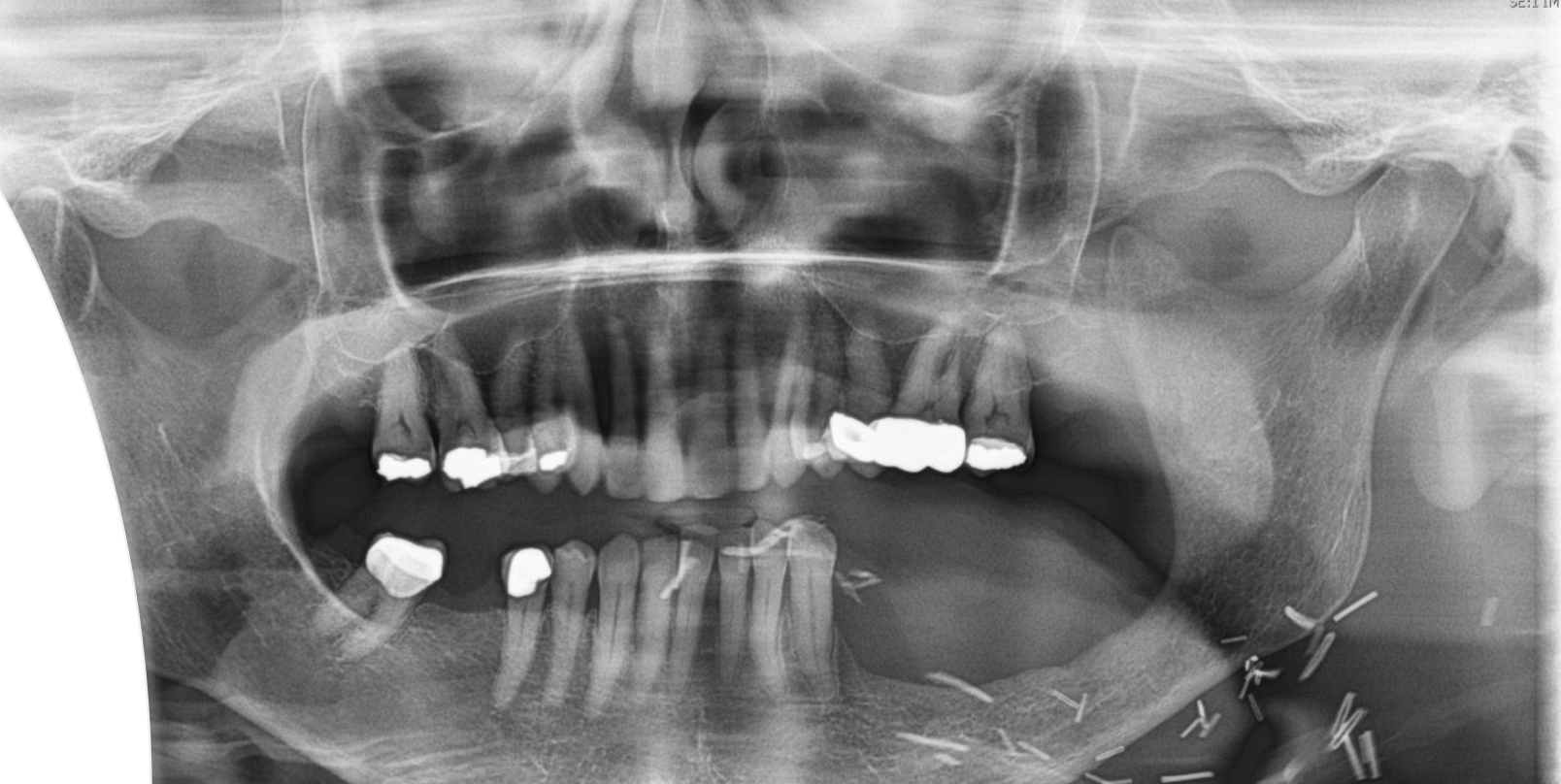
Case: Mandibular resection for STC from 2 to 2
Question
Scenario: A male patient who underwent resection of the anterior mandible (from tooth 2 to 2) for STC.
What’s shown: A close-up clinical photograph showing an open mouth, and a panoramic dental radiograph showing the maxilla and mandible.
Consider: How to restore the patient’s function and ability to chew again following the resection.
Answer
Observations:
- Resection of the mandible from tooth 2 to 2.
- Loss of anterior mandibular structure affecting oral function.
Reasoning:
- The patient had STC from 2 to 2, which required resection of that segment of the mandible.
- To improve his function and help him chew again, the clinician must evaluate restorative options, weighing a soft tissue graft against dentures or bone implants.
Outcome:
- The patient is planned to have a soft tissue graft, with further consideration for whether he will wear dentures or receive bone implants to restore his chewing function.
Takeaway:
- After mandibular resection for oral cancer, restoring function (such as chewing) requires evaluating and selecting appropriate reconstructive and prosthetic options like soft tissue grafts, dentures, or bone implants.
Footnotes
-
Original PDF page 1: W2 Oral Cancer Resection and Reconstruction - Foo, p.1 ↩
-
Original PDF page 2: W2 Oral Cancer Resection and Reconstruction - Foo, p.2 ↩
-
Original PDF page 3: W2 Oral Cancer Resection and Reconstruction - Foo, p.3 ↩
-
Original PDF page 5: W2 Oral Cancer Resection and Reconstruction - Foo, p.5 ↩
-
Original PDF page 4: W2 Oral Cancer Resection and Reconstruction - Foo, p.4 ↩
-
Original PDF page 6: W2 Oral Cancer Resection and Reconstruction - Foo, p.6 ↩
-
Original PDF page 7: W2 Oral Cancer Resection and Reconstruction - Foo, p.7 ↩
-
Original PDF page 8: W2 Oral Cancer Resection and Reconstruction - Foo, p.8 ↩
-
Original PDF page 9: W2 Oral Cancer Resection and Reconstruction - Foo, p.9 ↩
-
Original PDF page 10: W2 Oral Cancer Resection and Reconstruction - Foo, p.10 ↩
-
Original PDF page 11: W2 Oral Cancer Resection and Reconstruction - Foo, p.11 ↩
-
Original PDF page 12: W2 Oral Cancer Resection and Reconstruction - Foo, p.12 ↩
-
Original PDF page 13: W2 Oral Cancer Resection and Reconstruction - Foo, p.13 ↩
-
Original PDF page 14: W2 Oral Cancer Resection and Reconstruction - Foo, p.14 ↩
-
Original PDF page 15: W2 Oral Cancer Resection and Reconstruction - Foo, p.15 ↩
-
Original PDF page 16: W2 Oral Cancer Resection and Reconstruction - Foo, p.16 ↩
-
Original PDF page 17: W2 Oral Cancer Resection and Reconstruction - Foo, p.17 ↩
-
Original PDF page 18: W2 Oral Cancer Resection and Reconstruction - Foo, p.18 ↩
-
Original PDF page 19: W2 Oral Cancer Resection and Reconstruction - Foo, p.19 ↩
-
Original PDF page 20: W2 Oral Cancer Resection and Reconstruction - Foo, p.20 ↩
-
Original PDF page 21: W2 Oral Cancer Resection and Reconstruction - Foo, p.21 ↩
-
Original PDF page 22: W2 Oral Cancer Resection and Reconstruction - Foo, p.22 ↩
-
Original PDF page 23: W2 Oral Cancer Resection and Reconstruction - Foo, p.23 ↩
-
Original PDF page 25: W2 Oral Cancer Resection and Reconstruction - Foo, p.25 ↩
-
Original PDF page 26: W2 Oral Cancer Resection and Reconstruction - Foo, p.26 ↩
-
Original PDF page 27: W2 Oral Cancer Resection and Reconstruction - Foo, p.27 ↩
-
Original PDF page 29: W2 Oral Cancer Resection and Reconstruction - Foo, p.29 ↩
-
Original PDF page 30: W2 Oral Cancer Resection and Reconstruction - Foo, p.30 ↩
-
Original PDF page 31: W2 Oral Cancer Resection and Reconstruction - Foo, p.31 ↩
-
Original PDF page 32: W2 Oral Cancer Resection and Reconstruction - Foo, p.32 ↩
-
Original PDF page 33: W2 Oral Cancer Resection and Reconstruction - Foo, p.33 ↩
-
Original PDF page 34: W2 Oral Cancer Resection and Reconstruction - Foo, p.34 ↩
-
Original PDF page 35: W2 Oral Cancer Resection and Reconstruction - Foo, p.35 ↩
-
Original PDF page 36: W2 Oral Cancer Resection and Reconstruction - Foo, p.36 ↩
-
Original PDF page 37: W2 Oral Cancer Resection and Reconstruction - Foo, p.37 ↩
-
Original PDF page 38: W2 Oral Cancer Resection and Reconstruction - Foo, p.38 ↩
-
Original PDF page 39: W2 Oral Cancer Resection and Reconstruction - Foo, p.39 ↩
-
Original PDF page 40: W2 Oral Cancer Resection and Reconstruction - Foo, p.40 ↩
-
Original PDF page 41: W2 Oral Cancer Resection and Reconstruction - Foo, p.41 ↩
-
Original PDF page 42: W2 Oral Cancer Resection and Reconstruction - Foo, p.42 ↩
-
Original PDF page 43: W2 Oral Cancer Resection and Reconstruction - Foo, p.43 ↩
-
Original PDF page 44: W2 Oral Cancer Resection and Reconstruction - Foo, p.44 ↩
-
Original PDF page 45: W2 Oral Cancer Resection and Reconstruction - Foo, p.45 ↩
-
Original PDF page 46: W2 Oral Cancer Resection and Reconstruction - Foo, p.46 ↩
-
Original PDF page 47: W2 Oral Cancer Resection and Reconstruction - Foo, p.47 ↩
-
Original PDF page 48: W2 Oral Cancer Resection and Reconstruction - Foo, p.48 ↩
-
Original PDF page 49: W2 Oral Cancer Resection and Reconstruction - Foo, p.49 ↩
-
Original PDF page 50: W2 Oral Cancer Resection and Reconstruction - Foo, p.50 ↩
-
Original PDF page 51: W2 Oral Cancer Resection and Reconstruction - Foo, p.51 ↩
-
Original PDF page 52: W2 Oral Cancer Resection and Reconstruction - Foo, p.52 ↩